REVIEW 4 major objections 6 minor 50 references
ViG3D-UNet: Volumetric Vascular Connectivity-Aware Segmentation via 3D Vision Graph Representation
T0 review · 4 major / 6 minor · reviewed 2026-08-16 · deepseek-v4-flash
Pith's one-line read A 3D vision-graph U-Net produces coronary artery segmentations that stay connected while matching or beating four baselines on two CTA datasets.
desk verdict Architecturally plausible and honestly reported, but the connectivity headline is not actually measured: the evaluation uses precision and HD95 as proxies, and the gains over STUNet are within the reported noise. read the letter →
The pith
A machine-rendered reading of the paper's core claim, the machinery that carries it, and where it could break.
The reading
What carries the argument
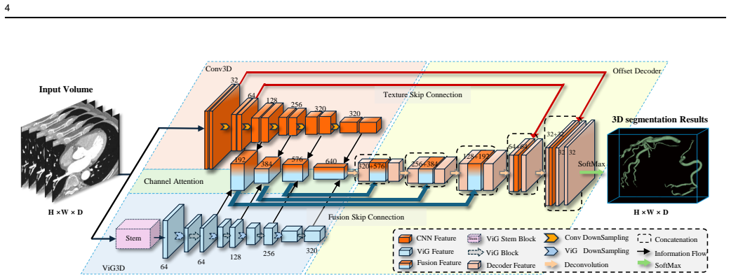
The load-bearing object is the ViG3D block, a volumetric graph-aggregation unit. A stem reduces an input patch to $\frac{H}{4} \times \frac{W}{4} \times \frac{D}{4}$, each voxel feature becomes a graph node, and each node is connected to its $K=7$ nearest neighbors; the block then applies max-relative graph convolution, $g(\cdot) = [x_i, \max(\{x_j - x_i \mid j \in \mathcal{N}(x_i)\})]$, followed by an MLP update and a feed-forward network, stacked in a pyramid of 2, 4, 16, and 2 blocks across scales. The graph aggregation is what transmits same-branch information between nearby vascular voxels, while the parallel CNN branch supplies texture; channel attention fuses the two, and the paperclip-shaped offset decoder concatenates early CNN texture features into the last decoder layers and fusion features into the first four layers to avoid redundant 3D computation.
What would settle it
Compute the number of connected components, the fraction of ground-truth branch endpoints covered by the prediction, or the centerline continuity on the ASOCA and ImageCAS test folds; if ViG3D-UNet does not improve these direct measures over the baselines at matched thresholds, the central connectivity claim is falsified.
Extended reading notes
Core claim
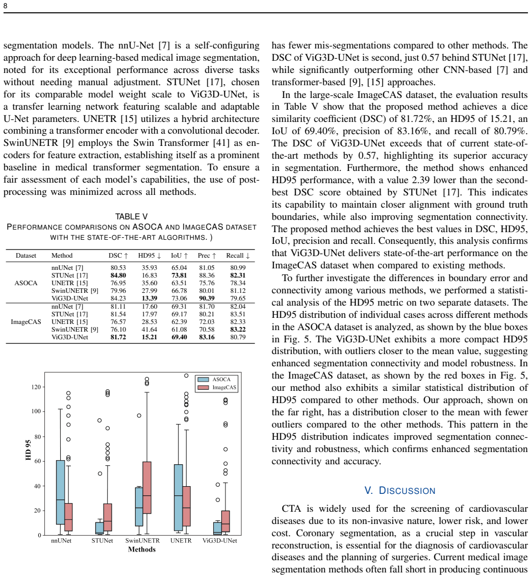
In the paper's own terms, the central discovery is that embedding a 3D Vision GNN module as one branch of a parallel encoder, fusing its topological features with CNN texture features through channel attention, and decoding with a paperclip-shaped offset decoder produces volumetric coronary artery segmentations that maintain connectivity across branches and at endpoints better than standard CNN and transformer baselines. The evidence is the score table: on ImageCAS the method has the best DSC, HD95, IoU, precision, and recall among the four compared methods, with DSC 81.72 against 81.54 for the strongest baseline and HD95 15.21 against 17.60 for the self-configuring U-Net baseline; on ASOCA it has the best HD95 and precision and second-best DSC (84.23, within 0.57 of the strongest baseline). The authors interpret the lower HD95 and higher precision, together with qualitative figures showing fewer breaks, as evidence of superior connectivity.
Load-bearing premise
The connectivity claim rests on treating precision and HD95 as measures of vascular connectivity; if those two metrics do not track true branch continuity, the paper has not actually shown better connectivity.
Editorial extensions
If this is right
- On ImageCAS, ViG3D-UNet reports the best DSC (81.72), HD95 (15.21), IoU (69.40), precision (83.16), and recall (80.79) among the four compared baselines.
- On ASOCA, it reports the best HD95 (13.39) and precision (90.39) and second-best DSC (84.23), suggesting the graph prior helps when training data are scarce.
- Ablation studies attribute the gain to all three components: removing the ViG3D module lowers DSC by 0.61 and raises HD95 by 2.39, removing channel attention lowers DSC by 0.51, and removing the offset decoder lowers DSC by 1.16 and raises HD95 by 3.13.
- Because the pipeline is end-to-end, it avoids the two-stage graph-construction-then-refinement cascade of earlier graph-based vessel segmentation, which should simplify training.
Reading between the lines
- A direct topological evaluation—connected-component count, fraction of ground-truth branch endpoints recovered, or centerline continuity—would be a sharper test of the connectivity claim, and the paper does not report such metrics.
- If the connectivity gain is real, the same graph-in-U-Net design should transfer to other sparse tubular structures such as airways, retinal vessels, and cerebral arteries, since the paper tests only coronary arteries.
- The optimal neighbor count $K=7$ and the stacked ViG3D configuration were tuned on ImageCAS; a testable extension is whether the best $K$ scales with voxel anisotropy or branch thickness across datasets.
- The lower standard deviation on the small ASOCA dataset hints that graph aggregation regularizes the model under limited training data, which could be checked by training on progressively smaller training subsets.
Signed reviews
Editorial analysis
A structured set of objections, weighed in public.
Referee Report
Summary. The paper proposes ViG3D-UNet, a 3D U-shaped segmentation network whose encoder combines a 3D CNN branch with a 3D vision graph neural network (ViG3D) branch, fuses the two feature streams with channel attention, and employs a 'paperclip-shaped' offset decoder. The method is evaluated on two public coronary CTA datasets, ASOCA and ImageCAS, against nnU-Net, STUNet, UNETR, and SwinUNETR. The authors claim that ViG3D-UNet achieves state-of-the-art accuracy and, as the central contribution, superior vascular connectivity, supported by precision, HD95, and visual examples. Ablation studies examine the graph neighbor count K, the number of ViG3D blocks per scale, and the contribution of each architectural module.
Significance. If the connectivity claim were established, this would be a useful empirical contribution: it offers an end-to-end graph-based alternative to two-stage graph segmentation pipelines and reports competitive accuracy on two public datasets against strong baselines. The architectural idea is clearly presented, and the use of public datasets plus a fair baseline setup is commendable. However, the paper's central claim—connectivity improvement—is supported only by proxy metrics, the reported accuracy gains over the strongest baseline are small, and no significance testing is provided. With direct topological metrics and proper statistical evaluation, the work could be a solid contribution; in its current form the headline claim is overstated.
major comments (4)
- [Section IV.A.3 and Section IV.D, Table V] The central claim that ViG3D-UNet 'surpassed competing methods in maintaining vascular segmentation connectivity' is not supported by the reported metrics. Precision (Eq. 11) counts voxel-level false positives, and HD95 (Eq. 9) measures surface distance; neither quantifies topology. A prediction can be highly precise and have small HD95 while still containing broken branches, disconnected components, or missing distal segments. The lower recall of ViG3D-UNet relative to STUNet on both datasets (ASOCA 79.65 vs 82.31; ImageCAS 80.79 vs 83.51) is consistent with omitted branches, which is a direct connectivity failure. I request direct connectivity metrics over the full test sets: number of connected components per coronary tree, fraction of ground-truth branch endpoints covered, centerline overlap or Dice on centerlines, and/or Betti-number error. The anecdotal visual evidence in Figures 6 and 7 should also be replaced or supplemented by these quantitative topology measurements.
- [Section IV.D, Table V and Fig. 5] No variance or significance testing is reported for the comparison against baselines. Table II shows the proposed method's standard deviations are large relative to the reported differences (e.g., ImageCAS DSC 81.72 ± 5.09 vs STUNet 81.54; HD95 15.21 ± 18.53 vs STUNet 17.97). Since the improvements over the strongest baseline are 0.57 DSC and 2.39 HD95 on ImageCAS, the reader cannot determine whether these differences are statistically reliable. Please report per-fold or per-case results and apply paired tests (e.g., Wilcoxon signed-rank or paired t-test on per-case DSC and HD95) across the five folds or on the test set, and make the figures show these comparisons explicitly.
- [Section IV.A.2 and Section IV.C] The hyperparameters K=7 and the per-scale ViG3D block counts (2, 4, 16, 2) are selected by experiments on the ImageCAS dataset, which is also the dataset used for the final head-to-head comparison. This makes the reported ImageCAS numbers a selection outcome rather than an unbiased evaluation. The authors should either perform nested validation on ImageCAS or demonstrate that the same hyperparameters are optimal on ASOCA before claiming generalizable connectivity improvement. Additionally, the paper's own limitation—'a blind search of feature distances is involved in the current spatial graph representation'—acknowledges that the graph construction is not informed by vessel structure, which weakens the mechanistic claim that the graph module specifically encodes vascular connectivity.
- [Section IV.A.1 and Section IV.A.2] The experimental protocol is described inconsistently. Section IV.A.1 states that ASOCA is 'strategically divided into 30 samples for training and 10 samples for testing' and ImageCAS into 800/200, while Section IV.A.2 says 'A five-fold cross-validation strategy was used for experiments on both datasets.' These statements cannot both describe the identical protocol. Please clarify whether the reported numbers are from a fixed split or from an aggregation of five folds, and if five-fold cross-validation was used, describe how the folds were constructed and why the dataset descriptions mention fixed splits.
minor comments (6)
- [Table V] The column header 'Recall↓' is incorrect; recall is a higher-is-better metric and should be marked with an upward arrow.
- [Section III.A, after Eq. (3)] In the sentence 'For each voxel vi, its neighboring voxels N (vj) were identified using the K-nearest neighbors method,' the notation N(vj) should be N(vi).
- [Section IV.C] The text says the impact of different numbers of ViG3D blocks is shown in Table IV, but the ViG3D unit scaling results are in Table III; Table IV contains the structural ablations.
- [Section IV.B] The sentence 'Experiments of a five-fold cross-validation strategy on the ASOCA and ImageCAS datasets, respectively' is grammatically incomplete.
- [Figures 6 and 7] The figure captions concatenate model names without spaces (e.g., 'nnUNet STU-Net Swin-UNETRUNETR ViG3D-UNetGT'); these should be separated for readability.
- [Table V title] The table title contains a stray closing parenthesis: 'WITH THE STATE-OF-THE-ART ALGORITHMS . )'.
Circularity Check
No circularity: all central claims rest on external benchmark evaluations; connectivity-proxy concerns are a validity issue, not a definitional reduction.
full rationale
ViG3D-UNet is an empirical architecture paper rather than a derivation. The central claim—superior vascular connectivity with high segmentation accuracy—is supported by five-fold cross-validated comparisons on two public datasets (ASOCA and ImageCAS) against external baselines (nnU-Net, STUNet, UNETR, SwinUNETR). The network components are built from standard graph-convolution and U-Net operations, and the reported metrics (DSC, IoU, precision, recall, ASSD, HD95) are defined independently of the method's inputs. No equation in the paper defines an input parameter in terms of an output metric, and no fitted parameter is later renamed as a prediction: the K and L ablations are hyperparameter selection, and the final comparison is an external benchmark result. The one overlapping-author citation ([20], used to support the general statement that CNN-based networks have improved segmentation accuracy) is not load-bearing for any of the paper's claims, so it does not constitute circularity under the stated rules. The concern that precision and HD95 are imperfect proxies for true vascular connectivity is a construct-validity or correctness issue, not a circularity reduction: the paper does not define connectivity as precision or HD95, nor define precision/HD95 in terms of connectivity. The paper's own limitations (small training sets; 'a blind search of feature distances' in graph construction) further indicate that the results are contingent empirical findings rather than consequences of the method's definitions. No self-definitional, fitted-input-called-prediction, self-citation-load-bearing, uniqueness-imported, ansatz-smuggled, or renaming step is present.
Assumptions & free parameters
free parameters (2)
- K-nearest neighbors K =
7
- ViG3D block counts per scale =
2, 4, 16, 2
assumptions (3)
- domain assumption Vascular topology can be captured by K-nearest-neighbor graphs built on dense voxel features.
- domain assumption Precision and HD95 are meaningful proxies for vascular connectivity.
- domain assumption All compared models are trained under equivalent protocols, making the comparisons fair.
Cite this review
Pith. "Pith review of ViG3D-UNet: Volumetric Vascular Connectivity-Aware Segmentation via 3D Vision Graph Representation." pith.science (2026). https://pith.science/paper/HXACCKKT
@misc{pith2026250413599,
  author       = {Pith},
  title        = {Pith review of: ViG3D-UNet: Volumetric Vascular Connectivity-Aware Segmentation via 3D Vision Graph Representation},
  year         = {2026},
  howpublished = {\url{https://pith.science/paper/HXACCKKT}},
  note         = {Machine review of arXiv:2504.13599}
}
read the original abstract
Accurate vascular segmentation is essential for coronary visualization and the diagnosis of coronary heart disease. This task involves the extraction of sparse tree-like vascular branches from the volumetric space. However, existing methods have faced significant challenges due to discontinuous vascular segmentation and missing endpoints. To address this issue, a 3D vision graph neural network framework, named ViG3D-UNet, was introduced. This method integrates 3D graph representation and aggregation within a U-shaped architecture to facilitate continuous vascular segmentation. The ViG3D module captures volumetric vascular connectivity and topology, while the convolutional module extracts fine vascular details. These two branches are combined through channel attention to form the encoder feature. Subsequently, a paperclip-shaped offset decoder minimizes redundant computations in the sparse feature space and restores the feature map size to match the original input dimensions. To evaluate the effectiveness of the proposed approach for continuous vascular segmentation, evaluations were performed on two public datasets, ASOCA and ImageCAS. The segmentation results show that the ViG3D-UNet surpassed competing methods in maintaining vascular segmentation connectivity while achieving high segmentation accuracy. Our code will be available soon.
Figures


Figures from the paper (4 more)



Reference graph
Works this paper leans on
-
[1]
Datasets and Description: The Automated Segmentation of Coronary Arteries (ASOCA) dataset, used in the MICCAI- 2020 Challenge [38], is a publicly accessible dataset tailored TABLE I DETAILED SETTINGS OF PARALLELIZED ENCODER Layer Output Size CNN Module ViG3D Module Enc 1 H 2 × W 2 × D 2 Conv3d C = 32 S = 1 Downsampling Conv3d C = 32 S = 2 × 1 Enc 2 H 4 × ...
work page 2020
-
[2]
Implementation Details: The optimal hyperparameters for the neighbor nodes in the ViG3D module were determined by primarily evaluating the performance of the method while adjusting the number of nodes for graph representation. Fur- thermore, the structure of ViG3D was verified through ablation studies, with specific details provided in the following secti...
-
[3]
The DSC metrics reflect how accurately the model captures coronary structures
Performance Metrics: The Dice Similarity Coefficient (DSC) is employed to measures the accuracy of segmentation results by evaluating the overlap between predicted and actual segmentation maps, defined as: DSC = 2×TP 2×TP +FP +FN , (7) where TP (True Positive) represents the number of pixels correctly identified as part of the coronary class, FP (False Po...
-
[4]
3d graph-connectivity constrained network for hepatic vessel segmen- tation,
R. Li, Y .-J. Huang, H. Chen, X. Liu, Y . Yu, D. Qian, and L. Wang, “3d graph-connectivity constrained network for hepatic vessel segmen- tation,” IEEE Journal of Biomedical and Health Informatics , vol. 26, no. 3, pp. 1251–1262, 2022
work page 2022
-
[5]
U-net: Convolutional networks for biomedical image segmentation,
O. Ronneberger, P. Fischer, and T. Brox, “U-net: Convolutional networks for biomedical image segmentation,” in Medical image computing and computer-assisted intervention–MICCAI 2015: 18th international con- ference, Munich, Germany, October 5-9, 2015, proceedings, part III 18 . Springer, 2015, pp. 234–241
2015
-
[6]
A review of deep-learning-based medical image segmentation methods,
X. Liu, L. Song, S. Liu, and Y . Zhang, “A review of deep-learning-based medical image segmentation methods,” Sustainability, vol. 13, no. 3, p. 1224, 2021
2021
-
[7]
nnu-net: a self-configuring method for deep learning-based biomedical image segmentation,
F. Isensee, P. F. Jaeger, S. A. Kohl, J. Petersen, and K. H. Maier-Hein, “nnu-net: a self-configuring method for deep learning-based biomedical image segmentation,” Nature methods, vol. 18, no. 2, pp. 203–211, 2021
2021
-
[9]
G. A. Roth, D. Abate, K. H. Abate, S. M. Abay, C. Abbafati, N. Abbasi, H. Abbastabar, F. Abd-Allah, J. Abdela, A. Abdelalim et al. , “Global, regional, and national age-sex-specific mortality for 282 causes of death in 195 countries and territories, 1980–2017: a systematic analysis for the global burden of disease study 2017,” The lancet, vol. 392, no. 10...
work page 1980
Show all 50 references
-
[10]
indicates that segmentation performance is significantly improved by utilizing the geometric structure of the target object. Recently, the achievements of large language models in medical image segmentation [11] have led to an enhancement in the ability of segmentation models ...
-
[11]
Cooper, J
R. Cooper, J. Cutler, P. Desvigne-Nickens, S. P. Fortmann, L. Friedman, R. Havlik, G. Hogelin, J. Marler, P. McGovern, G. Morosco et al. , “Trends and disparities in coronary heart disease, stroke, and other cardiovascular diseases in the united states: findings of the nationa...
2000
-
[12]
These limitations are exacerbated by conventional models’ inability to simul- taneously handle tubular geometry priors and microvascular contrast variations
, and (3) ill-defined boundaries [13]. These limitations are exacerbated by conventional models’ inability to simul- taneously handle tubular geometry priors and microvascular contrast variations. First, the U-Net framework and its variants struggle to capture all low-level fe...
2025 arXiv
-
[13]
Computed tomographic angiography in coronary artery disease,
P. W. Serruys, N. Kotoku, B. L. Nørgaard, S. Garg, K. Nieman, M. R. Dweck, J. J. Bax, J. Knuuti, J. Narula, D. Perera et al. , “Computed tomographic angiography in coronary artery disease,” EuroIntervention, vol. 18, no. 16, p. e1307, 2023
2023
-
[14]
Uctransnet: rethinking the skip connections in u-net from a channel-wise perspective with trans- former,
H. Wang, P. Cao, J. Wang, and O. R. Zaiane, “Uctransnet: rethinking the skip connections in u-net from a channel-wise perspective with trans- former,” in Proceedings of the AAAI conference on artificial intelligence, vol. 36, no. 3, 2022, pp. 2441–2449
2022
-
[15]
Unetr: Transformers for 3d medical image segmentation,
A. Hatamizadeh, Y . Tang, V . Nath, D. Yang, A. Myronenko, B. Land- man, H. R. Roth, and D. Xu, “Unetr: Transformers for 3d medical image segmentation,” in Proceedings of the IEEE/CVF winter conference on applications of computer vision , 2022, pp. 574–584
2022
-
[16]
In CNN-based networks, deeper architectures
have become the mainstream method for medical image segmentation. In CNN-based networks, deeper architectures
-
[17]
The attention mechanism [19] was introduced for more effective aggregation of contextual information
and denser connections between layers [18] are employed to improve the performance of segmentation algorithms. The attention mechanism [19] was introduced for more effective aggregation of contextual information. Although CNN-based networks have improved segmentation accuracy ...
-
[18]
Swin unetr: Swin transformers for semantic segmentation of brain tumors in mri images,
A. Hatamizadeh, V . Nath, Y . Tang, D. Yang, H. R. Roth, and D. Xu, “Swin unetr: Swin transformers for semantic segmentation of brain tumors in mri images,” in International MICCAI Brainlesion Workshop. Springer, 2021, pp. 272–284
2021
-
[20]
Segment anything model for medical image segmentation: Current applications and future directions,
Y . Zhang, Z. Shen, and R. Jiao, “Segment anything model for medical image segmentation: Current applications and future directions,” Com- puters in Biology and Medicine , vol. 171, p. 108238, 2024
2024
-
[21]
An energy matching vessel segmentation framework in 3-d medical images,
P. Liu, G. Huang, J. Jing, S. Bian, L. Cheng, X. Y . Lu, C. Rao, Y . Liu, Y . Hua, Y . Wang, and K. He, “An energy matching vessel segmentation framework in 3-d medical images,” IEEE Transactions on Medical Imaging, vol. 43, no. 4, pp. 1476–1488, 2024
2024
-
[22]
Improved topological preservation in 3d axon segmentation and centerline detec- tion using geometric assessment-driven topological smoothing (gats),
N. I. Shamsi, A. S. Xu, L. A. Gjesteby, and L. J. Brattain, “Improved topological preservation in 3d axon segmentation and centerline detec- tion using geometric assessment-driven topological smoothing (gats),” in Proceedings of the IEEE/CVF Winter Conference on Applications o...
2024
-
[23]
Coronary artery centerline extraction in cardiac ct angiography using a cnn-based orientation classifier,
J. M. Wolterink, R. W. van Hamersvelt, M. A. Viergever, T. Leiner, and I. I ˇsgum, “Coronary artery centerline extraction in cardiac ct angiography using a cnn-based orientation classifier,” Medical image analysis, vol. 51, pp. 46–60, 2019
2019
-
[24]
3d u-net: learning dense volumetric segmentation from sparse annotation,
¨O. C ¸ ic ¸ek, A. Abdulkadir, S. S. Lienkamp, T. Brox, and O. Ron- neberger, “3d u-net: learning dense volumetric segmentation from sparse annotation,” in Medical Image Computing and Computer-Assisted Intervention–MICCAI 2016: 19th International Conference, Athens, Greece, Oc...
2016
-
[25]
Stu-net: Scalable and transferable medical image segmentation models empowered by large-scale supervised pre-training,
Z. Huang, H. Wang, Z. Deng, J. Ye, Y . Su, H. Sun, J. He, Y . Gu, L. Gu, S. Zhang et al., “Stu-net: Scalable and transferable medical image segmentation models empowered by large-scale supervised pre-training,” arXiv preprint arXiv:2304.06716 , 2023
2023 arXiv
-
[26]
Unet++: Redesigning skip connections to exploit multiscale features in image segmentation,
Z. Zhou, M. M. R. Siddiquee, N. Tajbakhsh, and J. Liang, “Unet++: Redesigning skip connections to exploit multiscale features in image segmentation,” IEEE transactions on medical imaging , vol. 39, no. 6, pp. 1856–1867, 2019
2019
-
[27]
Adaptive context selection for polyp segmentation,
R. Zhang, G. Li, Z. Li, S. Cui, D. Qian, and Y . Yu, “Adaptive context selection for polyp segmentation,” in Medical Image Computing and Computer Assisted Intervention–MICCAI 2020: 23rd International Conference, Lima, Peru, October 4–8, 2020, Proceedings, Part VI 23 . Springer...
2020
-
[28]
An improved u-shape neural network for soft exudate segmentation,
H. Zhang, K. Lin, Y . Guan, Z. Gan, and C. Ouyang, “An improved u-shape neural network for soft exudate segmentation,” in Fourteenth International Conference on Digital Image Processing (ICDIP 2022) , vol. 12342. SPIE, 2022, pp. 395–401
2022
-
[29]
An image is worth 16x16 words: Transformers for image recognition at scale,
A. Dosovitskiy, L. Beyer, A. Kolesnikov, D. Weissenborn, X. Zhai, T. Unterthiner, M. Dehghani, M. Minderer, G. Heigold, and S. Gelly, “An image is worth 16x16 words: Transformers for image recognition at scale,” in International Conference on Learning Representations , 2021
2021
-
[30]
Medical transformer: Gated axial-attention for medical image segmentation,
J. M. J. Valanarasu, P. Oza, I. Hacihaliloglu, and V . M. Patel, “Medical transformer: Gated axial-attention for medical image segmentation,” in Medical Image Computing and Computer Assisted Intervention–MICCAI 2021: 24th International Conference, Strasbourg, France, September...
2021
-
[31]
Exfuse: Enhancing feature fusion for semantic segmentation,
Z. Zhang, X. Zhang, C. Peng, X. Xue, and J. Sun, “Exfuse: Enhancing feature fusion for semantic segmentation,” Springer, Cham, 2018
2018
-
[32]
Ori- net: Orientation-guided neural network for automated coronary arteries segmentation,
W. Jiang, Y . Li, Y . Jia, Y . Feng, Z. Yi, M. Chen, and J. Wang, “Ori- net: Orientation-guided neural network for automated coronary arteries segmentation,” Expert Systems with Applications , vol. 238, p. 121905, 2024
2024
-
[33]
Deep distance transform for tubular structure segmentation in ct scans,
Y . Wang, X. Wei, F. Liu, J. Chen, Y . Zhou, W. Shen, E. K. Fishman, and A. L. Yuille, “Deep distance transform for tubular structure segmentation in ct scans,” in Proceedings of the IEEE/CVF Conference on Computer Vision and Pattern Recognition , 2020, pp. 3833–3842. 11
2020
-
[34]
Cs2-net: Deep learning segmentation of curvilinear structures in medical imaging,
L. Mou, Y . Zhao, H. Fu, Y . Liu, J. Cheng, Y . Zheng, P. Su, J. Yang, L. Chen, A. F. Frangi et al. , “Cs2-net: Deep learning segmentation of curvilinear structures in medical imaging,” Medical image analysis , vol. 67, p. 101874, 2021
2021
-
[35]
Vision gnn: An image is worth graph of nodes,
K. Han, Y . Wang, J. Guo, Y . Tang, and E. Wu, “Vision gnn: An image is worth graph of nodes,” Advances in neural information processing systems, vol. 35, pp. 8291–8303, 2022
2022
-
[36]
Graph-based region and boundary aggregation for biomedical image segmentation,
Y . Meng, H. Zhang, Y . Zhao, X. Yang, Y . Qiao, I. J. MacCormick, X. Huang, and Y . Zheng, “Graph-based region and boundary aggregation for biomedical image segmentation,” IEEE transactions on medical imaging, vol. 41, no. 3, pp. 690–701, 2021
2021
-
[37]
A joint 3d unet-graph neural network-based method for airway segmen- tation from chest cts,
A. Garcia-Uceda Juarez, R. Selvan, Z. Saghir, and M. de Bruijne, “A joint 3d unet-graph neural network-based method for airway segmen- tation from chest cts,” in Machine Learning in Medical Imaging: 10th International Workshop, MLMI 2019, Held in Conjunction with MICCAI 2019, ...
2019
-
[38]
Graph convolution based cross-network multiscale feature fusion for deep vessel segmentation,
G. Zhao, K. Liang, C. Pan, F. Zhang, X. Wu, X. Hu, and Y . Yu, “Graph convolution based cross-network multiscale feature fusion for deep vessel segmentation,” IEEE Transactions on Medical Imaging , vol. 42, no. 1, pp. 183–195, 2022
2022
-
[39]
Imagecas: A large-scale dataset and benchmark for coronary artery segmentation based on computed tomography angiography images,
A. Zeng, C. Wu, G. Lin, W. Xie, J. Hong, M. Huang, J. Zhuang, S. Bi, D. Pan, N. Ullah et al. , “Imagecas: A large-scale dataset and benchmark for coronary artery segmentation based on computed tomography angiography images,” Computerized Medical Imaging and Graphics, vol. 109,...
2023
-
[40]
was applied to the last five decoders, with the loss weights successively halved from Decoder 6 to Decoder 2
-
[41]
Feature fusion for segmentation and classification of skin lesions,
Y . Zhang, Z. Chen, H. Yu, X. Yao, and H. Li, “Feature fusion for segmentation and classification of skin lesions,” in 2022 IEEE 19th international symposium on biomedical imaging (ISBI) . IEEE, 2022, pp. 1–5
2022
-
[42]
Scs-net: A scale and context sensitive network for retinal vessel segmentation,
H. Wu, W. Wang, J. Zhong, B. Lei, Z. Wen, and J. Qin, “Scs-net: A scale and context sensitive network for retinal vessel segmentation,” Medical Image Analysis , vol. 70, p. 102025, 2021. [Online]. Available: https://www.sciencedirect.com/science/article/pii/S1361841521000712
2021
-
[43]
Vigu: Vi- sion gnn u-net for fast mri,
J. Huang, A. I. Aviles-Rivero, C.-B. Sch ¨onlieb, and G. Yang, “Vigu: Vi- sion gnn u-net for fast mri,” in 2023 IEEE 20th International Symposium on Biomedical Imaging (ISBI) . IEEE, 2023, pp. 1–5
2023
-
[44]
Deepgcns: Can gcns go as deep as cnns?
G. Li, M. Muller, A. Thabet, and B. Ghanem, “Deepgcns: Can gcns go as deep as cnns?” in Proceedings of the IEEE/CVF international conference on computer vision , 2019, pp. 9267–9276
2019
-
[45]
Gaussian error linear units (gelus),
D. Hendrycks and K. Gimpel, “Gaussian error linear units (gelus),” arXiv preprint arXiv:1606.08415, 2016
2016 arXiv
-
[46]
Instance normalization: The missing ingredient for fast stylization,
D. Ulyanov, A. Vedaldi, and V . Lempitsky, “Instance normalization: The missing ingredient for fast stylization,”arXiv preprint arXiv:1607.08022, 2016
2016 arXiv
-
[47]
Automated segmen- tation of normal and diseased coronary arteries–the asoca challenge,
R. Gharleghi, D. Adikari, K. Ellenberger, S.-Y . Ooi, C. Ellis, C.-M. Chen, R. Gao, Y . He, R. Hussain, C.-Y . Leeet al., “Automated segmen- tation of normal and diseased coronary arteries–the asoca challenge,” Computerized Medical Imaging and Graphics, vol. 97, p. 102049, 2022
2022
-
[48]
Deeply-supervised nets,
C.-Y . Lee, S. Xie, P. Gallagher, Z. Zhang, and Z. Tu, “Deeply-supervised nets,” in Artificial intelligence and statistics . Pmlr, 2015, pp. 562–570
2015
-
[49]
Swin transformer: Hierarchical vision transformer using shifted windows,
Z. Liu, Y . Lin, Y . Cao, H. Hu, Y . Wei, Z. Zhang, S. Lin, and B. Guo, “Swin transformer: Hierarchical vision transformer using shifted windows,” in Proceedings of the IEEE/CVF international conference on computer vision, 2021, pp. 10 012–10 022
2021
-
[50]
Innovations in cardiac computed tomography: Imaging in coronary artery disease,
V . S. Manubolu, K. Ichikawa, and M. J. Budoff, “Innovations in cardiac computed tomography: Imaging in coronary artery disease,” Progress in Cardiovascular Diseases, 2024
2024
-
[51]
Non-invasive imaging of the coronary arteries,
P. D. Adamson and D. E. Newby, “Non-invasive imaging of the coronary arteries,” European Heart Journal , vol. 40, no. 29, pp. 2444–2454, 11 2018
2018
-
[52]
Fractal analysis of the vascular tree in the human retina
Masters and R. Barry, “Fractal analysis of the vascular tree in the human retina.” Annual Review of Biomedical Engineering , vol. 6, no. 1, pp. 427–452, 2004
2004
Reviewed August 16, 2026 · model on record in the stance chip above.
Discussion (0). Continue with ORCID to comment.