REVIEW 4 major objections 5 minor 30 references
A Novel Data Augmentation Tool for Enhancing Machine Learning Classification: A New Application of the Higher Order Dynamic Mode Decomposition for Improved Cardiac Disease Identification
T0 review · 4 major / 5 minor · reviewed 2026-08-12 · deepseek-v4-flash
Pith's one-line read A CNN trained on original echocardiography frames plus HODMD modes classifies unseen videos up to 22 points more accurately than one trained on frames alone.
desk verdict The idea is real but the headline comparison is confounded: no control for the 10% larger training set means the claimed HODMD benefit on unseen videos is not established. read the letter →
The pith
A machine-rendered reading of the paper's core claim, the machinery that carries it, and where it could break.
The reading
What carries the argument
The engine of the method is the higher order dynamic mode decomposition (HODMD), an extension of dynamic mode decomposition that represents a video as a sum of modes $v(x,t_k)\simeq\sum_{m=1}^{M}a_m u_m e^{(\delta_m+i\omega_m)t_k}$, where $u_m$ are spatial modes, $\omega_m$ frequencies, $\delta_m$ growth rates, and $a_m$ amplitudes. Dimension reduction via singular value decomposition is followed by a DMD-d step based on Takens delay embedding, yielding the modes that are then treated as images. The paper uses these modes as additional CNN training samples; the mechanism proposed is that the mode images expose underlying motion and structure that raw frames hide, forcing the network to learn more transferable features.
What would settle it
Train the identical CNN on 7700 original echocardiography images (the original 7000 plus 1000 additional original frames drawn from the same training samples) and test on the same 2700-image hold-out videos; if the accuracy matches or nearly matches the 67% and 74% obtained with DMD-mode augmentation, the modes are not the causal ingredient. A second check is to compare the confusion matrices on the hold-out set, since DMD modes specifically should alter which classes are confused.
Extended reading notes
Core claim
The central claim is that HODMD modes encode disease-specific dynamical structure in echocardiography sequences, and that training on these modes in addition to raw frames makes the CNN generalize to new subjects. The paper compares two training sets: dataset 01 with 7000 original images and dataset 02 with 7700 images comprising the same original frames plus 1000 selected DMD modes. Across all testcases the augmented model does better, most decisively on the unseen hold-out set, where accuracy moves from 42% to 67% for five classes and from 52% to 74% for four classes. The authors attribute the gain to the modes displaying enhanced patterns and details not perceptible in the original frames, and they report that increasing the number of modes beyond ten produced no additional improvement, which they interpret as evidence that mode quality, not quantity, is what matters.
Load-bearing premise
The load-bearing premise is that the accuracy jump comes from the DMD modes themselves rather than from having 1000 extra training images: the paper compares 7000 original frames against 7700 frames (7000 original plus 1000 modes) without a control trained on 7700 original frames.
Editorial extensions
If this is right
- On unseen hold-out videos, five-class accuracy rises from 42% to 67% and four-class accuracy from 52% to 74% when DMD modes are added.
- Validation accuracy stays essentially flat (99% to 98%) while hold-out accuracy jumps, suggesting the modes improve generalization rather than training fit.
- A small labeled dataset of 130 videos can be augmented with about 10% extra modal images without new data collection.
- The same HODMD-extraction and augmentation pipeline could extend to other temporal medical image modalities, since the method is data-driven and does not require disease-specific handcrafted features.
- The paper reports that mode quality rather than mode count drives the gain: more than ten modes per sample did not significantly help.
Reading between the lines
- Editorial inference: the reported comparison does not control for training-set size, so part of the gain may be a simple 10% increase in data; training a control model on 7700 original frames would isolate the contribution of the DMD modes.
- Editorial inference: the mode-selection step is based on visual inspection of pattern quality, amplitude, and noise; a quantitative selection rule would make the augmentation reproducible and testable across datasets.
- Editorial inference: since HODMD modes are computed from the same training videos, evaluating on videos from different centers or imaging systems would clarify whether the modes capture disease physiology or dataset-specific artifacts.
- Editorial inference: the success of this approach suggests a broader class of spectral or modal feature augmentation for small medical datasets; comparing HODMD against PCA modes or wavelet features would reveal how much of the gain is specific to the dynamical decomposition.
Signed reviews
Editorial analysis
A structured set of objections, weighed in public.
Referee Report
Summary. The manuscript proposes using higher order dynamic mode decomposition (HODMD) to extract DMD modes from echocardiography videos and then using those modes as additional training images for a CNN that classifies five cardiac conditions: healthy, diabetic cardiomyopathy, obesity, myocardial infarction, and TAC hypertrophy. The authors compare two testcases: training the CNN on 7,000 original frames versus training it on 7,000 original frames plus 1,000 DMD-mode images, and they repeat the comparison after excluding the healthy class. Accuracy is reported on a test split of the original dataset and on 2,700 frames from 30 held-out videos (testing I). The paper concludes that combining original images with DMD modes improves classification accuracy by up to 22%.
Significance. The core suggestion, that modal-decomposition features can serve as a data augmentation source when medical training data are scarce, is interesting and the authors take the commendable step of including a held-out-video test set. The four-class experiment is a useful robustness check. However, the present evidence does not isolate the contribution of HODMD modes: the augmented model is trained on 10% more images than the baseline, and the internal test set is composed of frames from the same training videos. These are correctable design issues rather than flaws in the method itself, but until they are fixed the headline accuracy gains are not established. I also note that no code or data are provided, which limits reproducibility.
major comments (4)
- [Section 3.1, Table 2] Testcase 01 is trained on 7,000 original images while testcase 02 is trained on 7,700 images (7,000 originals plus 1,000 DMD modes). The central comparison is therefore confounded: the higher accuracy on testing I (42% to 67% in the five-class experiment, 52% to 74% in the four-class experiment) could be produced by the additional 10% of training volume alone, without any DMD-specific effect. A control model trained on 7,700 original images, or a cross-validated comparison matched for training-set size, is required before the improvement can be attributed to HODMD.
- [Section 3.1, Figs. 3c-3f and 4c-4f] The 'testing' set in dataset 01 is extracted from the same 20 videos per class that supply the training and validation frames, because 100 successive frames from each sample are later split into training, validation, and testing. Consecutive frames from the same echocardiography sequence are nearly identical, so the reported internal testing accuracies (76% to 94% and 79% to 98%) are inflated by frame-level leakage and should not be used as evidence of generalization. Only the held-out-video data (testing I) give a valid generalization estimate, and that estimate still suffers from the training-set-size confound described above.
- [Section 4, Table 2] Testing I consists of 90 frames from each of only six videos per class, giving 30 videos in the five-class experiment and 24 in the four-class experiment. Reporting accuracy over 2,700 frames treats frames as independent observations when they are highly correlated within a video, and no confidence intervals or per-video accuracies are given. With this sample size, the 25-percentage-point improvement could be within run-to-run variability; repeated training runs and interval estimates are needed.
- [Section 3.2] The selection of DMD modes for augmentation is not a fixed algorithm: the authors state that modes were 'plotted and well observed' and that 'most representative modes (clear patterns), with high amplitudes and low noise levels' were chosen, and the number of modes per sample (10) was set accordingly. This manual, class-aware selection is not reproducible from the manuscript, and the concluding claim in Section 5 that quality rather than quantity of modes is the correct criterion relies on an unreported experiment. The preprocessing should be specified as a reproducible rule, such as a tolerance-based automatic selection, or the subjective component should be justified as part of the method.
minor comments (5)
- [Table 1] Table 1 is difficult to parse: the labels '4 classes classification' and '5 classes classification' appear as rows without values, and the healthy (H) row is listed even though Section 4.2 excludes healthy data from the four-class experiment. Please restructure the table to show the two experiments and their splits clearly.
- [Throughout] There are frequent typos and copyediting artifacts, including 'perfprmance' in the Figure 3 caption, 'di fferent' in the Introduction, 're presented' in Section 4.1, and 'the main scoop of this research' in Section 5, which should be 'main scope'.
- [Table 1 and text] The terminology is inconsistent: Table 1 uses 'TAC hypertension' while the rest of the paper says 'TAC hypertrophy'; please standardize the disease name.
- [Section 2.2] The hyperparameter tuning described in Section 2.2 reports only the learning rate; the search range and the final numbers of filters and dense units are not given, so the architecture choice is not fully reproducible.
- [Section 2.1, Eqs. (5) and (8)] The HODMD tolerances epsilon_SVD and epsilon_DMD in Eqs. (5) and (8) are never given numerical values, and the index d for the DMD-d algorithm is not reported, so the number of retained modes and the temporal embedding are not reproducible from the manuscript.
Circularity Check
No significant circularity found: the key result is evaluated on held-out videos, and the DMD modes are derived only from training data; the unequal training-set-size comparison is an experimental confound, not a circular derivation.
full rationale
The paper's central claim is that adding HODMD-derived modes to the training set improves CNN accuracy, with the headline evidence being the accuracy increase on testing I (held-out videos) from 42% to 67% for five classes and from 52% to 74% for four classes. The DMD modes are computed from the 20 training sequences per class (Section 3.2) and are added only to the training, validation, and testing partitions of dataset 02; the testing I videos are untouched by both training and mode extraction. Thus, the reported improvement on testing I is not forced by construction: no equation equates the predicted accuracy to a fitted parameter, and the HODMD tolerances and mode-selection criteria are not fit to the classification labels. The baseline and augmented experiments use the same CNN architecture and the same unseen testing I set. The main concern is the absence of a same-size control trained on 7,700 original images, which means the causal attribution of the gain to HODMD modes is not fully established; however, that is an experimental design limitation rather than a circularity. The self-citations, including Refs. [17] and [18], supply the HODMD algorithm and prior applications, not the classification result, and do not constitute a load-bearing self-citation chain. Therefore, no enumerated circular step is present.
Assumptions & free parameters
free parameters (3)
- Number of DMD modes per sample =
10
- HODMD tolerances (epsilon_SVD, epsilon_DMD)
- CNN hyperparameters =
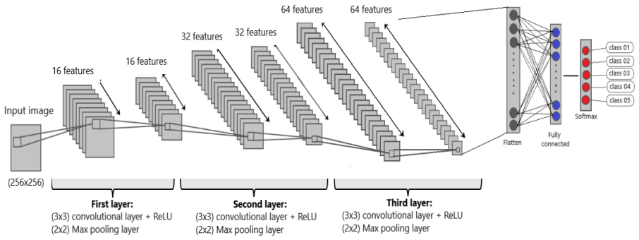
learning rate 1e-3, batch size 128, 3 conv layers
assumptions (3)
- domain assumption HODMD provides a valid modal decomposition of echocardiography sequences
- ad hoc to paper The manually selected DMD modes are representative disease-specific features
- domain assumption The CNN architecture described is sufficient for the classification task
Cite this review
Pith. "Pith review of A Novel Data Augmentation Tool for Enhancing Machine Learning Classification: A New Application of the Higher Order Dynamic Mode Decomposition for Improved Cardiac Disease Identification." pith.science (2026). https://pith.science/paper/I6WO4UEE
@misc{pith2026241115809,
  author       = {Pith},
  title        = {Pith review of: A Novel Data Augmentation Tool for Enhancing Machine Learning Classification: A New Application of the Higher Order Dynamic Mode Decomposition for Improved Cardiac Disease Identification},
  year         = {2026},
  howpublished = {\url{https://pith.science/paper/I6WO4UEE}},
  note         = {Machine review of arXiv:2411.15809}
}
read the original abstract
In this work, a data-driven, modal decomposition method, the higher order dynamic mode decomposition (HODMD), is combined with a convolutional neural network (CNN) in order to improve the classification accuracy of several cardiac diseases using echocardiography images. The HODMD algorithm is used first as feature extraction technique for the echocardiography datasets, taken from both healthy mice and mice afflicted by different cardiac diseases (Diabetic Cardiomyopathy, Obesity, TAC Hypertrophy and Myocardial Infarction). A total number of 130 echocardiography datasets are used in this work. The dominant features related to each cardiac disease were identified and represented by the HODMD algorithm as a set of DMD modes, which then are used as the input to the CNN. In a way, the database dimension was augmented, hence HODMD has been used, for the first time to the authors knowledge, for data augmentation in the machine learning framework. Six sets of the original echocardiography databases were hold out to be used as unseen data to test the performance of the CNN. In order to demonstrate the efficiency of the HODMD technique, two testcases are studied: the CNN is first trained using the original echocardiography images only, and second training the CNN using a combination of the original images and the DMD modes. The classification performance of the designed trained CNN shows that combining the original images with the DMD modes improves the results in all the testcases, as it improves the accuracy by up to 22%. These results show the great potential of using the HODMD algorithm as a data augmentation technique.
Figures


Figures from the paper (1 more)

Reference graph
Works this paper leans on
-
[1]
S. S. Virani, A. Alonso, H. J. Aparicio, E. J. Benjamin, M. S. Bitten- court, C. W. Callaway, A. P. Carson, A. M. Chamberlain, S. Cheng, F. N. Delling, et al., Heart disease and stroke statistics-2021 update: a report from the american heart association., Circulation 143 (8) (2021)
work page 2021
-
[2]
A. F. Parisi, P. F. Moynihan, E. D. Folland, C. L. Feldman, Quantitative detection of regional left ventricular contraction abnormalities by two- dimensional echocardiography. ii. accuracy in coronary artery disease., Circulation 63 (4) (1981) 761–767
work page 1981
-
[3]
K. Kusunose, T. Abe, A. Haga, D. Fukuda, H. Yamada, M. Harada, M. Sata, A deep learning approach for assessment of regional wall mo- tion abnormality from echocardiographic images, Cardiovascular Imag- ing 13 (2 Part 1) (2020) 374–381
work page 2020
-
[4]
A. Shalbaf, H. Behnam, Z. Alizade-Sani, M. Shojaifard, Automatic classification of left ventricular regional wall motion abnormalities in echocardiography images using nonrigid image registration, Journal of digital imaging 26 (2013) 909–919
work page 2013
-
[5]
X. Gao, W. Li, M. Loomes, L. Wang, A fused deep learning architecture for viewpoint classification of echocardiography, Information Fusion 36 (2017) 103–113
work page 2017
-
[6]
M. Yang, X. Xiao, Z. Liu, L. Sun, W. Guo, L. Cui, D. Sun, P. Zhang, G. Yang, Deep retinanet for dynamic left ventricle detection in multiview echocardiography classification, Scientific Programming 2020 (2020) 1– 6
work page 2020
- [7]
-
[8]
Y . Jang, Y . Hong, S. Ha, S. Kim, H.-J. Chang, Automatic segmentation of lv and rv in cardiac mri, in: Statistical Atlases and Computational Models of the Heart. ACDC and MMWHS Challenges: 8th International Work- shop, STACOM 2017, Held in Conjunction with MICCAI 2017, Que- bec City, Canada, September 10-14, 2017, Revised Selected Papers 8, Springer, 201...
work page 2017
Show all 30 references
-
[9]
Leclerc, E
S. Leclerc, E. Smistad, J. Pedrosa, A. Østvik, F. Cervenansky, F. Espinosa, T. Espeland, E. A. R. Berg, P.-M. Jodoin, T. Grenier, et al., Deep learning for segmentation using an open large-scale dataset in 2d echocardiogra- phy, IEEE transactions on medical imaging 38 (9) (201...
2019
-
[10]
J. M. Wolterink, T. Leiner, M. A. Viergever, I. I ˇsgum, Generative adver- sarial networks for noise reduction in low-dose ct, IEEE transactions on medical imaging 36 (12) (2017) 2536–2545
2017
-
[11]
E. Kang, H. J. Koo, D. H. Yang, J. B. Seo, J. C. Ye, Cycle-consistent adversarial denoising network for multiphase coronary ct angiography, Medical physics 46 (2) (2019) 550–562
2019
-
[12]
Green, E
M. Green, E. M. Marom, E. Konen, N. Kiryati, A. Mayer, 3-d neural de- noising for low-dose coronary ct angiography (ccta), Computerized Med- ical Imaging and Graphics 70 (2018) 185–191
2018
-
[13]
Wahlang, A
I. Wahlang, A. K. Maji, G. Saha, P. Chakrabarti, M. Jasinski, Z. Leonow- icz, E. Jasinska, Deep learning methods for classification of certain ab- normalities in echocardiography, Electronics 10 (4) (2021) 495
2021
-
[14]
Madani, J
A. Madani, J. R. Ong, A. Tibrewal, M. R. Mofrad, Deep echocardiogra- phy: data-efficient supervised and semi-supervised deep learning towards automated diagnosis of cardiac disease, NPJ digital medicine 1 (1) (2018) 59
2018
-
[15]
D.-Y . Tsai, Y . Lee, M. Sekiya, M. Ohkubo, Medical image classifica- tion using genetic-algorithm based fuzzy-logic approach, Journal of Elec- tronic Imaging 13 (4) (2004) 780–788
2004
-
[16]
Germain, A
P. Germain, A. Vardazaryan, N. Padoy, A. Labani, C. Roy, T. H. Schindler, S. El Ghannudi, Classification of cardiomyopathies from mr cine images using convolutional neural network with transfer learning, Diagnostics 11 (9) (2021) 1554
2021
-
[17]
Le Clainche, J
S. Le Clainche, J. M. Vega, Higher order dynamic mode decomposition, SIAM Journal on Applied Dynamical Systems 16 (2) (2017) 882–925
2017
-
[18]
Groun, M
N. Groun, M. Villalba-Orero, E. Lara-Pezzi, E. Valero, J. Garicano-Mena, S. Le Clainche, Higher order dynamic mode decomposition: From fluid dynamics to heart disease analysis, Computers in Biology and Medicine 144 (2022) 105384
2022
-
[19]
Groun, M
N. Groun, M. Villalba-Orero, E. Lara-Pezzi, E. Valero, J. Garicano-Mena, S. Le Clainche, A novel data-driven method for the analysis and recon- struction of cardiac cine mri, Computers in Biology and Medicine 151 (2022) 106317
2022
-
[20]
Groun, B
N. Groun, B. Begiashvili, E. Valero, J. Garicano-Mena, S. Le Clainche, Higher order dynamic mode decomposition beyond aerospace engineer- ing, Results in Engineering 20 (2023) 101471
2023
-
[21]
P. J. Schmid, Dynamic mode decomposition of numerical and experimen- tal data, Journal of fluid mechanics 656 (2010) 5–28
2010
-
[22]
F. Takens, Detecting strange attractors in turbulence, in: Dynamical Sys- tems and Turbulence, Warwick 1980: proceedings of a symposium held at the University of Warwick 1979/80, Springer, 2006, pp. 366–381
1980
-
[23]
J. M. Vega, S. Le Clainche, Higher order dynamic mode decomposition and its applications, Academic Press, 2020
2020
-
[24]
J.-G. Lee, S. Jun, Y .-W. Cho, H. Lee, G. B. Kim, J. B. Seo, N. Kim, Deep learning in medical imaging: general overview, Korean journal of radiology 18 (4) (2017) 570–584
2017
-
[25]
U. R. Acharya, S. L. Oh, Y . Hagiwara, J. H. Tan, H. Adeli, D. P. Subha, Automated eeg-based screening of depression using deep convolutional neural network, Computer methods and programs in biomedicine 161 (2018) 103–113
2018
-
[26]
Sharma, V
N. Sharma, V . Jain, A. Mishra, An analysis of convolutional neural net- works for image classification, Procedia computer science 132 (2018) 377–384
2018
-
[27]
L. Cai, J. Gao, D. Zhao, A review of the application of deep learning in medical image classification and segmentation, Annals of translational medicine 8 (11) (2020)
2020
-
[28]
Puttagunta, S
M. Puttagunta, S. Ravi, Medical image analysis based on deep learning approach, Multimedia tools and applications 80 (2021) 24365–24398
2021
-
[29]
MATLAB, 9.9.0.1538559 (R2020b) Update 3, The MathWorks Inc., Nat- ick, Massachusetts, 2020
2020
-
[30]
J. D. Novakovi ´c, A. Veljovi´c, S. S. Ili´c, ˇZ. Papi´c, M. Tomovi´c, Evaluation of classification models in machine learning, Theory and Applications of Mathematics & Computer Science 7 (1) (2017) 39. 12
2017
Reviewed August 12, 2026 · model on record in the stance chip above.
Discussion (0). Continue with ORCID to comment.