GLeVE: Graph-Guided Lesion Grounding with Proposal Verification in 3D CT
Pith reviewed 2026-05-22 06:38 UTC · model grok-4.3
The pith
GLeVE aligns each free-text lesion description to its exact location in a 3D CT scan by building a relation graph and verifying proposals against anatomy.
A machine-rendered reading of the paper's core claim, the machinery that carries it, and where it could break.
Core claim
GLeVE encodes each lesion description as an atomic semantic unit and runs relation-aware graph reasoning over organ attribution, attributes, and inter-lesion connections to create discriminative queries. Anatomy-aware proposal generation with region-level verification enforces strict one-to-one text-lesion alignment. Hierarchical octree refinement then progressively refines boundary delineation, yielding measurable improvements in segmentation and localization on AbdomenAtlas 3.0 compared with classical multimodal foundation models and report-supervised baselines.
What carries the argument
relation-aware graph reasoning that produces lesion-wise queries, paired with anatomy-aware proposal generation, region-level verification, and hierarchical octree autoregressive refinement
If this is right
- Lesion descriptions become atomic units that support direct one-to-one correspondence with image regions.
- Anatomy-aware verification reduces false alignments between text and nearby but unrelated structures.
- Octree refinement produces progressively tighter boundaries without retraining the entire model.
- The pipeline works with standard radiology reports and does not require additional dense annotations.
Where Pith is reading between the lines
- The same graph-plus-verification pattern could be tested on other 3D modalities such as MRI or PET to check whether the benefit is CT-specific.
- If the extracted relations prove reliable, the method might support automated checking of report completeness by flagging lesions that lack image matches.
- A natural next measurement would be how well the localized lesions support downstream tasks such as change detection across follow-up scans.
Load-bearing premise
Free-text radiology narratives contain enough structured relations about organs and lesions that graph reasoning can extract them reliably without dense pixel supervision or extra manual labels.
What would settle it
Remove the graph reasoning module and run the same experiments on AbdomenAtlas 3.0; if lesion-level localization accuracy drops to the level of plain vision-language baselines, the central claim does not hold.
Figures


read the original abstract
Grounding radiology report descriptions to 3D CT volumes is essential for verifiable clinical interpretation, yet remains challenging due to the semantic-spatial gap between free-text narratives and volumetric anatomy. Existing report-assisted and vision-language grounding methods typically rely on phrase-level alignment or dense pixel supervision, resulting in limited lesion-wise correspondence and suboptimal localization accuracy. We propose GLeVE, a graph-guided lesion grounding framework with anatomical prior verification and octree-based autoregressive refinement. GLeVE treats each lesion description as an atomic semantic unit and encodes organ attribution, attributes, and inter-lesion relations through relation-aware graph reasoning to produce discriminative lesion-wise queries. Anatomy-aware proposal generation with region-level verification enforces one-to-one text-lesion alignment, while hierarchical octree refinement progressively improves boundary delineation. Experiments on AbdomenAtlas 3.0 demonstrate consistent gains over classical multimodal foundation models and report-supervised baselines in both segmentation accuracy and lesion-level localization.
Editorial analysis
A structured set of objections, weighed in public.
Referee Report
Summary. The paper proposes GLeVE, a graph-guided lesion grounding framework for 3D CT that treats each lesion description as an atomic semantic unit. It encodes organ attribution, attributes, and inter-lesion relations via relation-aware graph reasoning to produce discriminative lesion-wise queries, applies anatomy-aware proposal generation with region-level verification to enforce one-to-one text-lesion alignment, and uses hierarchical octree refinement to improve boundary delineation. Experiments on AbdomenAtlas 3.0 report consistent gains over classical multimodal foundation models and report-supervised baselines in segmentation accuracy and lesion-level localization.
Significance. If the quantitative results and ablations hold, the work could advance report-supervised grounding in medical imaging by showing how graph-based relational modeling and hierarchical refinement can bridge the semantic-spatial gap without dense pixel supervision. This has potential clinical value for verifiable lesion localization from free-text narratives.
major comments (2)
- [§3 (Graph Reasoning and Proposal Verification)] The central assumption that free-text radiology reports reliably supply extractable organ attributions and inter-lesion relations for graph reasoning (without dense supervision or extra annotations) is load-bearing for the one-to-one alignment claim and subsequent proposal verification. The manuscript should include concrete examples from the dataset or failure-case analysis showing how ambiguous or implicit relations are handled, as this directly affects whether the graph module produces the promised discriminative queries.
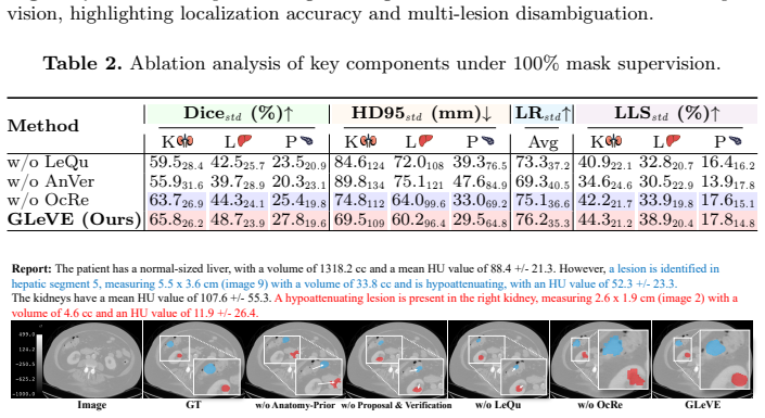
- [§4 (Experiments)] The abstract states 'consistent gains' on AbdomenAtlas 3.0 but the provided description lacks quantitative metrics, error bars, ablation studies on the graph and octree components, or discussion of failure modes. These details are required to substantiate the improvements in segmentation accuracy and lesion-level localization over baselines.
minor comments (2)
- [§3.1] Notation for the relation-aware graph (e.g., how nodes and edges are formally defined) could be clarified with a small diagram or pseudocode for readers unfamiliar with the specific graph construction.
- [Abstract] The abstract would benefit from naming the exact baseline methods and reporting at least one key metric (e.g., Dice or localization IoU) to give readers an immediate sense of the improvement magnitude.
Simulated Author's Rebuttal
We thank the referee for the thoughtful and constructive feedback. We address each major comment below and have revised the manuscript to incorporate additional examples, quantitative details, and clarifications where feasible.
read point-by-point responses
-
Referee: [§3 (Graph Reasoning and Proposal Verification)] The central assumption that free-text radiology reports reliably supply extractable organ attributions and inter-lesion relations for graph reasoning (without dense supervision or extra annotations) is load-bearing for the one-to-one alignment claim and subsequent proposal verification. The manuscript should include concrete examples from the dataset or failure-case analysis showing how ambiguous or implicit relations are handled, as this directly affects whether the graph module produces the promised discriminative queries.
Authors: We agree that explicit examples would help substantiate the graph reasoning module. In the revised version, we will add a dedicated subsection in §3 with concrete report excerpts from AbdomenAtlas 3.0, illustrating how organ attributions and inter-lesion relations (including implicit ones) are parsed and encoded. We will also include a short failure-case analysis highlighting cases where ambiguous phrasing leads to less discriminative queries and how the anatomy-aware verification mitigates this. revision: yes
-
Referee: [§4 (Experiments)] The abstract states 'consistent gains' on AbdomenAtlas 3.0 but the provided description lacks quantitative metrics, error bars, ablation studies on the graph and octree components, or discussion of failure modes. These details are required to substantiate the improvements in segmentation accuracy and lesion-level localization over baselines.
Authors: The full manuscript already reports quantitative metrics (Dice, localization accuracy), error bars from repeated runs, and ablations on the graph and octree modules in §4. However, we acknowledge the abstract is overly concise. We will revise the abstract to include key numerical improvements and add a dedicated paragraph on failure modes and limitations in the experiments section. Ablation tables will be expanded for clarity. revision: partial
Circularity Check
No significant circularity in derivation chain
full rationale
The paper introduces novel components including relation-aware graph reasoning for lesion-wise queries, anatomy-aware proposal generation with region-level verification, and hierarchical octree refinement. These are presented as independent methodological contributions that do not reduce by construction to fitted parameters, self-definitions, or self-citation chains in the provided abstract or description. No equations or steps are shown that equate predictions to inputs via renaming or ansatz smuggling. The framework is self-contained with gains demonstrated over external baselines, consistent with the default expectation that most papers exhibit no circularity.
Axiom & Free-Parameter Ledger
Lean theorems connected to this paper
-
IndisputableMonolith/Foundation/AlexanderDuality.leanalexander_duality_circle_linking echoes?
echoesECHOES: this paper passage has the same mathematical shape or conceptual pattern as the Recognition theorem, but is not a direct formal dependency.
hierarchical octree refinement progressively improves boundary delineation... octree-based autoregressive refinement
What do these tags mean?
- matches
- The paper's claim is directly supported by a theorem in the formal canon.
- supports
- The theorem supports part of the paper's argument, but the paper may add assumptions or extra steps.
- extends
- The paper goes beyond the formal theorem; the theorem is a base layer rather than the whole result.
- uses
- The paper appears to rely on the theorem as machinery.
- contradicts
- The paper's claim conflicts with a theorem or certificate in the canon.
- unclear
- Pith found a possible connection, but the passage is too broad, indirect, or ambiguous to say the theorem truly supports the claim.
Reference graph
Works this paper leans on
-
[1]
The Lancet Digital Health (2026)
Alabed, S., Anderson, A., Maiter, A., et al.: Large language models for simplifying radiology reports: A systematic review and meta-analysis of patient, public, and clinician evaluations. The Lancet Digital Health (2026)
work page 2026
-
[2]
Meng, M., Zhao, B.: M3D: Advancing 3D medical image analysis with multi-modal large language models
BAI, F., Du, Y., Huang, T., q.-h. Meng, M., Zhao, B.: M3D: Advancing 3D medical image analysis with multi-modal large language models. In: International Confer- ence on Learning Representations (2024)
work page 2024
-
[3]
In: Medical Imaging with Deep Learning (2025)
Bai, X., Liu, M., Chen, Y., Yang, H., Tian, Q.: Chest-OMDL: Organ-specific mul- tidisease detection and localization in chest computed tomography using weakly supervised deep learning from free-text radiology report. In: Medical Imaging with Deep Learning (2025)
work page 2025
-
[4]
EXACT: an explainable anomaly-aware vision foundation model for analysis of 3D chest CT
Bai, X., Liu, M., Song, T., et al.: EXACT: an explainable anomaly-aware vision foundation model for analysis of 3D chest CT. arXiv preprint arXiv:2604.24146 (2026)
work page internal anchor Pith review Pith/arXiv arXiv 2026
-
[5]
In: Proceedings of Medical Image Computing and Computer Assisted Intervention
Bassi, P.R., Li, W., Chen, J., et al.: Learning segmentation from radiology reports. In: Proceedings of Medical Image Computing and Computer Assisted Intervention. pp. 305–315 (2025)
work page 2025
-
[6]
In: Proceedings of the IEEE/CVF International Conference on Computer Vision
Bassi, P.R., Yavuz, M.C., Hamamci, I.E., Er, S., Chen, X., Li, W., Menze, B., Decherchi, S., Cavalli, A., Wang, K., et al.: RadGPT: Constructing 3D image-text tumor datasets. In: Proceedings of the IEEE/CVF International Conference on Computer Vision. pp. 23720–23730 (2025) 10 S. Jiang et al
work page 2025
-
[7]
Blankemeier, L., Cohen, J.P., Kumar, A., et al.: Merlin: A vision language foun- dation model for 3D computed tomography. Research Square pp. rs–3 (2024)
work page 2024
-
[8]
European Radiology35(5), 2589–2602 (2025)
Busch, F., Hoffmann, L., Dos Santos, D.P., et al.: Large language models for struc- tured reporting in radiology: Past, present, and future. European Radiology35(5), 2589–2602 (2025)
work page 2025
-
[9]
arXiv preprint arXiv:2503.12927 (2025)
Chen, H., Chen, Y., Yan, Z., Ding, M., Li, C., Zhu, Z., Qin, F.: MMLNB: Multi- modal learning for neuroblastoma subtyping classification assisted with textual description generation. arXiv preprint arXiv:2503.12927 (2025)
-
[10]
In: Proceedings of the IEEE/CVF Winter Conference on Applications of Computer Vision
Chen, Y., Zou, B., Guo, Z., et al.: SCUNet++: Swin-UNet and CNN bottleneck hy- brid architecture with multi-fusion dense skip connection for pulmonary embolism CT image segmentation. In: Proceedings of the IEEE/CVF Winter Conference on Applications of Computer Vision. pp. 7759–7767 (2024)
work page 2024
-
[11]
NPJ Digital Medicine8(1), 490 (2025)
Dong, F., Nie, S., Chen, M., et al.: Keyword-based AI assistance in the generation of radiology reports: A pilot study. NPJ Digital Medicine8(1), 490 (2025)
work page 2025
-
[12]
arXiv preprint arXiv:2203.00131 (2023)
Gao, Y., Zhou, M., Liu, D., Yan, Z., Zhang, S., Metaxas, D.N.: A data-scalable Transformer for medical image segmentation: Architecture, model efficiency, and benchmark. arXiv preprint arXiv:2203.00131 (2023)
-
[13]
Nature Biomedical Engineering pp
Hamamci, I.E., Er, S., Wang, C., Almas, F., et al.: Generalist foundation mod- els from a multimodal dataset for 3D computed tomography. Nature Biomedical Engineering pp. 1–19 (2026)
work page 2026
-
[14]
In: Proceedings of Medical Image Computing and Computer Assisted Intervention
Hao, Q., Yu, L., Tian, S., Ye, X., Zhang, L.: R1Seg-3D: Rethinking reasoning segmentation for medical 3D CTs. In: Proceedings of Medical Image Computing and Computer Assisted Intervention. pp. 415–425 (2025)
work page 2025
-
[15]
In: International Sym- posium on Biomedical Imaging
He, J., Li, P., Liu, G., Zhong, S.: Parameter-efficient fine-tuning medical multi- modal large language models for medical visual grounding. In: International Sym- posium on Biomedical Imaging. pp. 1–5. IEEE (2025)
work page 2025
- [16]
-
[17]
In: Proceedings of Medical Image Computing and Computer Assisted Intervention
Ichinose, A., Hatsutani, T., Nakamura, K., et al.: Visual grounding of whole radi- ology reports for 3D CT images. In: Proceedings of Medical Image Computing and Computer Assisted Intervention. pp. 611–621 (2023)
work page 2023
-
[18]
Nature Methods18(2), 203–211 (2021)
Isensee, F., Jaeger, P.F., Kohl, S.A., Petersen, J., Maier-Hein, K.H.: nnU-Net: a self-configuring method for deep learning-based biomedical image segmentation. Nature Methods18(2), 203–211 (2021)
work page 2021
-
[19]
IEEE Access 13, 112215–112254 (2025)
Kulsoom, U., Glavin, F.G., Bendechache, M.: Natural language processing and ma- chine learning for analysis of radiology reports-A systematic review. IEEE Access 13, 112215–112254 (2025)
work page 2025
-
[20]
Artificial Intelli- gence Review58(11), 344 (2025)
Li, Y., Kong, C., Zhao, G., Zhao, Z.: Automatic radiology report generation with deep learning: a comprehensive review of methods and advances. Artificial Intelli- gence Review58(11), 344 (2025)
work page 2025
-
[21]
arXiv preprint arXiv:2511.19046 (2025)
Liu, A., Xue, R., Cao, X.R., et al.: MedSAM3: Delving into segment anything with medical concepts. arXiv preprint arXiv:2511.19046 (2025)
-
[22]
In: International Conference on Learning Representations (2017)
Loshchilov, I., Hutter, F.: Decoupled weight decay regularization. In: International Conference on Learning Representations (2017)
work page 2017
-
[23]
In: Medical Imaging with Deep Learning (2025)
Nützel, F., Dombrowski, M., Kainz, B.: Generate to ground: Multimodal text con- ditioning boosts phrase grounding in medical vision-language models. In: Medical Imaging with Deep Learning (2025)
work page 2025
-
[24]
In: Proceedings of Medical Image Computing and Com- puter Assisted Intervention
Ronneberger, O., Fischer, P., Brox, T.: U-Net: Convolutional networks for biomed- ical image segmentation. In: Proceedings of Medical Image Computing and Com- puter Assisted Intervention. pp. 234–241 (2015) GLeVE: Graph-Guided Lesion Grounding with Proposal Verification 11
work page 2015
-
[25]
In: Proceedings of the IEEE/CVF Conference on Computer Vision and Pattern Recognition
Tang, Y., Yang, D., Li, W., Roth, H.R., Landman, B., Xu, D., Nath, V., Hatamizadeh, A.: Self-supervised pre-training of swin transformers for 3D med- ical image analysis. In: Proceedings of the IEEE/CVF Conference on Computer Vision and Pattern Recognition. pp. 20730–20740 (2022)
work page 2022
-
[26]
Radiology: Artificial Intelligence6(4), e240300 (2024)
Tejani, A.S., Klontzas, M.E., Gatti, A.A., et al.: Checklist for artificial intelligence in medical imaging (CLAIM): 2024 update. Radiology: Artificial Intelligence6(4), e240300 (2024)
work page 2024
-
[27]
IEEE Journal of Biomedical and Health Informatics29(12), 9051–9059 (2025)
Vilouras, K., Sanchez, P., O’Neil, A.Q., Tsaftaris, S.A.: Zero-shot medical phrase grounding with off-the-shelf diffusion models. IEEE Journal of Biomedical and Health Informatics29(12), 9051–9059 (2025)
work page 2025
-
[28]
Radiology: Artificial Intelligence 5(5), e230024 (2023)
Wasserthal, J., Breit, H.C., Meyer, M.T., et al.: TotalSegmentator: Robust segmen- tation of 104 anatomic structures in CT images. Radiology: Artificial Intelligence 5(5), e230024 (2023)
work page 2023
-
[29]
Yang, A., Li, A., Yang, B., et al.: Qwen3 technical report. arXiv preprint arXiv:2505.09388 (2025)
work page internal anchor Pith review Pith/arXiv arXiv 2025
-
[30]
Nature Biomedical Engineering pp
Yang, H., Zhou, H.Y., Liu, J., et al.: A multimodal vision–language model for gen- eralizable annotation-free pathology localization. Nature Biomedical Engineering pp. 1–15 (2026)
work page 2026
-
[31]
NPJ Digital Medicine8(1), 566 (2025)
Zhao, Z., Zhang, Y., Wu, C., Zhang, X., Zhou, X., Zhang, Y., Wang, Y., Xie, W.: Large-vocabulary segmentation for medical images with text prompts. NPJ Digital Medicine8(1), 566 (2025)
work page 2025
-
[32]
IEEE Transactions on Pattern Analysis and Machine Intelligence47(12), 11315–11329 (2025)
Zou, K., Bai, Y., Liu, B., et al.: Uncertainty-aware medical diagnostic phrase iden- tification and grounding. IEEE Transactions on Pattern Analysis and Machine Intelligence47(12), 11315–11329 (2025)
work page 2025
discussion (0)
Sign in with ORCID, Apple, or X to comment. Anyone can read and Pith papers without signing in.