REVIEW 3 major objections 6 minor 65 references
Optical fingerprint of bright and dark localized excitonic states in atomically thin 2D materials
T0 review · 3 major / 6 minor · reviewed 2026-08-14 · deepseek-v4-flash
Pith's one-line read Monolayer WSe2 with a Gaussian disorder potential should emit two families of localized exciton peaks below the bright line, with a temperature-controlled crossover near 20 K.
desk verdict A solid extension of the authors' PL framework to disorder-localized excitons, with interesting predictions for dark sidebands, but the 1s truncation and parameter fitting leave the key crossover under-validated. read the letter →
The pith
A machine-rendered reading of the paper's core claim, the machinery that carries it, and where it could break.
The reading
What carries the argument
The central object is the localized-exciton basis obtained by projecting the photon-assisted polarization onto eigenstates of a single-particle eigenvalue problem for the exciton center-of-mass motion inside the Gaussian disorder potential, Eq. (1). The disorder potential of depth $V_0$ and width $\sigma$ quantizes the center-of-mass momentum into bound states $\chi^{\mu n}$; free-exciton wavefunctions $\phi^\mu$ come from the Wannier equation with a Keldysh potential. To keep free and localized states orthogonal, the continuum is described with orthogonalized plane waves, Eq. (S5), and all capture, escape, and intervalley scattering rates are computed from exciton–phonon matrix elements in the second-order Born–Markov approximation. The photoluminescence intensity is then an Elliott-type sum over localized bright states plus a phonon-assisted sum over localized dark states, so temperature enters through phonon occupations and the computed scattering rates. The machinery's job is to convert a single disorder parameter pair $(V_0,\sigma)$ into a full PL spectrum with peak positions, widths, and the temperature crossover.
What would settle it
Measure the photoluminescence of a WSe$_2$ monolayer with engineered Gaussian-like strain traps (e.g., nanopillars) at temperatures from 4 K to 100 K and track the intensity ratio of the localized bright peak ($\approx$40 meV below $X$) to the dark side bands ($\approx$100–130 meV below): the model predicts a crossover near 20 K, with $X_{\mathrm{Loc}}$ dominating below and dark side bands above, and no localized peaks when the trap width falls below the exciton Bohr radius ($\approx$1 nm).
Extended reading notes
Core claim
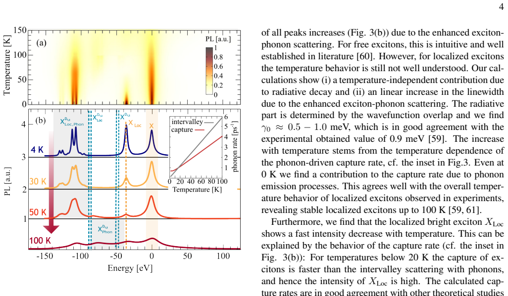
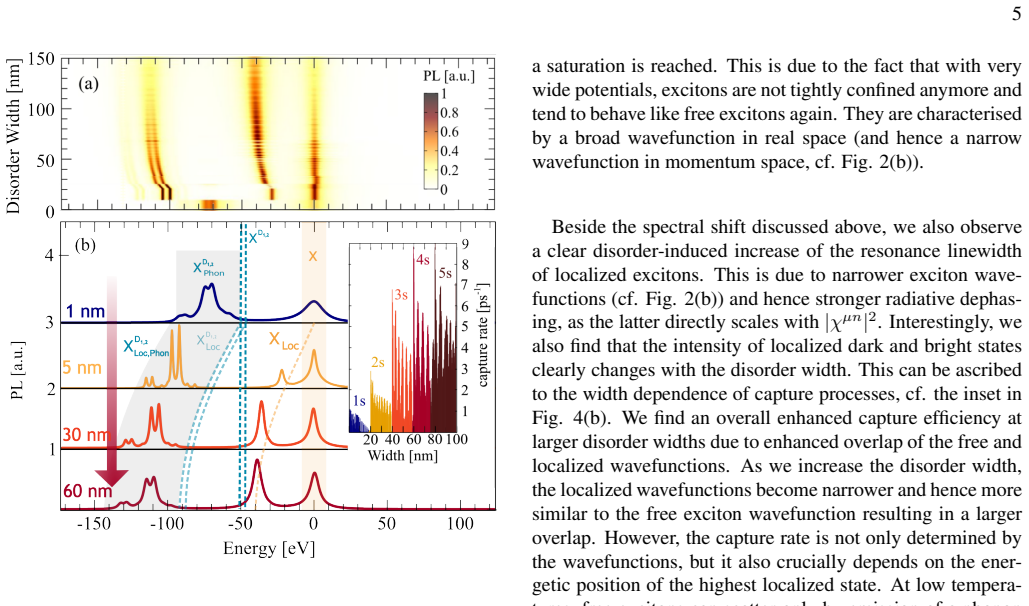
The central claim is that in WSe$_2$ monolayers with a sufficiently broad Gaussian disorder potential, both bright $KK$ excitons and momentum-dark $K\Lambda$ and $KK'$ excitons form localized 1s states at energies below their free counterparts, and these localized states leave distinct photoluminescence resonances—$X_{\mathrm{Loc}}$ for the bright state and phonon-assisted $X^{D_{1,2}}_{\mathrm{Loc}}$ side bands for the dark states. The localized bright peak sits about 40 meV below the free exciton $X$, and the dark side bands appear roughly 100–130 meV below. The competition between phonon-driven capture into the trap and phonon-assisted intervalley scattering out of the bright state controls the intensities: below about 20 K capture wins and $X_{\mathrm{Loc}}$ dominates; above that temperature excitons scatter to dark valleys before capture and the dark localized side bands take over. The same mechanism explains a temperature-independent radiative linewidth of about 0.5–1.0 meV plus a linearly growing phonon contribution, matching the experimental 0.9 meV value, and it predicts that the peaks shift to lower energies and saturate as the disorder width grows. The authors emphasize that this yields two spectral regimes—disorder-dominated at low $T$, phonon-dominated at higher $T$—and that the features are most pronounced in tungsten-based TMDs because their dark excitons lie below the bright one.
Load-bearing premise
The calculation assumes that excitons entering any higher localized state relax almost instantly to the 1s state, so only 1s localized states matter for the optical response; if that timescale separation fails, the computed capture rates, peak intensities, and the 20 K crossover would change.
Editorial extensions
If this is right
- Low-temperature PL of WSe$_2$ with strain or nanopillar traps should show a localized bright peak $X_{\mathrm{Loc}}$ about 40 meV below $X$ and phonon side bands from localized dark states about 100–130 meV below, as a fingerprint of trapping.
- The crossover temperature near 20 K separates a localization-dominated regime (bright localized peak dominant) from a phonon-dominated regime (dark localized side bands dominant).
- The radiative linewidth of localized excitons is about 0.5–1.0 meV and grows linearly with temperature from phonon scattering, reproducing the measured 0.9 meV.
- For disorder widths below the exciton Bohr radius (about 1 nm) no trapping occurs and only free-exciton phonon side bands appear; as the width grows, the localized peaks redshift and saturate near 60 nm.
- The intensity ratio between bright and dark localized resonances tracks the ratio of capture to intervalley scattering rates, so the spectrum can be used to infer trap characteristics.
Reading between the lines
- A temperature-dependent intensity ratio $I(X_{\mathrm{Loc}})/I(X^{D}_{\mathrm{Loc}})$ measured on a single sample with known trap width would directly test the predicted 20 K crossover—an experiment the paper does not report.
- Applied to other TMDs, the model predicts that the visibility of dark localized side bands depends on the energetic ordering of the $K\Lambda$ and $KK'$ dark states, so molybdenum-based monolayers (where dark states sit higher) should show a different fingerprint.
- The predicted capture-rate peaks whenever a new localized state enters the phonon window suggest that tuning $\sigma$ could resonantly enhance single-photon emission rates, a possible engineering lever.
- The strong dependence on the disorder-width–depth product $V_0\sigma^2$ implies that deterministic strain patterning could be used to write arrays of localized emitters with tailored energies.
Signed reviews
Editorial analysis
A structured set of objections, weighed in public.
Referee Report
Summary. This manuscript develops a microscopic density-matrix and cluster-expansion model for excitons in monolayer WSe2 in the presence of Gaussian disorder potentials. Free excitons are described by the Wannier equation, and localized excitons are obtained by diagonalizing the center-of-mass Hamiltonian of Eq. (1), yielding bound states whose binding energies and wavefunctions depend on disorder width and depth. The authors then compute photoluminescence spectra from bright KK excitons, localized bright excitons XLoc, and phonon-assisted sidebands of momentum-dark KΛ and KK' excitons, with capture/escape rates obtained from second-order Born-Markov exciton-phonon scattering in an orthogonalized-plane-wave basis. The central predictions are: a localized bright peak roughly 40 meV below the free bright exciton at low temperature, phonon-assisted sidebands of localized dark excitons about 100-130 meV below the bright exciton, and a temperature- and disorder-width-dependent crossover between localization-dominated and phonon-dominated PL regimes.
Significance. If the central claims hold, the paper provides a unified microscopic interpretation of the many low-temperature PL resonances below the bright exciton in WSe2, connecting them to bright and momentum-dark localized excitons and to the competition between disorder capture and intervalley phonon scattering. The model's strengths are that it treats free and localized excitons on the same footing, computes capture rates and spectra from microscopic exciton-phonon matrix elements, and makes explicit, testable predictions about temperature and disorder-width trends, including a predicted T≈20 K crossover and radiative linewidths of 0.5-1.0 meV. The main limitations are that the absolute position of XLoc is essentially set by the input disorder depth, that quantitative comparison with measured PL spectra is not provided, and that two load-bearing approximations, the restriction to 1s localized states and the orthogonalized-plane-wave treatment of the continuum, are asserted without controlled numerical checks.
major comments (3)
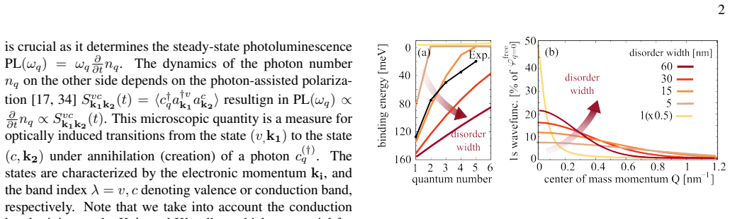
- [Main text, 'Photoluminescence of localized excitons', after Eq. (3), and Supplementary Material, Eq. (S6) discussion.] The restriction of the localized exciton manifold to the 1s state is load-bearing but is justified only by assertion. The main text states that 'intraexcitonic scattering of localized excitonic states (µn→µn′) appears on a much faster timescale than the capture processes itself,' and the Supplementary repeats that 'relaxation dynamics within the localized states happen on a much faster time scale than capture processes,' but no intra-localized relaxation rate is computed or compared with the capture and intervalley rates shown in the inset of Fig. 3. This matters directly for the central crossover claim: if excited localized states are not depopulated essentially instantaneously, they carry non-negligible occupation, produce additional bright PL peaks at smaller binding energies, and change the population of the 1s state that generates the predicted XLoc peak. The authors should provide a quantitative comparison of Γ(µn→µ1s) with Γcapture and Γintervalley for the parameters of Fig. 3, or include excited ns states explicitly and demonstrate that the spectra are converged.
- [Supplementary Material, Eq. (S5), and main text discussion of capture rates.] The orthogonalized-plane-wave treatment of the continuum is an uncontrolled approximation that enters directly into the computed capture rates and hence into the predicted crossover. The normalization factor N_Q in Eq. (S5) contains the overlap sum ∑|⟨χµm_Q|φPW_Q⟩|², but the manuscript does not report how large this sum is for the parameters used, nor does it check the δ-function idealization φPW_Q ≈ δ_{Q,QF} against a numerically orthogonalized continuum basis. Since the capture rates in the inset of Fig. 3 and the consequent visibility of XLoc versus dark sidebands depend on these overlaps, I request a convergence or consistency check, for example by comparing with continuum states obtained from the Wannier equation orthogonalized to the localized states, and by reporting the magnitude of the projection corrections.
- [Main text, results 'Photoluminescence of localized excitons' and 'Disorder-induced control of PL'.] The claim that 'calculated optical spectra agree well with recent experiments' is not quantitatively supported, and the position of the main XLoc peak is not an independent prediction. In Fig. 3 the XLoc peak appears approximately 40 meV below X because V0 = 40 meV was chosen from the nanopillar experiments of Ref. [21], so the peak position essentially follows from the input disorder depth. The more robust predictions are the relative intensity trends with temperature and disorder width, the linewidth behavior, and the dark-localized sidebands. I recommend that the authors explicitly separate input-determined features from emergent model predictions and, where possible, overlay calculated PL spectra on measured spectra (e.g., the data of Refs. [21, 58, 59]) to substantiate the claimed agreement.
minor comments (6)
- [Theoretical approach.] There is a typo 'resultign' in the sentence introducing the photon-assisted polarization; it should be 'resulting'.
- [Results, temperature-dependent PL.] The text 'phonon side[17, 32]bands' contains misplaced citation brackets; the phrase should read 'phonon sidebands' with citations following.
- [Disorder-induced control of PL.] In the paragraph on narrow disorder potentials, 'we are in the the phonon-dominated regime' contains a duplicated article and should be corrected.
- [Eq. (1).] The eigenvalue equation as printed contains 'εµnχµxn Q' on the right-hand side, which appears to be a typo for 'εµnχµn Q'; please check the equation.
- [Supplementary Material, Eq. (S5).] The notation ⟨χµm_Q|φPW_Q⟩ is ambiguous because the localized states carry the same Q index as the plane-wave state; please clarify the labeling of the basis states in the orthogonalization sum.
- [Figures 2 and 3.] The paper uses V0 = 120 meV for Fig. 2 but V0 = 40 meV for Figs. 3 and 4; a short remark explaining how the fixed-depth curves of Fig. 2 relate to the lower-depth spectra of Figs. 3-4 would improve readability.
Circularity Check
No circularity: localized-state energies and capture/intervalley rates are derived from the model parameters, not fitted to the predicted PL features.
full rationale
The paper's central predictions are derived rather than assumed. The XLoc peak position near 40 meV below X is obtained by solving the localized-exciton eigenvalue problem, Eq. (1), with a Gaussian disorder potential of depth V0 = 40 meV chosen from independent experimental reports on nanopillars and local strain ([21]); the eigenenergy is a computed function of V0 and width, not an identity or a fit to the PL spectrum. The temperature-dependent crossover between localization- and phonon-dominated regimes is controlled by microscopically computed capture and intervalley exciton-phonon scattering rates (Eqs. S3-S7), none of which are adjusted to reproduce the predicted spectra. The paper does cite previous work by the same authors ([17], [32], [63], [64]) for the underlying TMD Bloch equations and exciton-phonon matrix elements, but those citations supply the standard many-body formalism and are not invoked as external proof of the target result, so they are not circular. The 1s-only truncation of localized states, justified by an asserted timescale separation in the Supplementary, is an unvalidated approximation and a potential correctness risk, but it is not a circular step: it does not define the predicted quantities in terms of themselves. No step in the derivation reduces, by construction or by fitted parameter renaming, to its own input.
Assumptions & free parameters
free parameters (2)
- disorder depth V0 =
120 meV (Fig. 2), 40 meV (Figs. 3-4)
- disorder width sigma =
varied 1-60 nm; fixed 30 nm for temperature study
assumptions (6)
- standard math Wannier equation with Keldysh potential gives free-exciton wavefunctions and energies
- domain assumption Cluster expansion / density matrix formalism truncation at the excitonic two-particle level
- domain assumption Second-order Born-Markov approximation for exciton-phonon scattering and capture rates
- ad hoc to paper Orthogonalized plane waves for free continuum states
- ad hoc to paper Only the 1s localized state contributes to PL
- domain assumption Phonons are in a Bose equilibrium bath
Cite this review
Pith. "Pith review of Optical fingerprint of bright and dark localized excitonic states in atomically thin 2D materials." pith.science (2026). https://pith.science/paper/IRHZDZ7Z
@misc{pith2026190805071,
  author       = {Pith},
  title        = {Pith review of: Optical fingerprint of bright and dark localized excitonic states in atomically thin 2D materials},
  year         = {2026},
  howpublished = {\url{https://pith.science/paper/IRHZDZ7Z}},
  note         = {Machine review of arXiv:1908.05071}
}
read the original abstract
Point defects, local strain or impurities can crucially impact the optical response of atomically thin two-dimensional materials as they offer trapping potentials for excitons. These trapped excitons appear in photoluminescence spectra as new resonances below the bright exciton that can even be exploited for single photon emission. While large progress has been made in deterministically introducing defects, only little is known about their impact on the optical fingerprint of 2D materials. Here, based on a microscopic approach we reveal direct signatures of localized bright excitonic states as well as indirect phonon-assisted side bands of localized momentum-dark excitons. The visibility of localized excitons strongly depends on temperature and disorder potential width. This results in different regimes, where either the bright or dark localized states are dominant in optical spectra. We trace back this behavior to an interplay between disorder-induced exciton capture and intervalley exciton-phonon scattering processes.
Figures

Figures from the paper (1 more)
Reference graph
Works this paper leans on
-
[21]
C. Palacios-Berraquero, D. M. Kara, A. R.-P. Montblanch, M. Barbone, P. Latawiec, D. Yoon, A. K. Ott, M. Loncar, A. C. Ferrari, and M. Atat¨ure, Nature communications8, 15093 (2017)
work page 2017
-
[1]
T. Mueller and E. Malic, 2D Materials and Applications , ac- cepted (2018)
work page 2018
-
[2]
G. Wang, A. Chernikov, M. M. Glazov, T. F. Heinz, X. Marie, T. Amand, and B. Urbaszek, Rev. Mod. Phys. 90, 021001 (2018)
2018
-
[3]
K. F. Mak, C. Lee, J. Hone, J. Shan, and T. F. Heinz, Phys. Rev. Lett. 105, 136805 (2010)
2010
- [4]
-
[5]
J. O. Island, A. Kuc, E. H. Diependaal, R. Bratschitsch, H. S. van der Zant, T. Heine, and A. Castellanos-Gomez, Nanoscale 8, 2589 (2016)
work page 2016
-
[6]
H. J. Conley, B. Wang, J. I. Ziegler, R. F. Haglund Jr, S. T. Pantelides, and K. I. Bolotin, Nano letters 13, 3626 (2013)
work page 2013
-
[7]
J. S. Ross, P. Klement, A. M. Jones, N. J. Ghimire, J. Yan, D. Mandrus, T. Taniguchi, K. Watanabe, K. Kitamura, W. Yao, et al., Nature nanotechnology 9, 268 (2014)
work page 2014
Show all 65 references
-
[8]
Mouri, Y
S. Mouri, Y . Miyauchi, and K. Matsuda, Nano letters 13, 5944 (2013)
2013
-
[9]
Godde, D
T. Godde, D. Schmidt, J. Schmutzler, M. Aßmann, J. Debus, F. Withers, E. Alexeev, O. Del Pozo-Zamudio, O. Skrypka, K. Novoselov, et al., Physical Review B 94, 165301 (2016)
2016
-
[10]
Jadczak, J
J. Jadczak, J. Kutrowska-Girzycka, P. Kapu ´sci´nski, Y . Huang, A. W´ojs, and L. Bryja, Nanotechnology 28, 395702 (2017)
2017
-
[11]
Nagler, M
P. Nagler, M. V . Ballottin, A. A. Mitioglu, M. V . Durnev, T. Taniguchi, K. Watanabe, A. Chernikov, C. Sch ¨uller, M. M. Glazov, P. C. Christianen, et al. , Physical review letters 121, 057402 (2018)
2018
-
[12]
Malic, M
E. Malic, M. Selig, M. Feierabend, S. Brem, D. Christiansen, F. Wendler, A. Knorr, and G. Bergh¨auser, Physical Review Ma- terials 2, 014002 (2018)
2018
-
[13]
Molas, C
M. Molas, C. Faugeras, A. Slobodeniuk, K. Nogajewski, M. Bartos, D. Basko, and M. Potemski, 2D Materials4, 021003 (2017)
2017
-
[14]
Y . Zhou, G. Scuri, D. S. Wild, A. A. High, A. Dibos, L. A. Jauregui, C. Shu, K. De Greve, K. Pistunova, A. Y . Joe, et al., Nature nanotechnology 12, 856 (2017)
2017
-
[15]
Lindlau, C
J. Lindlau, C. Robert, V . Funk, J. F¨orste, M. F¨org, L. Colombier, A. Neumann, E. Courtade, S. Shree, T. Taniguchi, et al., arXiv preprint arXiv:1710.00988 (2017)
2017 arXiv
-
[16]
Zhang, Y
X.-X. Zhang, Y . You, S. Y . F. Zhao, and T. F. Heinz, Physical review letters 115, 257403 (2015)
2015
-
[17]
S. Brem, A. Ekman, D. Christiansen, F. Katsch, M. Selig, C. Robert, X. Marie, B. Urbaszek, A. Knorr, and E. Malic, arXiv preprint arXiv:1904.04711 (2019)
2019 arXiv
-
[18]
Kumar, A
S. Kumar, A. Kaczmarczyk, and B. D. Gerardot, Nano letters 15, 7567 (2015)
2015
-
[19]
M. R. Rosenberger, C. K. Dass, H.-J. Chuang, S. V . Sivaram, K. M. McCreary, J. R. Hendrickson, and B. T. Jonker, ACS nano 13, 904 (2019)
2019
-
[20]
Koperski, K
M. Koperski, K. Nogajewski, A. Arora, V . Cherkez, P. Mallet, J.-Y . Veuillen, J. Marcus, P. Kossacki, and M. Potemski, Nature nanotechnology 10, 503 (2015)
2015
-
[22]
J. Kern, I. Niehues, P. Tonndorf, R. Schmidt, D. Wigger, R. Schneider, T. Stiehm, S. Michaelis de Vasconcellos, D. E. Reiter, T. Kuhn, et al., Advanced materials 28, 7101 (2016)
2016
-
[23]
G. D. Shepard, O. A. Ajayi, X. Li, X. Zhu, J. Hone, and S. Strauf, 2D Materials 4, 021019 (2017)
2017
-
[24]
Chakraborty, L
C. Chakraborty, L. Kinnischtzke, K. M. Goodfellow, R. Beams, and A. N. Vamivakas, Nature Nanotechnology10, 507 (2015)
2015
-
[25]
Y .-M. He, G. Clark, J. R. Schaibley, Y . He, M.-C. Chen, Y .-J. Wei, X. Ding, Q. Zhang, W. Yao, X. Xu, et al. , Nature Nan- otechnology 10, 497 (2015)
2015
-
[26]
Srivastava, M
A. Srivastava, M. Sidler, A. V . Allain, D. S. Lembke, A. Kis, and A. Imamo˘glu, Nature Nanotechnology 10, 491 (2015)
2015
-
[27]
Tonndorf, R
P. Tonndorf, R. Schmidt, R. Schneider, J. Kern, M. Buscema, G. A. Steele, A. Castellanos-Gomez, H. S. van der Zant, S. M. de Vasconcellos, and R. Bratschitsch, Optica 2, 347 (2015)
2015
-
[28]
Branny, S
A. Branny, S. Kumar, R. Proux, and B. D. Gerardot, Nature communications 8, 15053 (2017)
2017
-
[29]
Haug and S
H. Haug and S. W. Koch, Quantum Theory of the Optical and Electronic Properties of Semiconductors (5th ed. (World Scien- tific Publishing Co. Pre. Ltd., Singapore, 2004).)
2004
-
[30]
Kira and S
M. Kira and S. Koch, Progress in Quantum Electronics 30, 155 (2006)
2006
-
[31]
Malic and A
E. Malic and A. Knorr, Graphene and Carbon Nanotubes: Ul- trafast Optics and Relaxation Dynamics (John Wiley & Sons, 2013)
2013
-
[32]
Selig, G
M. Selig, G. Bergh ¨auser, M. Richter, R. Bratschitsch, A. Knorr, and E. Malic, 2D Materials (2018)
2018
-
[33]
S. Brem, M. Selig, G. Berghaeuser, and E. Malic, Scientific reports 8, 8238 (2018)
2018
-
[34]
Thr ¨anhardt, S
A. Thr ¨anhardt, S. Kuckenburg, A. Knorr, T. Meier, and S. Koch, Physical Review B 62, 2706 (2000)
2000
-
[35]
Chernikov, T
A. Chernikov, T. C. Berkelbach, H. M. Hill, A. Rigosi, Y . Li, O. B. Aslan, D. R. Reichman, M. S. Hybertsen, and T. F. Heinz, Phys. Rev. Lett. 113, 076802 (2014)
2014
-
[36]
Arora, M
A. Arora, M. Koperski, K. Nogajewski, J. Marcus, C. Faugeras, and M. Potemski, Nanoscale 7, 10421 (2015)
2015
-
[37]
29, 658 (1978)
Keldysh, JETP Lett. 29, 658 (1978)
1978
-
[38]
Cudazzo, I
P. Cudazzo, I. V . Tokatly, and A. Rubio, Phys. Rev. B 84, 085406 (2011)
2011
-
[39]
Malic, A
E. Malic, A. Setaro, P. Bluemmel, C. F. Sanz-Navarro, P. Or- dejn, S. Reich, and A. Knorr, Journal of Physics: Condensed Matter 24, 394006 (2012)
2012
-
[40]
Kormanyos, G
A. Kormanyos, G. Burkard, M. Gmitra, J. Fabian, V . Zoly- omi, N. D. Drummond, and V . Falko, 2D Materials 2, 022001 (2015)
2015
-
[41]
Wen-Fang, Chinese Physics Letters 22, 1768 (2005)
X. Wen-Fang, Chinese Physics Letters 22, 1768 (2005)
2005
-
[42]
Hichri, I
A. Hichri, I. Ben Amara, S. Ayari, and S. Jaziri, Journal of Applied Physics 121, 235702 (2017)
2017
-
[43]
Adamowski, M
J. Adamowski, M. Sobkowicz, B. Szafran, and S. Bednarek, Physical Review B 62, 4234 (2000)
2000
-
[44]
Rosati, F
R. Rosati, F. Lengers, D. E. Reiter, and T. Kuhn, Physical Re- view B 98, 195411 (2018)
2018
-
[45]
Brooks and G
M. Brooks and G. Burkard, Physical Review B 97, 195454 (2018)
2018
-
[46]
T. T. Tran, S. Choi, J. A. Scott, Z.-Q. Xu, C. Zheng, G. Seniuti- nas, A. Bendavid, M. S. Fuhrer, M. Toth, and I. Aharonovich, Advanced Optical Materials 5, 1600939 (2017)
2017
-
[47]
Bergh ¨auser, P
G. Bergh ¨auser, P. Steinleitner, P. Merkl, R. Huber, A. Knorr, and E. Malic, Physical Review B 98, 020301 (2018)
2018
-
[48]
Salehi and A
S. Salehi and A. Saffarzadeh, Surface Science 651, 215 (2016)
2016
-
[49]
Zhang, C.-G
S. Zhang, C.-G. Wang, M.-Y . Li, D. Huang, L.-J. Li, W. Ji, and S. Wu, Physical review letters 119, 046101 (2017)
2017
-
[50]
Refaely-Abramson, D
S. Refaely-Abramson, D. Y . Qiu, S. G. Louie, and J. B. Neaton, Physical review letters 121, 167402 (2018)
2018
-
[51]
T. C. Berkelbach, M. S. Hybertsen, and D. R. Reichman, Phys- ical Review B 88, 045318 (2013)
2013
-
[52]
Hoyer, C
W. Hoyer, C. Ell, M. Kira, S. Koch, S. Chatterjee, S. Mosor, G. Khitrova, H. Gibbs, and H. Stolz, Physical Review B 72, 075324 (2005). 7
2005
-
[53]
D. F. Walls and G. J. Milburn, Quantum optics (Springer Sci- ence & Business Media, 2007)
2007
-
[54]
Herring, Physical Review 57, 1169 (1940)
C. Herring, Physical Review 57, 1169 (1940)
1940
-
[55]
Schneider, W
H. Schneider, W. Chow, and S. W. Koch, physica status solidi (b) 238, 589 (2003)
2003
-
[56]
Cadiz, E
F. Cadiz, E. Courtade, C. Robert, G. Wang, Y . Shen, H. Cai, T. Taniguchi, K. Watanabe, H. Carrere, D. Lagarde,et al., Phys. Rev. X 7, 021026 (2017)
2017
-
[57]
V . Axt, K. Victor, and A. Stahl, Physical Review B 53, 7244 (1996)
1996
-
[58]
L. N. Tripathi, O. Iff, S. Betzold, Ł. Dusanowski, M. Em- merling, K. Moon, Y . J. Lee, S.-H. Kwon, S. Hofling, and C. Schneider, ACS Photonics 5, 1919 (2018)
2018
-
[59]
Y .-M. He, S. H ¨ofling, and C. Schneider, Optics express 24, 8066 (2016)
2016
-
[60]
Selig, G
M. Selig, G. Bergh ¨auser, A. Raja, P. Nagler, C. Sch ¨uller, T. F. Heinz, T. Korn, A. Chernikov, E. Malic, and A. Knorr, Nature communications 7, 13279 (2016)
2016
-
[61]
Klein, M
J. Klein, M. Lorke, M. Florian, F. Sigger, J. Wierzbowski, J. Cerne, K. M¨uller, T. Taniguchi, K. Watanabe, U. Wurstbauer, et al., arXiv preprint arXiv:1901.01042 (2019)
2019 arXiv
-
[62]
Ayari and S
S. Ayari and S. Jaziri, physica status solidi (b) 256, 1800682 (2019)
2019
-
[63]
Feierabend, G
M. Feierabend, G. Bergh ¨auser, M. Selig, S. Brem, T. Shegai, S. Eigler, and E. Malic, Physical Review Materials 2, 014004 (2018)
2018
-
[64]
S. Brem, J. Zipfel, M. Selig, A. Raja, L. Waldecker, J. D. Ziegler, T. Taniguchi, K. Watanabe, A. Chernikov, and E. Malic, Nanoscale (2019)
2019
-
[65]
Mali ´c, K
E. Mali ´c, K. J. Ahn, M. J. Bormann, P. H ¨ovel, E. Sch ¨oll, A. Knorr, M. Kuntz, and D. Bimberg, Applied physics letters 89, 101107 (2006). 1 Optical fingerprint of bright and dark localized excitonic states in atomically thin 2D materials Maja Feierabend, Samuel Brem and Erm...
2006
Reviewed August 14, 2026 · model on record in the stance chip above.
Discussion (0). Continue with ORCID to comment.