REVIEW 4 major objections 6 minor 2 references
The Next Layer: Augmenting Foundation Models with Structure-Preserving and Attention-Guided Learning for Local Patches to Global Context Awareness in Computational Pathology
T0 review · 4 major / 6 minor · reviewed 2026-08-05 · deepseek-v4-flash
Pith's one-line read The paper claims that adding learned absolute spatial encodings and neighborhood-aware attention to foundation-model patch features improves slide-level classification and survival prediction across cancer types.
desk verdict Solid MIL architecture and interpretability study, but the headline survival results rest on an unstated training loss and a likely patient-leakage problem. read the letter →
The pith
A machine-rendered reading of the paper's core claim, the machinery that carries it, and where it could break.
The reading
What carries the argument
The load-bearing object is MASE (Multi-scale Absolute Spatial Encoding), a module that rearranges patch embeddings into the exact 3D grid of their original slide coordinates and runs two-stage convolutions—multi-scale kernels of size 1x1, 3x3, 5x5, and 7x7 followed by a 1x1 convolution—to produce position-aware features that keep global tissue architecture intact. It works with two loss terms: a top-K neighborhood-aware loss that finds the highest-attention patches, pulls their local neighbors in the grid into an attention-weighted cross-entropy term, and a background-suppression loss that penalizes attention on non-tissue patches. Together they convert a bag of independent patch embeddings
What would settle it
Permute patch coordinates within each slide before MASE, keeping all folds, backbones, and hyperparameters fixed, on the seven survival cohorts; if the C-index does not fall below the intact-coordinate version, the claim that absolute spatial encoding drives the gains is falsified.
Extended reading notes
Core claim
EAGLE-Net's central claim is that absolute spatial layout and the local neighborhoods around high-attention patches carry information that patch-level foundation embeddings leave on the table. The model makes that concrete in three steps: MASE rebuilds the patch feature set into a position-preserving grid and applies two-stage convolutions (kernels 1, 3, 5, 7 followed by 1x1) to learn multi-scale absolute positional encodings; a top-K neighborhood-aware loss pulls the spatial neighbors of the most-attended patches into the classification loss, weighted by their attention scores; and a background-suppression term drives attention away from non-tissue patches. Benchmarking against AbMIL, Gated
Load-bearing premise
The survival benchmark rests on an unstated training choice: the paper never specifies the loss, event-time handling, or censoring weighting used to train EAGLE-Net for survival, even though the loss equations written out are for one-hot classification.
Editorial extensions
If this is right
- Histology foundation models can stay frozen while a spatial MIL layer on top adds layout awareness, so the approach transfers across different backbones without retraining them.
- Survival models built from attention-pooled embeddings become more prognostic across diverse cancer types, with the paper reporting top C-indices in six of seven cohorts.
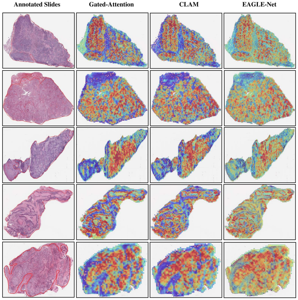
- Attention maps become a more trustworthy evidence channel: they concentrate on tumor, necrosis, and immune infiltrates and reduce false-positive highlighting of benign tissue.
- The same position-preserving pooling and neighborhood-aware loss can be applied to other gigapixel imaging tasks where tile layout matters, not just pathology.
- Packing multiple slides from one patient into a single canvas lets the model see inter-slide heterogeneity, capturing micro-lesions alongside dominant morphology.
Reading between the lines
- The paper leaves the survival training objective unspecified; a natural next test is to replace the classification-style loss with a proper Cox or ranking loss with censoring weights and measure whether the reported C-index gains grow or shrink.
- The neighborhood-aware loss probably enforces spatial coherence of attention as a side effect; ablating L2 and L3 separately while measuring Dice and frequency-domain metrics would separate the coherence effect from the absolute-positioning effect.
- The frequency-domain metrics (angular energy dispersion and radial profile) are used only for evaluation; using them as training regularizers could sharpen boundary delineation further.
- A shuffle-coordinate sanity check—permuting patch positions before MASE—would directly test whether the reported gains come from true spatial structure rather than from the extra convolutional parameters.
Editorial analysis
A structured set of objections, weighed in public.
Referee Report
Summary. The paper proposes EAGLE-Net, a multiple-instance learning (MIL) framework for whole-slide images that augments pretrained foundation-model features with (i) a Multi-scale Absolute Spatial Encoding (MASE) module to preserve global tissue layout, (ii) a top-K neighborhood-aware loss L2 that reinforces attention around highly attended patches, and (iii) a background-suppression loss L3. The method is evaluated on three classification tasks (TCGA/CPTAC lung subtyping and PANDA ISUP grading) and seven survival cohorts (six TCGA types plus CPTAC-LUAD), using three histology foundation backbones (REMEDIES, Uni-V1, Uni2-h). The paper reports up to 3% higher classification accuracy, top concordance indices in 6 of 7 survival cohorts, and attention maps that align with expert-annotated tumor regions, with quantitative interpretability metrics on 300 annotated LUAD slides.
Significance. If the results are reproducible, the paper makes a useful engineering contribution to computational pathology: the benchmarking scale is large (14,432 WSIs), the evaluation across three foundation backbones is commendable, and the interpretability analysis with pathologist annotations is a practical step beyond qualitative heatmaps. The MASE idea of encoding absolute spatial coordinates via multi-scale convolutions is simple and could generalize. However, the headline survival claim, which is the main quantitative result, is currently not verifiable because the training objective for survival is never specified; the loss equations define only classification. The paper cannot be fully assessed until this central ambiguity is resolved, along with the patient-level leakage risk in cross-validation.
major comments (4)
- [Methods, 'Total Loss and Ablation Experiments'; Results, 'EAGLE-Net Achieves Superior Performance on Pan-Cancer Prognost] The survival benchmark (Fig. 2a and Extended Data Fig. 8) is the paper's headline, but no survival loss is defined. L1, L2, and L3 are all written for classification with a known class k (one-hot bag label), and there is no Cox partial likelihood, ranking loss, or censoring weighting anywhere in Methods. The sentence 'applied Cox proportional-hazards models to attention-pooled embeddings' is compatible with at least three protocols: (i) end-to-end training with a Cox loss, (ii) training with the classification losses and then fitting Cox on the pooled embedding, or (iii) using EAGLE-Net only as a feature extractor. Each protocol gives different C-indices and assigns different roles to L2/L3. Please specify the exact survival objective, censoring handling, and hyperparameters; if a Cox loss replaces L1, the equations are incomplete; if classification is used as a surrogate, state this and
- [Methods, 'Pan-Cancer Datasets' and 'Slide Processing and Patient-Level Tissue Packing'] The tissue-packing step aggregates multiple slides from the same patient into one canvas, making the patient the natural unit of analysis. The paper states '5-fold Monte Carlo cross-validation' but does not state that the folds are stratified by patient. If slides are split without grouping by patient ID, the same patient can appear in both training and test sets, inflating the reported C-index. Please report explicit patient-level splitting, the number of patients per fold, and clarify that all metrics (notably C-index) are computed over distinct patients rather than slides.
- [Results, 'EAGLE-Net Reaches Robust Classification Performance'] Classification accuracies are reported as point estimates without confidence intervals or significance tests. For example, TCGA accuracy is 0.980 for both EAGLE-Net and CLAM, and the PANDA kappa advantage is 0.984 vs 0.970 on Radboud but 0.985 vs 0.985 on Karolinska. The 'up to 3%' claim rests on a single CPTAC comparison. Please report fold-wise distributions, confidence intervals, and paired tests (e.g., Wilcoxon across folds or patients) for all classification and survival comparisons, and indicate which differences are statistically significant after multiple-testing correction.
- [Methods, 'Multi-scale Absolute Spatial Encoding'] In the MASE equations, P_i = Conv(V_stack, W_{1×1}, b5, pad=3). With a 1×1 kernel, padding of 3 changes the spatial dimensions, so H_i ⊕ P_i is ill-defined unless H_i is padded correspondingly; the intended padding is likely 0. This is a concrete reproducibility issue in a core architectural component. Also, the text refers to 'top-k' instances but the equations use K; please define K explicitly and state how it is chosen (the ablation in Extended Data Fig. 8b is useful but should be tied to a concrete default value and selection procedure).
minor comments (6)
- [Throughout] Typos and wording: 'Cohan’s kappa' should be 'Cohen’s kappa'; 'archived' should be 'achieved'; 'Monte-Carolo' should be 'Monte Carlo'; 'metrices' should be 'metrics'; 'FDR' in Fig. 6b appears to mean 'FPR'. Please proofread.
- [Results, Fig. 3 caption] The caption says 'ten-fold cross-validated concordance indices,' but Methods specifies 5-fold Monte Carlo cross-validation. Please make the number of folds consistent.
- [Extended Data Fig. 1c] The caption mentions 'clear-cell renal-cell carcinoma subtyping' and 'lymph-node-metastasis detection,' but these tasks are not reported in Results. Either add the results or correct the caption.
- [Methods, 'Attention-based Neighborhood-aware Loss'] The neighborhood-aware loss L2 is described as 'self-supervised' in the Introduction and Discussion, but it uses the slide-level bag label and the learned attention weights; this is better described as weakly supervised or self-guided. Please adjust the terminology.
- [Results, 'EAGLE-Net’s Generated Attention Maps Align with Tumor Masks'] The Dice/FPR evaluation thresholds attention maps into tumor masks, but the thresholding procedure is not described. Please specify how the binary mask is derived from continuous attention scores.
- [Code availability] The repository is listed as 'will be available'; given the unresolved survival training ambiguity, releasing the code (or at least the training configuration) is essential for reproducibility.
Circularity Check
No significant circularity: the paper's claims are empirical benchmark results against external MIL baselines, and the cited self-work is background or data, not load-bearing.
full rationale
The paper's central claims are empirical benchmark results against external MIL baselines (CLAM, TransMIL, ILRA, DSMIL, etc.) on held-out TCGA/CPTAC/PANDA cohorts. The loss equations (L1-L3) define a training objective; they are not derived from the reported C-indices or accuracies. The MASE positional encoding and neighborhood-aware loss are architectural/objective design choices whose effects are measured, not assumed. No step reduces a prediction to a fitted parameter or to a self-citation: the cited prior work by the authors (refs 4, 10-13) is used as background/survey context, and the annotation reference [32] is a data resource; neither is load-bearing for the stated performance gains. The survival evaluation is under-specified—only a Cox model on attention-pooled embeddings is described and no survival loss is given—but this is a reproducibility/completeness gap, not circular reasoning: the survival outcome is external clinical data, and the C-index is computed against that data, not constructed from the model's inputs. Similarly, using attention weights to supervise a neighborhood loss is self-referential training dynamics, but it does not make the reported benchmark results equivalent to the loss definition. Therefore no significant circularity was found.
Assumptions & free parameters
free parameters (6)
- lambda (L2 loss weight)
- beta (L3 loss weight)
- K (number of top-attended patches)
- r (neighborhood radius)
- d (embedding dimension after fully connected layer)
- Tissue packing thresholds (slideThr=0.01, patientThr=0.25) =
0.01, 0.25
assumptions (3)
- domain assumption CNN convolutions on the patch grid learn meaningful absolute positional encodings for histopathology
- domain assumption Top-K attention instances and their local neighborhoods are diagnostically relevant and can serve as self-supervision
- standard math Attention pooling and linear transformations behave as standard in MIL
Cite this review
Pith. "Pith review of The Next Layer: Augmenting Foundation Models with Structure-Preserving and Attention-Guided Learning for Local Patches to Global Context Awareness in Computational Pathology." pith.science (2026). https://pith.science/paper/JWFNFYPK
@misc{pith2026250819914,
  author       = {Pith},
  title        = {Pith review of: The Next Layer: Augmenting Foundation Models with Structure-Preserving and Attention-Guided Learning for Local Patches to Global Context Awareness in Computational Pathology},
  year         = {2026},
  howpublished = {\url{https://pith.science/paper/JWFNFYPK}},
  note         = {Machine review of arXiv:2508.19914}
}
read the original abstract
Foundation models have recently emerged as powerful feature extractors in computational pathology, yet they typically omit mechanisms for leveraging the global spatial structure of tissues and the local contextual relationships among diagnostically relevant regions - key elements for understanding the tumor microenvironment. Multiple instance learning (MIL) remains an essential next step following foundation model, designing a framework to aggregate patch-level features into slide-level predictions. We present EAGLE-Net, a structure-preserving, attention-guided MIL architecture designed to augment prediction and interpretability. EAGLE-Net integrates multi-scale absolute spatial encoding to capture global tissue architecture, a top-K neighborhood-aware loss to focus attention on local microenvironments, and background suppression loss to minimize false positives. We benchmarked EAGLE-Net on large pan-cancer datasets, including three cancer types for classification (10,260 slides) and seven cancer types for survival prediction (4,172 slides), using three distinct histology foundation backbones (REMEDIES, Uni-V1, Uni2-h). Across tasks, EAGLE-Net achieved up to 3% higher classification accuracy and the top concordance indices in 6 of 7 cancer types, producing smooth, biologically coherent attention maps that aligned with expert annotations and highlighted invasive fronts, necrosis, and immune infiltration. These results position EAGLE-Net as a generalizable, interpretable framework that complements foundation models, enabling improved biomarker discovery, prognostic modeling, and clinical decision support
Figures


Figures from the paper (8 more)


Reference graph
Works this paper leans on
-
[1]
The tumor microenvironment and its role in promoting tumor growth
1 Whiteside, T. The tumor microenvironment and its role in promoting tumor growth. Oncogene 27, 5904-5912 (2008). 2 Rybinski, B. & Yun, K. Addressing intra -tumoral heterogeneity and therapy resistance. Oncotarget 7, 72322 (2016). 3 Wu, T. & Dai, Y. Tumor microenvironment and therapeutic response. Cancer letters 387, 61-68 (2017). 4 Waqas, M., Tahir, M. A...
arXiv 2008
-
[24]
186-195 (Springer). 23 Vu, Q. D., Rajpoot, K., Raza, S. E. A. & Rajpoot, N. Handcrafted histological transformer (h2t): Unsupervised representation of whole slide images. Medical image analysis 85, 102743 (2023). 24 Shao, Z. et al. Transmil: Transformer based correlated multiple instance learning for whole slide image classification. Advances in neural in...
arXiv 2023
Reviewed August 5, 2026 · model on record in the stance chip above.
Discussion (0). Sign in to comment.