REVIEW 4 major objections 6 minor 56 references
Modeling the prion protein-mediated transport of extracellular vesicles on the neuron surface
T0 review · 4 major / 6 minor · reviewed 2026-08-09 · deepseek-v4-flash
Pith's one-line read The paper claims that extracellular vesicle transport along neuronal surfaces is captured by a flashing Brownian ratchet with an asymmetric sawtooth potential for passive, cytoskeleton-driven motion and a symmetric sawtooth potential for…
desk verdict A mostly honest, useful modeling paper for EV transport whose central drift signal may be an artifact of sign-flipping the data before the skewness test. read the letter →
The pith
A machine-rendered reading of the paper's core claim, the machinery that carries it, and where it could break.
The reading
What carries the argument
The central object is the flashing Brownian ratchet, a stochastic process that alternates between free diffusion and motion in a periodic potential. For passive transport the potential is an asymmetric sawtooth $V_1$ of period $L_1$, which generates a directed drift; for active transport the potential is a symmetric sawtooth $V_2$, which by construction produces no net drift and models receptor-to-receptor rolling. The vesicle and receptor are coupled by a linear elastic spring, interpreted as the prion protein, and a four-state Markov chain controls switching between bound and unbound states and between passive and active mechanisms. This machinery carries the argument because the asymmetry of $V_1$ is the sole source of directionality in the model, while the symmetric $V_2$ accounts for undirected active motion.
What would settle it
Track vesicles after treating neurons with Cytochalasin D and simultaneously blocking PrP–receptor interactions with a competing antibody: the passive-ratchet model predicts that the drift seen in CytoD-EV should vanish, and a persistent directed component would falsify the claim. Alternatively, a long-time measurement of CytoD-HN trajectories would falsify the model's assignment if it reveals a net drift, since the symmetric active potential cannot generate one.
Extended reading notes
Core claim
The central claim is that a data-driven stochastic model, built from a flashing Brownian ratchet with an asymmetric sawtooth potential for passive transport and a flashing ratchet with a symmetric sawtooth potential for active rolling, captures the key features of extracellular vesicle motion on neuronal surfaces. The model assigns each experimental condition a distinct mechanism: untreated control combines passive and active transport; vesicles treated with Cytochalasin D move purely passively; neurons treated with Cytochalasin D support purely active rolling. Under this assignment, simulations reproduce the experimental skewness finding that Ctrl and CytoD-EV displacements contain a drift component while CytoD-HN displacements do not, and the simulated mean squared displacement curves and displacement histograms agree qualitatively with experiment.
Load-bearing premise
The load-bearing premise is that Cytochalasin D cleanly separates the two mechanisms—vesicle treatment disabling only the active one and neuron treatment disabling only the passive one—so each fitted parameter keeps its mechanistic meaning; if the drug also perturbs receptor diffusion or adhesion, the agreement between simulation and experiment becomes curve-fitting.
Editorial extensions
If this is right
- If the model is correct, the directed component of vesicle motion on neurons comes from the passive, cytoskeleton-driven ratchet, so treatments that disable the neuronal actin network should remove drift while treatments that disable vesicle actin should not.
- The model predicts that increasing the stiffness of the PrP–receptor link increases vesicle displacements, because the receptor can pull the vesicle more effectively against thermal fluctuations.
- The model predicts that lowering the surface density of neuronal receptors impairs vesicle motility, because the vesicle must diffuse farther to reach the next binding site.
- The model predicts that in the purely active regime, vesicle motion is enhanced diffusion without net direction, consistent with the experimentally observed absence of a drift component in CytoD-HN.
- Because the control condition combines both mechanisms, the model attributes the higher zero-velocity rate and lower mobility of control vesicles to the passive state dominating their motion.
Reading between the lines
- An implicit consequence is that 'active transport' in this model is not directional: the symmetric active potential can only enhance undirected motion, so any net displacement in the control must be attributed entirely to the passive ratchet.
- A testable extension: treating neurons with Cytochalasin D and simultaneously blocking PrP–receptor binding should abolish the drift seen in CytoD-EV; if a directed component survives, the passive mechanism is not the sole source of directionality.
- The model's one-mechanism-per-treatment mapping suggests sorting individual vesicle trajectories by kinematic indicators to check whether control trajectories naturally cluster into a passive-like and an active-like subpopulation, as the two-state switching would predict.
- Because the control condition requires switching between passive and active states, the fitted transition rates imply a prediction for the statistics of pauses and jumps in single trajectories, which could be checked against longer recordings.
Signed reviews
Editorial analysis
A structured set of objections, weighed in public.
Referee Report
Summary. The paper proposes a data-driven stochastic model for the motion of prion-protein-bearing extracellular vesicles (EVs) on neuronal surfaces. It combines an overdamped Langevin description of the EV with Markov-switching flashing ratchet potentials: an asymmetric sawtooth potential V1 for passive, cytoskeleton-driven transport and a symmetric sawtooth potential V2 for active, actin-based rolling. The model is specialized to three experimental conditions, Ctrl (both mechanisms), CytoD-HN (active only), and CytoD-EV (passive only), and its parameters are calibrated to experimental trajectories from D'Arrigo et al. The authors report qualitative agreement between simulations and experiments in terms of mean squared displacement curves, displacement histograms, kinematic indicators, and a skewness-based classification of which conditions display directed transport, with only CytoD-HN lacking a drifting component.
Significance. If the central claim holds, the paper would provide a useful quantitative framework for decomposing EV motion on neurons into passive and active mechanisms, with potential implications for how prion-like pathogenic cargo spreads in neurodegenerative disease. The manuscript is clearly written in terms of stochastic differential equations and Markov processes, and it has strengths worth acknowledging: the code and data are promised on Zenodo, the one-dimensional reduction is empirically motivated (about 80% of the motion is tangent to the neuron surface), and a sensitivity analysis is included. However, the significance is currently conditional because the experimental evidence for directed transport rests on a per-trajectory sign-flipping procedure that may itself create the reported skewness, and because the model validation is largely in-sample, with mechanism assignments assumed rather than independently tested.
major comments (4)
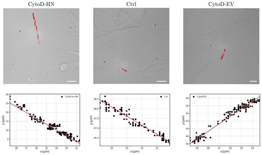
- [Section 2 (Data post-processing); Section 3 (Normality and Skewness tests)] The per-trajectory sign standardization described in Section 2 invalidates the skewness tests used as evidence for directed transport. The authors state that 'we standardized the preferential direction of the EVs towards positive values by inverting the sign of the displacements in samples where the linear regression slope computed on the tangent displacement was negative.' A purely diffusive finite trajectory almost always has a nonzero regression slope by chance, and flipping every trajectory with a negative slope mechanically creates a pooled displacement distribution with positive skew. The p-values reported in Section 3 (Ctrl p=0.00296, CytoD-EV p=0.0275, CytoD-HN p=0.731) therefore cannot be interpreted as evidence of a physical drift component. Because the model's central success criterion is reproducing the presence and absence of drift across conditions, the drift analysis must be rerun on unflipped tangent displacements, or with a reflection-invariant statistical test, and the paper must report whether the skewness result survives this correction.
- [Section 2 (Numerical method); Section 3 (Numerical vs in-vitro results)] The assignment of transport mechanisms to experimental conditions is assumed rather than tested. The numerical model hard-codes CytoD-HN as purely active and CytoD-EV as purely passive, with the text stating that 'these scenarios are distinguished by the presence of different vesicle transport mechanisms,' while the parameters in Table 4 are calibrated to the same experimental data used for comparison. Consequently the skewness agreement in 'Numerical vs in-vitro results' is partly manufactured: the drift in simulated CytoD-EV arises from the fitted asymmetry alpha=0.2 in V1, and the absence of drift in CytoD-HN is built into the spatially symmetric V2. The paper should validate the mechanism assignments against independent data, for example through the ATP-depletion or myosin-inhibition experiments already mentioned in the Conclusions, or explicitly reframe the results as a conditional demonstration of a hypothesis rather than a confirmation.
- [Section 3 (Parameters settings, Sensitivity analysis)] No identifiability or uncertainty analysis is provided for the calibrated parameters in Tables 3 and 4. The sensitivity analysis is one-at-a-time, so it does not address whether different parameter combinations could produce similar skewness, MSD, and histogram outputs, nor the effect of the limited sample sizes (n=15, 13, 14 after quality selection). Without confidence intervals, profile likelihoods, or an identifiability check, the mechanistic interpretation attached to individual fitted values, especially alpha, L1, and the switching rates, is not quantitatively supported. Please add an identifiability analysis and report parameter uncertainty, or clearly label the fitted values as representative rather than inferred.
- [Section 2 (Mathematical model, Eq. (3b))] The active transport equation (3b) is asserted rather than derived from a physical balance; the manuscript itself notes that 'despite its appearance, Eq.(3b) is not a momentum balance equation.' Since active transport is one of the two central mechanisms, the specific form of the symmetric sawtooth potential and the use of the effective friction xi_eff need a mechanistic derivation or, at minimum, a clear statement of which microphysical ingredients (for example, actin filament elongation rates or receptor detachment kinetics) produce each term. Moreover, because V2 is symmetric, the active mechanism cannot generate net drift by construction; the CytoD-HN case is therefore only a model of undirected rolling, and the absence of drift in CytoD-HN does not discriminate this active mechanism from a passive symmetric diffusive process.
minor comments (6)
- [Table 2] The unit for net mean velocity is listed as 'microm/s^-1' or 'um^-1' in the printed table; it should be microm/s.
- [Figure 1 caption] The caption writes 'Cyto-EV' while the text consistently uses 'CytoD-EV'; please unify the terminology.
- [Main text and Supplementary Information] Several cross-references appear as unresolved 'Section ??', both in the main text and in the supplementary material; these need to be fixed before publication.
- [Section 2 (Mathematical model, Eq. (3b))] The quantity Dr in Eq. (3b) is called a 'rotational diffusion coefficient' but it is assigned the translational value k_B T / xi_eff; please correct the terminology or use a distinct symbol.
- [Figure 9] The sensitivity axes for h2 are labeled in units of k_B T (for example, 'h2 = 1e6 kB T') while Table 4 reports h2 in joules; please make the units consistent or clearly state the conversion.
- [Table 3] The entry for the cytosol viscosity, 'x[10 - 1500] eta_w depending on the protein size', is garbled and should be rewritten as a clear range with a supporting reference.
Circularity Check
The paper's drift 'prediction' is built from fitted inputs: the experimental skewness evidence follows a sign-flipping preprocessing step, the simulated drift in Ctrl/CytoD-EV is produced by the calibrated asymmetric ratchet α=0.2, and the absence of drift in CytoD-HN is guaranteed by the symmetric active potential.
-
self definitional
[Section 2, 'Data post-processing'; used in Section 3, 'Normality and Skewness tests']
"As last step before the data analysis, in order to identify directed transport phenomena, we standardized the preferential direction of the EVs towards positive values by inverting the sign of the displacements in samples where the linear regression slope computed on the tangent displacement was negative."
The skewness test that later establishes a 'directed transport component' (Ctrl p=0.00296, CytoD-EV p=0.0275, CytoD-HN p=0.731) is applied to displacements that were sign-flipped whenever the trajectory's regression slope was negative. Since even a purely Brownian trajectory has a nonzero regression slope with high probability, this sign standardization makes the pooled displacement distribution positively biased by construction. The drift signal that the model is then designed to reproduce is therefore at least partly manufactured by the preprocessing step, rather than being independent evidence of a physical directed component.
-
fitted input called prediction
[Section 2, 'Mathematical model' (Eq. 2), Table 4, and Section 3, 'Sensitivity analysis' / 'Numerical vs in-vitro results']
"On the other hand, other parameters of the model, such as those that determine the shape of the ratchet potentials or the transition rates of the Markov processes, cannot be directly related to experimentally measured physical quantities and are therefore at best calibrated to match the experimental data, see Table 4. [...] We observe that for α <1/2, the histograms are shifted to the right, meaning that the EV transport is biased towards positive values. On the contrary, for α >1/2 the drift shifts towards negative values."
Table 4 calibrates α=0.2 for the passive ratchet potential V1, i.e. an asymmetric potential that produces positive drift by the flashing-ratchet mechanism cited in the paper. CytoD-HN, by contrast, is simulated with the spatially symmetric potential V2, which cannot produce net drift by symmetry. The subsequent numerical skewness 'agreement' — drift in Ctrl/CytoD-EV and no drift in CytoD-HN — therefore follows from the chosen fitted potential shapes and mechanism assignments rather than from an independent prediction. The sensitivity analysis explicitly shows that α controls the direction of drift, confirming that the simulated skewness is an output of a fitted input, not a free-of-fit result.
full rationale
The central validation claim — that the model reproduces the presence of a drifting component in Ctrl and CytoD-EV and its absence in CytoD-HN — reduces to fitted inputs by construction. On the experimental side, the skewness test is run after sign-flipping trajectories with negative regression slopes, which biases the pooled displacement distribution toward positive skew. On the modeling side, the simulated drift pattern is dictated by the calibrated asymmetric potential (α=0.2) for the passive ratchet and by the symmetric potential V2 for the active mechanism. The paper itself describes the relevant parameters as 'calibrated to match the experimental data' and acknowledges that 'further experimental validation with larger datasets would be necessary to fully confirm the proposed mechanistic interpretations.' This is the classic pattern of a fitted input being presented as a successful prediction, so the paper earns a partial-circularity score rather than a clean bill. There is independent content in the model — literature-based physical constants, comparisons of kinematic indicators, and the sensitivity analysis — and the mechanism assignment borrowed from D'Arrigo et al. (2021) is experimental rather than a self-defined uniqueness theorem. Those features prevent a higher score, but the load-bearing drift/no-drift comparison is not an independent test of the model.
Assumptions & free parameters
free parameters (12)
- k =
8e-9 N/m
- h1 =
1.7128e-14 J
- h2 =
2.141e-14 J
- alpha =
0.2
- pi1_off =
500 Hz
- pi1_on =
180 Hz
- pi2_off =
10 Hz
- pi2_on =
600 Hz
- eta_PA =
100 Hz
- eta_AP =
200 Hz
- L1 =
5e-6 m
- L2 =
6.9e-7 m
assumptions (6)
- domain assumption EV and receptor dynamics are overdamped Brownian with friction from Stokes law and Einstein relation.
- domain assumption EV and receptor are coupled by a Hookean spring force fel = k(y-x) with constant k.
- ad hoc to paper CytoD-EV is purely passive, CytoD-HN is purely active, Ctrl combines both mechanisms.
- ad hoc to paper Active rolling can be represented by a flashing symmetric sawtooth potential V2 with zero mean force.
- domain assumption EV motion is essentially one-dimensional along the neurite axis.
- domain assumption Binding, unbinding, and passive-active switching are Markov processes with constant rates.
Cite this review
Pith. "Pith review of Modeling the prion protein-mediated transport of extracellular vesicles on the neuron surface." pith.science (2026). https://pith.science/paper/KEO45I7L
@misc{pith2026250203610,
  author       = {Pith},
  title        = {Pith review of: Modeling the prion protein-mediated transport of extracellular vesicles on the neuron surface},
  year         = {2026},
  howpublished = {\url{https://pith.science/paper/KEO45I7L}},
  note         = {Machine review of arXiv:2502.03610}
}
read the original abstract
Neurodegenerative diseases are among the leading causes of global mortality, characterized by the progressive deterioration of specific neuron populations, ultimately leading to cognitive decline and dementia. Extracellular vesicles (EVs) are believed to play a role in the early stages of these diseases, acting as carriers of pathogens and contributing to neuroinflammation and disease propagation. This study presents a mathematical model aimed at characterizing the movement of EVs bearing prion protein (PrP) on their surface along neuronal surfaces. The model, informed by experimental data, investigates the influence of PrP and actin polymerization on EV transport dynamics and explores the possible interplay between passive and active mechanisms. EVs isolated from non-human astrocytes were analyzed under three conditions: untreated control (Ctrl), neurons treated with Cytochalasin D (CytoD-HN), and EVs treated with Cytochalasin D (CytoD-EV). The mathematical model is data-driven, testing different hypotheses regarding the underlying transport mechanisms. In the CytoD-EV dataset, EV movement was modeled using a flashing Brownian ratchet to represent directed motion. For active transport in the CytoD-HN set, a symmetric periodic potential was used to describe EV rolling along the neuron surface. The Ctrl scenario incorporates both mechanisms, reflecting a more complex transport behavior. A sensitivity analysis and comparison between numerical predictions and experimental data suggest that the model effectively captures key features of EV motion, providing a quantitative framework to interpret different transport regimes. While some variability remains, the approach offers a promising basis for future investigations into the role of cytoskeletal dynamics in EV-mediated disease propagation.
Figures


Figures from the paper (7 more)


Reference graph
Works this paper leans on
-
[1]
R. Ait-Haddou and W. Herzog. Brownian ratchet models of molecular motors. Cell biochemistry and biophysics , 38:191–213, 2003
work page 2003
-
[2]
A. Ajdari and J. Prost. Drift induced by a spatially periodic potential of low symmetry-pulsed dielectrophoresis. Comptes rendus de l academie des sciences serie II , 315(13):1635–1639, 1992
work page 1992
-
[3]
D. Andrini, V. Balbi, G. Bevilacqua, G. Lucci, G. Pozzi, and D. Riccobelli. Mathematical modelling of axonal cortex contractility. Brain Multiphysics, 3:100060, 2022
work page 2022
-
[4]
2012 alzheimer’s disease facts and figures
Alzheimer’s Association et al. 2012 alzheimer’s disease facts and figures. Alzheimer’s & dementia , 8(2):131–168, 2012
work page 2012
-
[5]
R. D. Astumian and M. Bier. Fluctuation driven ratchets: molecular motors. Physical review letters, 72(11):1766, 1994
work page 1994
-
[6]
J. J. Blum and M. C. Reed. A model for slow axonal transport and its application to neurofilamentous neuropathies. Cell motility and the cytoskeleton , 12(1):53–65, 1989
work page 1989
-
[7]
P. C. Bressloff. Stochastic processes in cell biology , volume 41. Springer, 2014
work page 2014
-
[8]
A. Brown. Slow axonal transport: stop and go traffic in the axon. Nature Reviews Molecular Cell Biology, 1(2):153–156, 2000
work page 2000
Show all 56 references
-
[9]
A. Brown. Slow axonal transport. In Larry R. Squire, editor, Encyclopedia of Neuroscience , pages 1–9. Academic Press, Oxford, 2009
2009
-
[10]
stop-and-go
A. Brown, L. Wang, and P. Jung. Stochastic simulation of neurofilament transport in axons: the “stop-and-go” hypothesis. Molecular biology of the cell , 16(9):4243–4255, 2005
2005
-
[11]
F. F. Damberger, B. Christen, D. R. P´ erez, S. Hornemann, and K. W¨ uthrich. Cellular prion protein conformation and function. Proceedings of the National Academy of Sciences , 108(42):17308–17313, 2011
2011
-
[12]
D’Arrigo, M
G. D’Arrigo, M. Gabrielli, F. Scaroni, P. Swuec, L. Amin, A. Pegoraro, E. Adinolfi, F. Di Virgilio, D. Cojoc, G. Legname, et al. Astrocytes-derived extracellular vesicles in motion at the neuron surface: Involvement of the prion protein. Journal of Extracellular Vesicles , 10(...
2021
-
[13]
De Rooij and E
R. De Rooij and E. Kuhl. Physical biology of axonal damage. Frontiers in cellular neuroscience , 12:144, 2018
2018
-
[14]
Drago, M
F. Drago, M. Lombardi, I. Prada, M. Gabrielli, P. Joshi, D. Cojoc, J. Franck, I. Fournier, J. Vizioli, and C. Verderio. Atp modifies the proteome of extracellular vesicles released by microglia and influences their action on astrocytes. Frontiers in pharmacology, 8:910, 2017
2017
-
[15]
B. N. Dugger and D. W. Dickson. Pathology of neurodegenerative diseases. Cold Spring Harbor perspectives in biology, 9(7):a028035, 2017
2017
-
[16]
S. N. Ethier and J. Lee. The flashing brownian ratchet and parrondo’s paradox. Royal Society Open Science, 5(1):171685, 2018
2018
-
[17]
R. P. Feynman, R. B. Leighton, M. Sands, et al. The Feynman lectures on physics , volume 1. Addison-Wesley Reading, MA, 1971
1971
-
[18]
Frost and M.I
B. Frost and M.I. Diamond. Prion-like mechanisms in neurodegenerative diseases. Nature Reviews Neuroscience, 11(3):155–159, 2010
2010
-
[19]
Gabrielli, I
M. Gabrielli, I. Prada, P. Joshi, C. Falcicchia, G. D’Arrigo, G. Rutigliano, E. Battocchio, R. Zenatelli, F. Tozzi, A. Radeghieri, et al. Microglial large extracellular vesicles propagate early synaptic dys- function in alzheimer’s disease. Brain, 145(8):2849–2868, 2022
2022
-
[20]
Gallart-Palau, X
X. Gallart-Palau, X. Guo, A. Serra, and S. K. Sze. Alzheimer’s disease progression characterized by alterations in the molecular profiles and biogenesis of brain extracellular vesicles. Alzheimer’s research & therapy, 12(1):1–15, 2020
2020
-
[21]
Garc´ ıa-Garc´ ıa, P
R. Garc´ ıa-Garc´ ıa, P. Collet, and L. Truskinovsky. Guided active particles. Physical Review E , 100(4):042608, 2019
2019
-
[22]
Goychuk, V
I. Goychuk, V. O. Kharchenko, and R. Metzler. Molecular motors pulling cargos in the viscoelastic cytosol: how power strokes beat subdiffusion. Physical Chemistry Chemical Physics , 16(31):16524– 16535, 2014
2014
-
[23]
S.V. Gupta. Units of measurement . Springer, 2020. 30
2020
-
[24]
Hardy and H
J. Hardy and H. Orr. The genetics of neurodegenerative diseases. Journal of neurochemistry , 97(6):1690–1699, 2006
2006
-
[25]
A. F. Hill. Extracellular vesicles and neurodegenerative diseases. Journal of Neuroscience , 39(47):9269–9273, 2019
2019
-
[26]
M. M. Holm, J. Kaiser, and M. E. Schwab. Extracellular vesicles: multimodal envoys in neural maintenance and repair. Trends in Neurosciences, 41(6):360–372, 2018
2018
-
[27]
Matlab version: 9.13.0 (r2022b), 2022
The MathWorks Inc. Matlab version: 9.13.0 (r2022b), 2022
2022
-
[28]
Jucker and L
M. Jucker and L. C. Walker. Propagation and spread of pathogenic protein assemblies in neurode- generative diseases. Nature neuroscience, 21(10):1341–1349, 2018
2018
-
[29]
Keller and C
D. Keller and C. Bustamante. The mechanochemistry of molecular motors. Biophysical journal , 78(2):541–556, 2000
2000
-
[30]
Korson, W
L. Korson, W. Drost-Hansen, and F. J. Millero. Viscosity of water at various temperatures. The Journal of Physical Chemistry , 73(1):34–39, 1969
1969
-
[31]
G. Legname. Elucidating the function of the prion protein. PLoS pathogens, 13(8):e1006458, 2017
2017
-
[32]
Lombardi, M
M. Lombardi, M. Gabrielli, E. Adinolfi, and C. Verderio. Role of atp in extracellular vesicle biogenesis and dynamics. Frontiers in Pharmacology, 12:654023, 2021
2021
-
[33]
M. O. Magnasco. Forced thermal ratchets. Physical Review Letters , 71(10):1477, 1993
1993
-
[34]
Marbach, J
S. Marbach, J. A. Zheng, and M. Holmes-Cerfon. The nanocaterpillar’s random walk: diffusion with ligand–receptor contacts. Soft Matter , 18(16):3130–3146, 2022
2022
-
[35]
McKinney et al
W. McKinney et al. Data structures for statistical computing in python. In Proceedings of the 9th Python in Science Conference , volume 445, pages 51–56. Austin, TX, 2010
2010
-
[36]
C. S. Peskin, G. B. Ermentrout, and G. F. Oster. The correlation ratchet: a novel mechanism for generating directed motion by atp hydrolysis. In Cell Mechanics and Cellular Engineering , pages 479–489. Springer, 1994. 31
1994
-
[37]
J. C. Polanco, C. Li, N. Durisic, R. Sullivan, and J. G¨ otz. Exosomes taken up by neurons hijack the endosomal pathway to spread to interconnected neurons. Acta neuropathologica communications, 6(1):1–14, 2018
2018
-
[38]
S. B. Prusiner. Biology and genetics of prions causing neurodegeneration. Annual review of genetics , 47:601–623, 2013
2013
-
[39]
H. Qian. The mathematical theory of molecular motor movement and chemomechanical energy transduction. Journal of Mathematical Chemistry , 27:219–234, 2000
2000
-
[40]
Riccobelli
D. Riccobelli. Active elasticity drives the formation of periodic beading in damaged axons. Physical Review E, 104(2):024417, 2021
2021
-
[41]
Riesner, K
D. Riesner, K. Kellings, K. Post, H. Wille, H. Serban, D. Groth, M. A. Baldwin, and S. B. Prusiner. Disruption of prion rods generates 10-nm spherical particles having high alpha-helical content and lacking scrapie infectivity. Journal of virology , 70(3):1714–1722, 1996
1996
-
[42]
Ruan, J.C
Z. Ruan, J.C. Delpech, S. Venkatesan Kalavai, A. A. Van Enoo, J. Hu, S. Ikezu, and T. Ikezu. P2rx7 inhibitor suppresses exosome secretion and disease phenotype in p301s tau transgenic mice. Molecular neurodegeneration, 15:1–14, 2020
2020
-
[43]
Sampaoli, A
S. Sampaoli, A. Agosti, G. Pozzi, and P. Ciarletta. A toy model of misfolded protein aggregation and neural damage propagation in neurodegenerative diseases. International Journal of Non-Linear Mechanics, 144:104083, 2022
2022
-
[44]
Sardar Sinha, A
M. Sardar Sinha, A. Ansell-Schultz, L. Civitelli, C. Hildesj¨ o, M. Larsson, L. Lannfelt, M. Ingelsson, and M. Hallbeck. Alzheimer’s disease pathology propagation by exosomes containing toxic amyloid- beta oligomers. Acta neuropathologica, 136:41–56, 2018
2018
-
[45]
Sarnataro, A
D. Sarnataro, A. Pepe, and C. Zurzolo. Cell biology of prion protein. Progress in molecular biology and translational science , 150:57–82, 2017
2017
-
[46]
Stephenson, E
J. Stephenson, E. Nutma, P. van der Valk, and S. Amor. Inflammation in cns neurodegenerative diseases. Immunology, 154(2):204–219, 2018
2018
-
[47]
Stokes et al
G.G. Stokes et al. On the effect of the internal friction of fluids on the motion of pendulums. 1851. 32
-
[48]
W. K. Surewicz and M. I. Apostol. Prion protein and its conformational conversion: a structural perspective. Prion Proteins, pages 135–167, 2011
2011
-
[49]
Toral and P
R. Toral and P. Colet. Stochastic numerical methods: an introduction for students and scientists . John Wiley & Sons, 2014
2014
-
[50]
Van den Bedem and E
H. Van den Bedem and E. Kuhl. Tau-ism: the yin and yang of microtubule sliding, detachment, and rupture. Biophysical journal, 109(11):2215–2217, 2015
2015
-
[51]
Van Rossum and F
G. Van Rossum and F. L. Drake Jr. Python reference manual . Centrum voor Wiskunde en Infor- matica Amsterdam, 1995
1995
-
[52]
Vilela, N
M. Vilela, N. Halidi, S. Besson, H. Elliott, K. Hahn, J. Tytell, and G. Danuser. Fluctuation analysis of activity biosensor images for the study of information flow in signaling pathways. In Methods in enzymology, volume 519, pages 253–276. Elsevier, 2013
2013
-
[53]
Virtanen, R
P. Virtanen, R. Gommers, T. E. Oliphant, M. Haberland, T. Reddy, D. Cournapeau, E. Burovski, P. Peterson, W. Weckesser, J. Bright, et al. Scipy 1.0: fundamental algorithms for scientific com- puting in python. Nature methods, 17(3):261–272, 2020
2020
-
[54]
Y. Wang, V. Balaji, S. Kaniyappan, L. Kr¨ uger, S. Irsen, K. Tepper, R. Chandupatla, W. Maetzler, A. Schneider, E. Mandelkow, et al. The release and trans-synaptic transmission of tau via exosomes. Molecular neurodegeneration, 12(1):1–25, 2017
2017
-
[55]
Weickenmeier, M
J. Weickenmeier, M. Jucker, A. Goriely, and E. Kuhl. A physics-based model explains the prion-like features of neurodegeneration in alzheimer’s disease, parkinson’s disease, and amyotrophic lateral sclerosis. Journal of the Mechanics and Physics of Solids , 124:264–281, 2019
2019
-
[56]
Weickenmeier, E
J. Weickenmeier, E. Kuhl, and A. Goriely. Multiphysics of prionlike diseases: Progression and atrophy. Physical review letters , 121(15):158101, 2018. 33
2018
Reviewed August 9, 2026 · model on record in the stance chip above.
Discussion (0). Continue with ORCID to comment.