REVIEW 5 major objections 6 minor 62 references
Learnable Prompting SAM-induced Knowledge Distillation for Semi-supervised Medical Image Segmentation
T0 review · 5 major / 6 minor · reviewed 2026-08-11 · deepseek-v4-flash
Pith's one-line read KnowSAM reports that distilling a SAM teacher's softened predictions into two co-trained subnets beats prior semi-supervised medical segmentation methods on five benchmarks.
desk verdict A broad, cleanly written SAM-distillation SSL paper; the empirical sweep is impressive but the teacher is fed the students' fused predictions, so the key novelty is unverified. read the letter →
The pith
A machine-rendered reading of the paper's core claim, the machinery that carries it, and where it could break.
The reading
What carries the argument
The load-bearing mechanism is a teacher-student loop closed through SAM's decoder. Two subnets produce predictions $\hat{Y}_a$ and $\hat{Y}_b$; their entropy maps $\mathcal{H}_a, \mathcal{H}_b$ and an XOR dissimilarity map $\mathcal{M}$ are fused into an aggregated map $\hat{Y}_f$, which is fed to SAM as a mask prompt. A lightweight network $\psi(\cdot)$ maps SAM's image embedding $Z$ to a learnable dense prompt $P_b$, and SAM's decoder computes $\hat{Y}_s = F_s(P_b, \hat{Y}_f; \Theta_s)$. The distillation loss is $L_{kd} = KL(\hat{Y}^a_T, \hat{Y}^s_T) + KL(\hat{Y}^b_T, \hat{Y}^s_T)$ with temperature-softened probability maps, and gradients flow only into the two subnets, so the students learn to reproduce SAM's refined predictions while SAM itself is supervised only by ground-truth labels.
What would settle it
On a held-out labeled subset, compute SAM's prediction when prompted by the fused student mask and by a mask from an independent source; if SAM's output with the fused prompt is not measurably closer to the ground truth than the students' fused output is, the distillation gain cannot be coming from new SAM knowledge.
Extended reading notes
Core claim
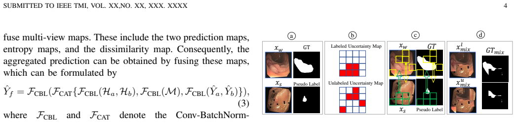
The central discovery is that a foundation segmenter can be turned into an effective teacher for semi-supervised medical segmentation if its prompt is the fused prediction of two student networks and its softened outputs are distilled back into those students. The authors call this SAM-induced Knowledge Distillation (SKD). Around that loop, a learnable prompt strategy generates dense feature prompts through a lightweight network instead of hard box coordinates, and an adapter fine-tunes SAM for medical data; a multi-view co-training module fuses the two subnets' predictions with entropy and dissimilarity maps; and an uncertainty-guided copy-paste augmentation creates mixed training samples. On the BCSS pathology benchmark with 10% labeled data, KnowSAM reaches 72.69 Dice compared with 66.00 for BCP, and the paper reports consistent improvements across all five evaluated medical segmentation tasks.
Load-bearing premise
The framework assumes SAM's prediction is better than the students' fused output, so that distilling it back into the students adds real new information instead of amplifying their existing mistakes.
Editorial extensions
If this is right
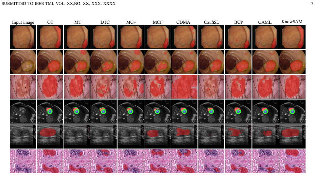
- On the BCSS pathology benchmark with 10% labeled data, KnowSAM reaches 72.69 Dice versus 66.00 for BCP, and the pattern of improvement repeats on colonoscopy, ISIC-2018, thyroid ultrasound, and ACDC.
- The SAM distillation component alone, without the entropy or mutual-consistency losses, already produces large gains, so the KD loop carries much of the reported improvement.
- At inference time only the two lightweight subnets are needed; the SAM teacher can be discarded, so the accuracy gain does not add runtime cost.
- Because the design is presented as a general distillation wrapper, other semi-supervised segmentation methods can adopt the same SAM-teacher loop, which the paper states explicitly.
- Replacing hard box coordinates with a learnable dense prompt avoids the sharp performance drops that incorrect box prompts cause with SAM; the ablation shows the learnable prompt strategy consistently beats box prompting for SAM on all four tested datasets.
Reading between the lines
- The reported gains are all measured with the students' own fused mask as SAM's prompt; an untested consequence is that the loop may be partly self-confirming. Comparing against an independently prompted SAM would separate genuine transfer from echo.
- The method's reliance on SAM means its performance on out-of-distribution medical images is tied to how well SAM generalizes there; a domain-shift stress test with unseen scanners or stain types would reveal whether the distillation adds robustness or only in-distribution accuracy.
- Because the KL distillation is applied to unlabeled data with no supervised term, the benefit should scale with the amount and diversity of unlabeled data; the paper does not report a scaling curve with unlabeled count varied independently, which would be a natural follow-up.
Editorial analysis
A structured set of objections, weighed in public.
Referee Report
Summary. The paper proposes KnowSAM, a semi-supervised medical image segmentation framework that combines a multi-view co-training scheme with two sub-networks, a learnable prompt strategy for SAM that feeds a fused mask prompt and learned dense feature prompts into SAM's decoder, a SAM-induced knowledge distillation loss that pushes the two sub-networks toward SAM's softened predictions, and an uncertainty-guided copy-paste data augmentation. The method is evaluated on five colonoscopy datasets, ISIC-2018, thyroid ultrasound datasets, ACDC, and BCSS at 10% and 30% labeled ratios, reporting Dice/IoU/95HD improvements over several existing semi-supervised methods. The paper also presents ablations for the hybrid aggregation module, the prompt strategy, the data augmentation, and the loss components.
Significance. If the SAM-knowledge-distillation mechanism genuinely transfers external knowledge beyond the sub-networks' own consensus, the framework would be a valuable contribution to semi-supervised medical image segmentation, and the reported gains over strong baselines such as BCP and CAML are potentially interesting. The paper deserves credit for a broad experimental scope, a fairly detailed ablation study, and a planned code release. However, the central claim that SAM provides independent knowledge is not convincingly established, because the mask prompt given to SAM is the students' own fused prediction, making the distillation loop partly self-referential. The absence of error bars or repeated label splits further weakens confidence in the point-estimate improvements. The paper is well structured and clearly written, but the load-bearing experimental evidence needs strengthening.
major comments (5)
- [Section III-C, Eqs. (7) and (10)] The knowledge-distillation loop is self-referential: the mask prompt for SAM is the fused prediction Yf of the two subnets (Eq. 3), and Eq. (7) produces Ys = Fs(Pb, Yf; Θs). The distillation loss Lkd in Eq. (10) then trains each subnet to match Ys. Since SAM's decoder is strongly conditioned on the mask prompt, Ys may be a near-copy of Yf, making Lkd essentially a consistency loss between each subnet and their own fused output rather than a transfer of external knowledge. The paper reports no control with an independent teacher (e.g., SAM prompted by fixed coordinates or by ground-truth masks on labeled data) and no comparison with a prompt-free SAM variant. Please add such ablations and quantify the agreement between Ys and Yf on unlabeled data to demonstrate that SAM injects information beyond the subnets' own consensus.
- [Section IV-D, Table V] The LPS ablation is internally inconsistent with the distillation claim. On Kvasir-10%, SAM with LPS alone reaches 87.29 Dice, while the final KnowSAM student with LPS reaches only 85.98; on ISIC-10% the corresponding numbers are 87.90 for SAM and 86.51 for KnowSAM. Moreover, KnowSAM with LPS is worse than KnowSAM with Box prompting on Kvasir-10% (85.98 vs 86.32). If SKD successfully transferred SAM's knowledge, the students should approach or exceed the teacher's performance; the observed gap instead suggests that Lkd acts as a weak regularizer. Please reconcile these numbers, clarify whether the final inference uses Yf or a single subnet output, and discuss why LPS improves SAM but does not translate into an improvement of the final KnowSAM student on these datasets.
- [Section IV-C, Tables I-III] All reported results are single point estimates from one labeled/unlabeled split, without error bars, multiple random splits, or statistical significance tests. This is especially consequential at the 10% labeled ratio, where the ACDC experiment uses only 7 labeled scans and the reported gain over the second-best method is as small as 0.51 Dice points (TN3K-10%). Please provide mean ± std over at least three random splits, or otherwise justify that the reported differences are stable and not artifacts of a particular split.
- [Section IV-A and Table III] The BCSS experimental setup is inconsistent. The dataset description states that 3,888 images are divided into training, validation, and test sets in the proportions 70%, 10%, and 20%, which implies roughly 2,721 training images and about 272 labeled images at a 10% labeled ratio. However, Table III reports '7(10%) 63(90%)', which appears to be the ACDC scan counts rather than the BCSS image counts. Please correct the label count or clarify the actual data split used; as written, the table is not interpretable and the '10% labeled' claim for BCSS is ambiguous.
- [Abstract and Section V] The paper repeatedly claims that the SAM distillation framework 'can be seamlessly integrated into other semi-supervised segmentation methods' and 'easily extended' to other tasks, but no experiment demonstrates such integration. The only comparison is between the proposed MC-seg baseline and the full KnowSAM; no experiment plugs SKD into an existing method such as BCP or UA-MT. Either provide integration experiments that substantiate this claim or remove/weaken it in the abstract and conclusion.
minor comments (6)
- [Eq. (13)] Equation (13) sums over classes c=0 to C, while Eq. (1) sums over c=0 to C-1; please make the index range consistent and clarify whether the background class is included in the entropy loss.
- [Section IV-C] The text says the comparison includes 12 SOTA methods but then enumerates only nine (MT, UA-MT, DTC, MC-Net, MC-Net+, URPC, MCF, BCP, CAML). CDMA, CauSSL, and BS-Net appear in the tables but are missing from the enumeration; please correct the list.
- [Fig. 2 caption] The caption refers to 'two types of prompts (Yf and the learnable feature prompt)', but the learnable feature prompt is denoted Pb in the text; please unify the notation between the figure and the equations.
- [Table V] The table rows labeled 'SAM Box', 'SAM LPS', 'KnowSAM Box', and 'KnowSAM LPS' should explicitly state whether the numbers are SAM's own predictions or the final fused inference of the subnets; the accompanying text says 'we report the predicted results of SAM and the fused predictions Yf', but the row labels do not make this distinction clear.
- [Section IV-D.2] The sentence 'This is attributed to a learnable feature prompt provided by the LPS' is vague; it should specify the mechanism by which the learned dense prompt tokens improve SAM's predictions compared with hard coordinate prompts.
- [Section III-C] Please clarify whether the SAM teacher is fine-tuned during training and whether the gradient of Lkd is blocked from flowing into SAM; Eq. (8) shows a supervised loss on SAM, but the interaction between Lsam and Lkd is not fully described.
Circularity Check
No significant circularity: the SAM teacher is student-conditioned, but the equations do not reduce the teacher prediction to the students' own outputs by construction.
full rationale
The central mechanism is Eq. 7, Ys = Fs(Pb, Yf; Theta_s), where Yf (Eq. 3) is a learned fusion of the two subnets' outputs. This does make the SAM teacher partly student-conditioned, and Lkd (Eq. 10) therefore contains a self-consistency component. However, Ys is not equal to Yf by construction: it also depends on SAM's image encoder, the learnable feature prompt Pb = psi(Z; Theta_m), and adapter parameters fine-tuned with Lsam (Eq. 8) on labeled ground truth. The paper's ablations even quantify an information gap: in Table V, SAM with LPS reaches 87.29 Dice on Kvasir-10% while the final KnowSAM student reaches 85.98, so the teacher's output is not merely an echo of the student fusion. The absence of a control with a teacher whose prompt is independent of the subnets weakens attribution of the gains to SAM's external knowledge, but that is an experimental-design limitation rather than a definitional reduction. No load-bearing self-citation or imported uniqueness theorem is used; the cited adapter, mutual-supervision, and baseline methods are external. The empirical comparisons are against outside benchmarks. Therefore no step in the claimed derivation is equivalent to its inputs by construction.
Assumptions & free parameters
free parameters (5)
- lambda_e (entropy loss weight) =
0.9
- beta and Gaussian warm-up decay factor for lambda_m =
beta=1, decay factor=5
- Number of patches N and top-k in UGDA =
N=16, top-5
- KD temperature T =
unspecified
- Number of feature prompts Nb and dimension L =
unspecified
assumptions (5)
- domain assumption Entropy minimization on unlabeled predictions improves semi-supervised segmentation.
- domain assumption SAM's pre-trained features, after adapter tuning, are useful for medical image segmentation.
- domain assumption The fused prediction Yf is a reliable mask prompt for SAM.
- ad hoc to paper Distilling SAM's output Ys into the subnets is beneficial even though Ys depends on the subnets' own fused prediction.
- domain assumption The random 10% and 30% labeled splits are representative and do not need repeated sampling.
Cite this review
Pith. "Pith review of Learnable Prompting SAM-induced Knowledge Distillation for Semi-supervised Medical Image Segmentation." pith.science (2026). https://pith.science/paper/KG4JCNJE
@misc{pith2026241213742,
  author       = {Pith},
  title        = {Pith review of: Learnable Prompting SAM-induced Knowledge Distillation for Semi-supervised Medical Image Segmentation},
  year         = {2026},
  howpublished = {\url{https://pith.science/paper/KG4JCNJE}},
  note         = {Machine review of arXiv:2412.13742}
}
read the original abstract
The limited availability of labeled data has driven advancements in semi-supervised learning for medical image segmentation. Modern large-scale models tailored for general segmentation, such as the Segment Anything Model (SAM), have revealed robust generalization capabilities. However, applying these models directly to medical image segmentation still exposes performance degradation. In this paper, we propose a learnable prompting SAM-induced Knowledge distillation framework (KnowSAM) for semi-supervised medical image segmentation. Firstly, we propose a Multi-view Co-training (MC) strategy that employs two distinct sub-networks to employ a co-teaching paradigm, resulting in more robust outcomes. Secondly, we present a Learnable Prompt Strategy (LPS) to dynamically produce dense prompts and integrate an adapter to fine-tune SAM specifically for medical image segmentation tasks. Moreover, we propose SAM-induced Knowledge Distillation (SKD) to transfer useful knowledge from SAM to two sub-networks, enabling them to learn from SAM's predictions and alleviate the effects of incorrect pseudo-labels during training. Notably, the predictions generated by our subnets are used to produce mask prompts for SAM, facilitating effective inter-module information exchange. Extensive experimental results on various medical segmentation tasks demonstrate that our model outperforms the state-of-the-art semi-supervised segmentation approaches. Crucially, our SAM distillation framework can be seamlessly integrated into other semi-supervised segmentation methods to enhance performance. The code will be released upon acceptance of this manuscript at: https://github.com/taozh2017/KnowSAM
Figures


Figures from the paper (4 more)


Reference graph
Works this paper leans on
-
[1]
Deep adversarial networks for biomedical image segmentation utilizing unannotated images,
Y . Zhang, L. Yang, J. Chen, M. Fredericksen, D. P. Hughes, and D. Z. Chen, “Deep adversarial networks for biomedical image segmentation utilizing unannotated images,” in Proc. MICCAI. Springer, 2017, pp. 408–416
work page 2017
-
[2]
Uncertainty-aware hierarchical aggregation network for medical image segmentation,
T. Zhou, Y . Zhou, G. Li, G. Chen, and J. Shen, “Uncertainty-aware hierarchical aggregation network for medical image segmentation,” IEEE TCSVT , vol. 34, no. 8, pp. 7440–7453, 2024
work page 2024
-
[3]
Embracing imperfect datasets: A review of deep learning solutions for medical image segmentation,
N. Tajbakhsh, L. Jeyaseelan, Q. Li, J. N. Chiang, Z. Wu, and X. Ding, “Embracing imperfect datasets: A review of deep learning solutions for medical image segmentation,” Medical Image Analysis , vol. 63, p. 101693, 2020. SUBMITTED TO IEEE TMI, VOL. XX,NO. XX, XXX. XXXX 11
work page 2020
-
[4]
Bidirectional copy- paste for semi-supervised medical image segmentation,
Y . Bai, D. Chen, Q. Li, W. Shen, and Y . Wang, “Bidirectional copy- paste for semi-supervised medical image segmentation,” in Proc. CVPR, 2023, pp. 11 514–11 524
work page 2023
-
[5]
Correlation-aware mutual learning for semi-supervised medical image segmentation,
S. Gao, Z. Zhang, J. Ma, Z. Li, and S. Zhang, “Correlation-aware mutual learning for semi-supervised medical image segmentation,” in Proc. MICCAI. Springer, 2023, pp. 98–108
work page 2023
-
[6]
Semi-supervised medical image segmentation through dual-task consistency,
X. Luo, J. Chen, T. Song, and G. Wang, “Semi-supervised medical image segmentation through dual-task consistency,” in Proc. AAAI , vol. 35, no. 10, 2021, pp. 8801–8809
work page 2021
-
[7]
Semi-supervised medical image segmentation via uncertainty rectified pyramid consistency,
X. Luo, G. Wang, W. Liao, J. Chen, T. Song, Y . Chen, S. Zhang, D. N. Metaxas, and S. Zhang, “Semi-supervised medical image segmentation via uncertainty rectified pyramid consistency,” Medical Image Analysis , vol. 80, p. 102517, 2022
work page 2022
-
[8]
Semi-supervised left atrium segmentation with mutual consistency training,
Y . Wu, M. Xu, Z. Ge, J. Cai, and L. Zhang, “Semi-supervised left atrium segmentation with mutual consistency training,” in Proc. MICCAI . Springer, 2021, pp. 297–306
work page 2021
Show all 62 references
-
[9]
Mcf: Mutual correction framework for semi-supervised medical image segmentation,
Y . Wang, B. Xiao, X. Bi, W. Li, and X. Gao, “Mcf: Mutual correction framework for semi-supervised medical image segmentation,” in Proc. CVPR, 2023, pp. 15 651–15 660
2023
-
[10]
Mean teachers are better role models: Weight-averaged consistency targets improve semi-supervised deep learning results,
A. Tarvainen and H. Valpola, “Mean teachers are better role models: Weight-averaged consistency targets improve semi-supervised deep learning results,” Proc. NeurIPS, vol. 30, 2017
2017
-
[11]
Ambiguity-selective consistency regularization for mean-teacher semi-supervised medical image segmentation,
Z. Xu, Y . Wang, D. Lu, X. Luo, J. Yan, Y . Zheng, and R. K.-y. Tong, “Ambiguity-selective consistency regularization for mean-teacher semi-supervised medical image segmentation,” Medical Image Analysis, vol. 88, p. 102880, 2023
2023
-
[12]
Uncertainty-aware self-ensembling model for semi-supervised 3D left atrium segmenta- tion,
L. Yu, S. Wang, X. Li, C.-W. Fu, and P.-A. Heng, “Uncertainty-aware self-ensembling model for semi-supervised 3D left atrium segmenta- tion,” in Proc. MICCAI. Springer, 2019, pp. 605–613
2019
-
[13]
Semi-supervised medical image segmentation via a tripled-uncertainty guided mean teacher model with contrastive learning,
K. Wang, B. Zhan, C. Zu, X. Wu, J. Zhou, L. Zhou, and Y . Wang, “Semi-supervised medical image segmentation via a tripled-uncertainty guided mean teacher model with contrastive learning,” Medical Image Analysis, vol. 79, p. 102447, 2022
2022
-
[14]
Co- training with high-confidence pseudo labels for semi-supervised medical image segmentation,
Z. Shen, P. Cao, H. Yang, X. Liu, J. Yang, and O. R. Zaiane, “Co- training with high-confidence pseudo labels for semi-supervised medical image segmentation,” arXiv preprint arXiv:2301.04465 , 2023
2023 arXiv
-
[15]
Semi-supervised contrastive learn- ing for label-efficient medical image segmentation,
X. Hu, D. Zeng, X. Xu, and Y . Shi, “Semi-supervised contrastive learn- ing for label-efficient medical image segmentation,” in Proc. MICCAI. Springer, 2021, pp. 481–490
2021
-
[16]
Dense biased networks with deep priori anatomy and hard region adaptation: Semi- supervised learning for fine renal artery segmentation,
Y . He, G. Yang, J. Yang, Y . Chen, Y . Kong, J. Wuet al., “Dense biased networks with deep priori anatomy and hard region adaptation: Semi- supervised learning for fine renal artery segmentation,” Medical Image Analysis, vol. 63, p. 101722, 2020
2020
-
[17]
Segment anything,
A. Kirillov, E. Mintun, N. Ravi, H. Mao, C. Rolland, L. Gustafson, T. Xiao, S. Whitehead, A. C. Berg, W.-Y . Loet al., “Segment anything,” arXiv preprint arXiv:2304.02643 , 2023
2023 arXiv
-
[18]
SAM fails to segment anything?–sam-adapter: Adapting sam in un- derperformed scenes: Camouflage, shadow, and more,
T. Chen, L. Zhu, C. Ding, R. Cao, S. Zhang, Y . Wang et al. , “SAM fails to segment anything?–sam-adapter: Adapting sam in un- derperformed scenes: Camouflage, shadow, and more,” arXiv preprint arXiv:2304.09148, 2023
2023 arXiv
-
[19]
Medical sam adapter: Adapting segment anything model for medical image segmentation,
J. Wu, R. Fu, H. Fang, Y . Liu, Z. Wang, Y . Xu, Y . Jin, and T. Arbel, “Medical sam adapter: Adapting segment anything model for medical image segmentation,” arXiv preprint arXiv:2304.12620 , 2023
2023 arXiv
-
[20]
Concatenate, fine- tuning, re-training: A SAM-enabled framework for semi-supervised 3d medical image segmentation,
S. Li, L. Qi, Q. Yu, J. Huo, Y . Shi, and Y . Gao, “Concatenate, fine- tuning, re-training: A SAM-enabled framework for semi-supervised 3d medical image segmentation,” arXiv preprint arXiv:2403.11229 , 2024
2024 arXiv
-
[21]
Esp-medsam: Efficient self-prompting sam for universal domain-generalized medical image segmentation,
Q. Xu, J. Li, X. He, Z. Liu, Z. Chen, W. Duan et al. , “Esp-medsam: Efficient self-prompting sam for universal domain-generalized medical image segmentation,” arXiv preprint arXiv:2407.14153 , 2024
2024 arXiv
-
[22]
Semisam: Exploring sam for enhancing semi-supervised medical image segmentation with extremely limited annotations,
Y . Zhang, Y . Cheng, and Y . Qi, “Semisam: Exploring sam for enhancing semi-supervised medical image segmentation with extremely limited annotations,” arXiv preprint arXiv:2312.06316 , 2023
2023 arXiv
-
[23]
Segment anything model with uncertainty rectification for auto-prompting medical image segmentation,
Y . Zhang, S. Hu, C. Jiang, Y . Cheng, and Y . Qi, “Segment anything model with uncertainty rectification for auto-prompting medical image segmentation,” arXiv preprint arXiv:2311.10529 , 2023
2023 arXiv
-
[24]
SS-TBN: A semi-supervised tri-branch network for covid-19 screening and lesion segmentation,
L.-L. Zeng, K. Gao, D. Hu, Z. Feng, C. Hou, P. Rong, and W. Wang, “SS-TBN: A semi-supervised tri-branch network for covid-19 screening and lesion segmentation,” IEEE TPAMI , vol. 45, no. 8, pp. 10 427– 10 442, 2023
2023
-
[25]
Enhancing pseudo label quality for semi- supervised domain-generalized medical image segmentation,
H. Yao, X. Hu, and X. Li, “Enhancing pseudo label quality for semi- supervised domain-generalized medical image segmentation,” in Proc. AAAI, vol. 36, no. 3, 2022, pp. 3099–3107
2022
-
[26]
Learning with limited annotations: a survey on deep semi-supervised learning for medical image segmentation,
R. Jiao, Y . Zhang, L. Ding, B. Xue, J. Zhang, R. Cai, and C. Jin, “Learning with limited annotations: a survey on deep semi-supervised learning for medical image segmentation,” CIBM, p. 107840, 2023
2023
-
[27]
SSA-Net: Spatial self-attention network for COVID- 19 pneumonia infection segmentation with semi-supervised few-shot learning,
X. Wang, Y . Yuan, D. Guo, X. Huang, Y . Cui, M. Xia, Z. Wang, C. Bai, and S. Chen, “SSA-Net: Spatial self-attention network for COVID- 19 pneumonia infection segmentation with semi-supervised few-shot learning,” Medical Image Analysis , vol. 79, p. 102459, 2022
2022
-
[28]
Semi-supervised neuron segmentation via reinforced consistency learn- ing,
W. Huang, C. Chen, Z. Xiong, Y . Zhang, X. Chen, X. Sun, and F. Wu, “Semi-supervised neuron segmentation via reinforced consistency learn- ing,” IEEE TMI , vol. 41, no. 11, pp. 3016–3028, 2022
2022
-
[29]
Double noise mean teacher self- ensembling model for semi-supervised tumor segmentation,
K. Zheng, J. Xu, and J. Wei, “Double noise mean teacher self- ensembling model for semi-supervised tumor segmentation,” in Proc. ICASSP. IEEE, 2022, pp. 1446–1450
2022
-
[30]
Revisiting weak-to- strong consistency in semi-supervised semantic segmentation,
L. Yang, L. Qi, L. Feng, W. Zhang, and Y . Shi, “Revisiting weak-to- strong consistency in semi-supervised semantic segmentation,” in Proc. CVPR, 2023, pp. 7236–7246
2023
-
[31]
MTANS: multi-scale mean teacher combined adversarial network with shape-aware embedding for semi-supervised brain lesion segmentation,
G. Chen, J. Ru, Y . Zhou, I. Rekik, Z. Pan, X. Liu, Y . Lin, B. Lu, and J. Shi, “MTANS: multi-scale mean teacher combined adversarial network with shape-aware embedding for semi-supervised brain lesion segmentation,” NeuroImage, vol. 244, p. 118568, 2021
2021
-
[32]
Uncertainty- guided dual-views for semi-supervised volumetric medical image seg- mentation,
H. Peiris, M. Hayat, Z. Chen, G. Egan, and M. Harandi, “Uncertainty- guided dual-views for semi-supervised volumetric medical image seg- mentation,” Nature Machine Intelligence , vol. 5, no. 7, pp. 724–738, 2023
2023
-
[33]
Collaborative and adversarial learning of focused and dispersive representations for semi-supervised polyp segmentation,
H. Wu, G. Chen, Z. Wen, and J. Qin, “Collaborative and adversarial learning of focused and dispersive representations for semi-supervised polyp segmentation,” in Proc. ICCV, 2021, pp. 3489–3498
2021
-
[34]
Deep mutual distillation for semi- supervised medical image segmentation,
Y . Xie, Y . Yin, Q. Li, and Y . Wang, “Deep mutual distillation for semi- supervised medical image segmentation,” in Proc. MICCAI. Springer, 2023, pp. 540–550
2023
-
[35]
Simcvd: Simple contrastive voxel-wise representation distillation for semi-supervised medical image segmentation,
C. You, Y . Zhou, R. Zhao, L. Staib, and J. S. Duncan, “Simcvd: Simple contrastive voxel-wise representation distillation for semi-supervised medical image segmentation,” IEEE TMI , vol. 41, no. 9, pp. 2228– 2237, 2022
2022
-
[36]
Bootstrapping semi-supervised medical image segmentation with anatomical-aware contrastive distillation,
C. You, W. Dai, Y . Min, L. Staib, and J. S. Duncan, “Bootstrapping semi-supervised medical image segmentation with anatomical-aware contrastive distillation,” in Proc. IPMI. Springer, 2023, pp. 641–653
2023
-
[37]
Cross-mix monitoring for medical image segmentation with limited supervision,
Y . Shu, H. Li, B. Xiao, X. Bi, and W. Li, “Cross-mix monitoring for medical image segmentation with limited supervision,” IEEE TMM , vol. 25, pp. 1700–1712, 2022
2022
-
[38]
S2me: Spatial- spectral mutual teaching and ensemble learning for scribble-supervised polyp segmentation,
A. Wang, M. Xu, Y . Zhang, M. Islam, and H. Ren, “S2me: Spatial- spectral mutual teaching and ensemble learning for scribble-supervised polyp segmentation,” arXiv preprint arXiv:2306.00451 , 2023
2023 arXiv
-
[39]
Semi-supervised semantic segmentation with cross pseudo supervision,
X. Chen, Y . Yuan, G. Zeng, and J. Wang, “Semi-supervised semantic segmentation with cross pseudo supervision,” in Proc. CVPR, 2021, pp. 2613–2622
2021
-
[40]
Semi-supervised patho- logical image segmentation via cross distillation of multiple attentions,
L. Zhong, X. Liao, S. Zhang, and G. Wang, “Semi-supervised patho- logical image segmentation via cross distillation of multiple attentions,” arXiv preprint arXiv:2305.18830 , 2023
2023 arXiv
-
[41]
Distilling the knowledge in a neural network,
G. Hinton, O. Vinyals, and J. Dean, “Distilling the knowledge in a neural network,” arXiv preprint arXiv:1503.02531 , 2015
2015 arXiv
-
[42]
Cutmix: Reg- ularization strategy to train strong classifiers with localizable features,
S. Yun, D. Han, S. J. Oh, S. Chun, J. Choe, and Y . Yoo, “Cutmix: Reg- ularization strategy to train strong classifiers with localizable features,” in Proc. ICCV, 2019, pp. 6023–6032
2019
-
[43]
Constructing and exploring intermediate domains in mixed domain semi-supervised medical image segmentation,
Q. Ma, J. Zhang, L. Qi, Q. Yu, Y . Shi, and Y . Gao, “Constructing and exploring intermediate domains in mixed domain semi-supervised medical image segmentation,” in Proc. CVPR, 2024, pp. 11 642–11 651
2024
-
[44]
Mutual consistency learning for semi-supervised medical image segmentation,
Y . Wu, Z. Ge, D. Zhang, M. Xu, L. Zhang, Y . Xia, and J. Cai, “Mutual consistency learning for semi-supervised medical image segmentation,” Medical Image Analysis , vol. 81, p. 102530, 2022
2022
-
[45]
Caussl: Causality- inspired semi-supervised learning for medical image segmentation,
J. Miao, C. Chen, F. Liu, H. Wei, and P.-A. Heng, “Caussl: Causality- inspired semi-supervised learning for medical image segmentation,” in Proc. ICCV, 2023, pp. 21 426–21 437
2023
-
[46]
Bilateral supervision network for semi-supervised medical image segmentation,
A. He, T. Li, J. Yan, K. Wang, and H. Fu, “Bilateral supervision network for semi-supervised medical image segmentation,” IEEE TMI , vol. 43, no. 5, pp. 1715–1726, 2024
2024
-
[47]
Toward embedded detection of polyps in wce images for early diagnosis of colorectal cancer,
J. Silva, A. Histace, O. Romain, X. Dray, and B. Granado, “Toward embedded detection of polyps in wce images for early diagnosis of colorectal cancer,” IJCARS, vol. 9, pp. 283–293, 2014
2014
-
[48]
WM-DOV A maps for accurate polyp highlighting in colonoscopy: Validation vs. saliency maps from physicians,
J. Bernal, F. J. S ´anchez, G. Fern ´andez-Esparrach, D. Gil, C. Rodr ´ıguez, and F. Vilari ˜no, “WM-DOV A maps for accurate polyp highlighting in colonoscopy: Validation vs. saliency maps from physicians,” CMIG, vol. 43, pp. 99–111, 2015
2015
-
[49]
Automated polyp detection in colonoscopy videos using shape and context information,
N. Tajbakhsh, S. R. Gurudu, and J. Liang, “Automated polyp detection in colonoscopy videos using shape and context information,” IEEE TMI, vol. 35, no. 2, pp. 630–644, 2015
2015
-
[50]
A benchmark for endoluminal scene segmentation of colonoscopy images,
D. V ´azquez, J. Bernal, F. J. S ´anchez, G. Fern ´andez-Esparrach, A. M. L´opez, A. Romero, M. Drozdzal, A. Courville et al. , “A benchmark for endoluminal scene segmentation of colonoscopy images,” Journal of Healthcare Engineering , vol. 2017, 2017
2017
-
[51]
Kvasir-seg: A segmented polyp dataset,
D. Jha, P. H. Smedsrud, M. A. Riegler, P. Halvorsen, T. de Lange, D. Johansen, and H. D. Johansen, “Kvasir-seg: A segmented polyp dataset,” in MMM. Springer, 2020, pp. 451–462
2020
-
[52]
Pranet: Parallel reverse attention network for polyp segmentation,
D.-P. Fan, G.-P. Ji, T. Zhou, G. Chen, H. Fu, J. Shen, and L. Shao, “Pranet: Parallel reverse attention network for polyp segmentation,” in Proc. MICCAI. Springer, 2020, pp. 263–273. SUBMITTED TO IEEE TMI, VOL. XX,NO. XX, XXX. XXXX 12
2020
-
[53]
Thyroid region prior guided attention for ultrasound segmentation of thyroid nodules,
H. Gong, J. Chen, G. Chen, H. Li, G. Li, and F. Chen, “Thyroid region prior guided attention for ultrasound segmentation of thyroid nodules,” CIBM, vol. 155, p. 106389, 2023
2023
-
[54]
Comparison of thyroid segmentation techniques for 3D ultrasound,
T. Wunderling, B. Golla, P. Poudel, C. Arens, M. Friebe, and C. Hansen, “Comparison of thyroid segmentation techniques for 3D ultrasound,” in Medical Imaging: Image Processing , vol. 10133, 2017, pp. 346–352
2017
-
[55]
An open access thyroid ultrasound image database,
L. Pedraza, C. Vargas, F. Narv ´aez, O. Dur´an, E. Mu˜noz, and E. Romero, “An open access thyroid ultrasound image database,” in Proc. ISMIPA, vol. 9287. SPIE, 2015, pp. 188–193
2015
-
[56]
Skin lesion analysis toward melanoma detection 2018: A challenge hosted by the international skin imaging collaboration (isic),
N. Codella, V . Rotemberg, P. Tschandl, M. E. Celebi, S. Dusza, D. Gut- man et al. , “Skin lesion analysis toward melanoma detection 2018: A challenge hosted by the international skin imaging collaboration (isic),” arXiv preprint arXiv:1902.03368 , 2019
2018 arXiv
-
[57]
Deep learning techniques for automatic mri cardiac multi-structures segmentation and diagnosis: is the problem solved?
O. Bernard, A. Lalande, C. Zotti, F. Cervenansky, X. Yang, P.-A. Heng, I. Cetin, K. Lekadir, O. Camara, M. A. G. Ballester et al. , “Deep learning techniques for automatic mri cardiac multi-structures segmentation and diagnosis: is the problem solved?” IEEE TMI, vol. 37, no. 1...
2018
-
[58]
Structured crowdsourcing enables convolutional segmentation of histology images,
M. Amgad, H. Elfandy, H. Hussein, L. A. Atteya, M. A. Elsebaie, L. S. Abo Elnasr, R. A. Sakr, H. S. Salem, A. F. Ismail, A. M. Saad et al. , “Structured crowdsourcing enables convolutional segmentation of histology images,” Bioinformatics, vol. 35, no. 18, pp. 3461–3467, 2019
2019
-
[59]
U-net: Convolutional net- works for biomedical image segmentation,
O. Ronneberger, P. Fischer, and T. Brox, “U-net: Convolutional net- works for biomedical image segmentation,” inProc. MICCAI. Springer, 2015, pp. 234–241
2015
-
[60]
V-net: Fully convolutional neural networks for volumetric medical image segmentation,
F. Milletari, N. Navab, and S.-A. Ahmadi, “V-net: Fully convolutional neural networks for volumetric medical image segmentation,” in Proc. 3DV, 2016, pp. 565–571
2016
-
[61]
Cross prompting consistency with segment anything model for semi- supervised medical image segmentation,
J. Miao, C. Chen, K. Zhang, J. Chuai, Q. Li, and P.-A. Heng, “Cross prompting consistency with segment anything model for semi- supervised medical image segmentation,” in Proc. MICCAI. Springer, 2024, pp. 167–177
2024
-
[62]
Segment anything in medical images,
J. Ma, Y . He, F. Li, L. Han, C. You, and B. Wang, “Segment anything in medical images,” Nature Communications , vol. 15, no. 1, p. 654, 2024
2024
Reviewed August 11, 2026 · model on record in the stance chip above.
Discussion (0). Continue with ORCID to comment.