REVIEW 4 major objections 5 minor 15 references
Deep Learning Framework for Early Detection of Pancreatic Cancer Using Multi-Modal Medical Imaging Analysis
T0 review · 4 major / 5 minor · reviewed 2026-08-05 · deepseek-v4-flash
Pith's one-line read A dual-modality deep learning model distinguishes pancreatic cancer from non-cancer tissue with 96.4% sensitivity and specificity.
desk verdict The headline metrics are probably inflated by image-level cross-validation leakage and conflict with the paper's own confusion matrix, but the new dual-modality dataset and transparent reporting make it worth a referee. read the letter →
The pith
A machine-rendered reading of the paper's core claim, the machinery that carries it, and where it could break.
The reading
What carries the argument
The load-bearing object is a dual-channel image representation: autofluorescence is placed in the blue channel and the SHG signal in the green channel (scaled by 1.3 relative to autofluorescence), normalized to [0,255], then augmented with rotations up to ±30 degrees. This representation is fed into a modified ResNet-18: earlier experiments froze pretrained backbones to fight overfitting, and the final binary model is fully trainable with dropout 0.1, a 512-unit hidden layer, mixup, label smoothing, weight decay, gradient clipping, and class-weighted loss. Five-fold cross-validation generates the reported performance estimates. The mechanism lets the network combine metabolic fluorescence cu
What would settle it
Regroup the 5-fold cross-validation by patient so each patient's images appear in only one fold, then retrain; if sensitivity, specificity, and AUC drop materially below 0.964, 0.964, and 0.989, the original numbers were inflated by patient leakage. An external cohort test would further settle true generalization.
Extended reading notes
Core claim
The paper reports a binary ResNet-18 classifier that takes paired autofluorescence and second-harmonic-generation (SHG) images of pancreatic tissue and labels each image as cancer or non-cancer. Trained on 239 images from 40 patients with class weighting, mixup, label smoothing, and a fully trainable head (after earlier frozen-backbone experiments), the model reaches sensitivity 0.964, specificity 0.964, ROC AUC 0.989 for both classes, precision 0.960 for cancer, and an F1 score of 0.941 in 5-fold cross-validation. The confusion matrix shows 24 of 26 cancer images and 27 of 28 non-cancer images classified correctly. The authors present this as near-perfect separation, supported by Grad-CAM v
Load-bearing premise
The reported accuracy assumes the cross-validation split keeps all images from the same patient in the same fold; the paper never states this, so the near-perfect scores could partly come from patient-specific features rather than general cancer patterns.
Editorial extensions
If this is right
- If the reported performance holds outside this cohort, the model could serve as an automated second reader for PDAC on optical biopsy images, reducing reliance on manual histopathology review.
- The pipeline—frozen pretrained backbones, class-weighted losses, modest augmentation, and a fully trainable smaller network—gives a workable recipe for deep learning on small medical datasets where standard fine-tuning overfits.
- Because precision stays above 0.9 across recall levels, the classifier can be threshold-tuned to favor sensitivity or specificity for different clinical contexts without retraining.
- The Grad-CAM maps indicate the model attends to collagen structure and relevant tissue regions, which is the kind of explanation that could help pathologists trust and verify automated findings.
- The same dual-modality framework is positioned to be extended to other cancer types and to prospective or endoscopic imaging settings.
Reading between the lines
- The paper does not report patient-level grouping in the 5-fold split; if images from the same patient appear in both training and validation, the 0.964/0.989 figures may drop. A patient-level split is the natural first test.
- Ablation studies that run the same pipeline on autofluorescence-only and SHG-only images would reveal which modality carries the discriminative signal; the paper does not isolate their contributions.
- Pairing this classifier with existing fiber-optic SHG and autofluorescence endoscopes is a plausible next step, but in-vivo motion and lower signal quality would likely require retraining on endoscopic data.
- The binary design sidesteps the model's weaker normal-versus-fibrosis separation; a deployable three-way diagnostic would need more normal-tissue samples or additional features.
Editorial analysis
A structured set of objections, weighed in public.
Referee Report
Summary. The paper develops a deep learning framework to classify pancreatic tissue images into normal, fibrotic, and cancerous categories using combined autofluorescence and second harmonic generation (SHG) microscopy. The authors compare six CNN/ViT architectures, adopt transfer learning with frozen backbones to combat overfitting, apply class-weighted training for imbalance, and use 5-fold cross-validation. They then build a binary ResNet-18 classifier for cancer vs. non-cancer, reporting sensitivity and specificity of 0.964 and ROC AUC of 0.989. The manuscript includes Grad-CAM interpretability analysis and makes code publicly available. The central claim is that this framework achieves near-perfect classification performance and has potential for clinical deployment in early PDAC detection.
Significance. If the reported results are reliable, this would be a clinically valuable contribution: an automated, interpretable pipeline for PDAC tissue classification using optical imaging, with strong sensitivity and specificity on a limited dataset. The paper's strengths include a systematic comparison of architectures, explicit handling of class imbalance, public code release, and interpretability analysis. However, the evaluation protocol has a potentially fatal methodological gap: the cross-validation is not described as patient-grouped, despite multiple images per patient, so the headline metrics may reflect patient-specific leakage rather than generalization to new patients. Additional internal inconsistencies further undermine confidence. The significance is therefore conditional on a rigorous re-analysis.
major comments (4)
- [Section 2.6 (with Section 2.1)] The dataset contains multiple images per patient (Section 2.1: eight ductal structures per tissue category; 239 images from 40 patients), yet the 5-fold cross-validation in Section 2.6 is described only as 'dividing the dataset' into five portions. No patient-level grouping is mentioned. If images from the same patient appear in both training and validation folds, the model can exploit patient-specific artifacts or biological idiosyncrasies, artificially inflating sensitivity, specificity, and AUC. This is the load-bearing assumption for the central claim. The authors must specify whether folds are stratified by patient and, if not, re-run the evaluation with patient-independent splits and report the resulting metrics.
- [Abstract vs. Section 3.4.1] The abstract states that the final optimized framework uses 'frozen pre-trained layers', but Section 3.4.1 describes the final binary implementation as a 'fully trainable network' with dropout and a 512-unit hidden layer. This direct contradiction affects reproducibility and the description of the method. Please clarify which configuration produced the reported binary classification results, and ensure the abstract matches the actual final architecture.
- [Section 3.4.2 and Figure 13] The text claims 'both sensitivity and specificity reaching 0.964', but the confusion matrix in Figure 13 shows cancer detection at 24/26 (92.3%) and non-cancer detection at 27/28 (96.4%). The cancer sensitivity is 92.3%, not 96.4%. The Conclusion repeats '96.4% sensitivity and specificity'. This is a numerical inconsistency in the headline result; it must be reconciled or corrected.
- [Section 2.3 and Figure 3] The paper reports that all architectures plateaued at around 13% validation accuracy in the initial transfer-learning experiments. For a 3-class problem, random guessing would give about 33% accuracy, so 13% is below chance. This surprising result is not explained. Was this a misreported metric, a consequence of severe class imbalance, or a training procedure issue? Without clarification, the motivating narrative of 'severe overfitting' is not credible.
minor comments (5)
- [Abstract] Typo: 'Pacreatic ductal adenocarcinoma' should be 'Pancreatic ductal adenocarcinoma'; also 'an demonstrates' should be 'and demonstrates'.
- [Section 3.4.2] The phrase 'ROC AUC of 0.989 for both classes' is imprecise; ROC AUC is a per-class or one-vs-rest measure. Clarify whether this is macro-average or a single one-vs-rest result.
- [General] No confidence intervals are reported for sensitivity, specificity, or AUC. Given the small sample (239 images, 40 patients), interval estimates are essential for assessing the precision of the claimed performance.
- [Section 2.6 and Table 2] The multi-class results in Table 2 are reported for the 'best fold' (accuracy 0.947). Selecting the best fold for headline reporting is optimistic; report mean and standard deviation across the five folds.
- [Title and Introduction] The paper's title and framing emphasize 'early detection' of PDAC, but the experiments classify already-acquired pathology images into tissue categories, not detection in asymptomatic patients. Please temper the wording to match the actual task.
Circularity Check
No significant circularity: the reported accuracies are held-out cross-validation metrics; no prediction reduces by construction to a fitted parameter or self-citation.
full rationale
The paper's central claim is an empirical classification result (Section 3.4.2, Figures 12-15). The binary classifier was trained with class-weighted loss, label smoothing, mixup, and a fixed ResNet-18 architecture, and performance was measured on 5-fold cross-validation folds. The paper does not define any derived quantity in terms of the target metric, does not fit a parameter and then call a related quantity a prediction, and contains no self-citations that carry the argument. Transfer learning from ImageNet is cited to external work (He et al.; Dosovitskiy et al.) and is used as a training initialization, not as a way to force the reported test-set numbers. The class weights and channel scaling factors are training/input preprocessing choices; the held-out fold metrics are not computed from those choices by an equation. The main validity caveat is that the cross-validation is not described as patient-grouped, so image-level leakage could inflate generalization estimates; however, that is a data-splitting concern rather than a circularity of the paper's derivation. Similarly, the internal inconsistency between the reported 0.964 sensitivity/specificity and the 92.3% cancer sensitivity in Figure 13 is a correctness issue, not a circularity. No circular step can be exhibited with a quote, so the appropriate finding is no significant circularity.
Assumptions & free parameters
free parameters (8)
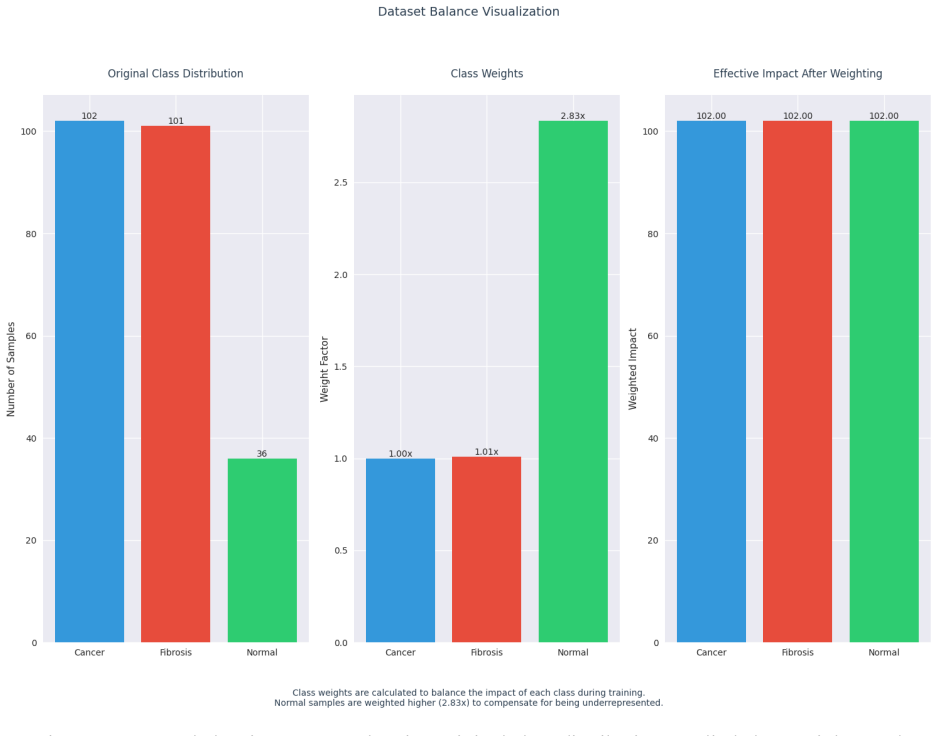
- class weighting factor for normal tissue =
2.83
- SHG channel scaling factor =
1.3
- autofluorescence channel scaling factor =
1.0
- dropout rate =
0.1
- hidden layer dimension =
512
- learning rate =
1e-4
- weight decay =
0.01
- batch size =
16
assumptions (4)
- domain assumption Pathologist annotations of normal, fibrotic, and cancerous regions are correct ground truth.
- domain assumption Images from the same patient are statistically independent.
- domain assumption ImageNet-pretrained features transfer to SHG and autofluorescence microscopy images.
- ad hoc to paper The combination of autofluorescence and SHG channels into a single RGB image preserves the diagnostically relevant information.
Cite this review
Pith. "Pith review of Deep Learning Framework for Early Detection of Pancreatic Cancer Using Multi-Modal Medical Imaging Analysis." pith.science (2026). https://pith.science/paper/KZ6N47Y2
@misc{pith2026250820877,
  author       = {Pith},
  title        = {Pith review of: Deep Learning Framework for Early Detection of Pancreatic Cancer Using Multi-Modal Medical Imaging Analysis},
  year         = {2026},
  howpublished = {\url{https://pith.science/paper/KZ6N47Y2}},
  note         = {Machine review of arXiv:2508.20877}
}
read the original abstract
Pacreatic ductal adenocarcinoma (PDAC) remains one of the most lethal forms of cancer, with a five-year survival rate below 10% primarily due to late detection. This research develops and validates a deep learning framework for early PDAC detection through analysis of dual-modality imaging: autofluorescence and second harmonic generation (SHG). We analyzed 40 unique patient samples to create a specialized neural network capable of distinguishing between normal, fibrotic, and cancerous tissue. Our methodology evaluated six distinct deep learning architectures, comparing traditional Convolutional Neural Networks (CNNs) with modern Vision Transformers (ViTs). Through systematic experimentation, we identified and overcome significant challenges in medical image analysis, including limited dataset size and class imbalance. The final optimized framework, based on a modified ResNet architecture with frozen pre-trained layers and class-weighted training, achieved over 90% accuracy in cancer detection. This represents a significant improvement over current manual analysis methods an demonstrates potential for clinical deployment. This work establishes a robust pipeline for automated PDAC detection that can augment pathologists' capabilities while providing a foundation for future expansion to other cancer types. The developed methodology also offers valuable insights for applying deep learning to limited-size medical imaging datasets, a common challenge in clinical applications.
Figures


Reference graph
Works this paper leans on
-
[1]
(1) Sarantis, P.; Koustas, E.; Papadimitropoulou, A.; Papavassiliou, A. G.; Karamouzis, M. V. Pancreatic Ductal Adenocarcinoma: Treatment Hurdles, Tumor Microenvironment and Immunotherapy. World J Gastrointest Oncol 2020, 12 (2), 173–181. https://doi.org/10.4251/wjgo.v12.i2.173. (2) Renjifo-Correa, M. E.; Fanni, S. C.; Bustamante -Cristancho, L. A.; Cuiba...
-
[7]
https://doi.org/10.3389/fcvm.2020.00025. (16) Enriquez, J. S.; Chu, Y.; Pudakalakatti, S.; Hsieh, K. L.; Salmon, D.; Dutta, P.; Millward, N. Z.; Lurie, E.; Millward, S.; McAllister, F.; Maitra, A.; Sen, S.; Killary, A.; Zhang, J.; Jiang, X.; Bhattacharya, P. K.; Shams, S. Hyperpolarized Magnetic Resonance and Artificial Intelligence: Frontiers of Imaging ...
arXiv 2020
-
[12]
R.; Tilbury, K.; Nadia rnykh, O.; Brewer, M
(19) Wen, B.; Campbell, K. R.; Tilbury, K.; Nadia rnykh, O.; Brewer, M. A.; Patankar, M.; Singh, V.; Eliceiri, K. W.; Campagnola, P. J. 3D Texture Analysis for Classification of Second Harmonic Generation Images of Human Ovarian Cancer. Sci Rep 2016, 6 (1), 35734. https://doi.org/10.1038/srep35734. (20) Tilbury, K.; Han, X.; Brooks, P. C.; Khalil, A. Mult...
-
[129]
(4) Yao, L.; Zhang, Z.; Keles, E.; Yazici, C.; Tirkes, T.; Bagci, U
https://doi.org/10.4103/ejcrp.eJCRP -D-23-00002. (4) Yao, L.; Zhang, Z.; Keles, E.; Yazici, C.; Tirkes, T.; Bagci, U. A Review of Deep Learning and Radiomics Approaches for Pancreatic Cancer Diagnosis from Medical Imaging. Current Opinion in Gastroenterology 2023, 39 (5),
-
[220]
https://doi.org/10.1007/s10462- 024-11050-4. (6) Hayashi, H.; Uemura, N.; Matsumura, K.; Zhao, L.; Sato, H.; Shiraishi, Y.; Yamashita, Y.; Baba, H. Recent Advances in Artificial Intelligence for Pancreatic Ductal Adenocarcinoma. World Journal of Gastroenterology 2021, 27 (43), 7480–7496. https://doi.org/10.3748/wjg.v27.i43.7480. (7) Anghel, C.; Grasu, M. ...
doi:10.1007/s10462- 2021
-
[436]
(5) Moglia, A.; Cavicchioli, M.; Mainardi, L.; Cerveri, P
https://doi.org/10.1097/MOG.0000000000000966. (5) Moglia, A.; Cavicchioli, M.; Mainardi, L.; Cerveri, P. Deep Learning for Pancreas Segm entation on Computed Tomography: A Systematic Review. Artif Intell Rev 2025, 58 (8),
-
[438]
(8) Alves, N.; Schuurmans, M.; Litjens, G.; Bosma, J
https://doi.org/10.3390/diagnostics14040438. (8) Alves, N.; Schuurmans, M.; Litjens, G.; Bosma, J. S.; Hermans, J.; Huisman, H. Fully Automatic Deep Learning Framework for Pancreatic Ductal Adenocarcinoma Detection on Computed Tomography. arXiv December 2,
-
[778]
An Image Is Worth 16x16 Words: Transformers for Image Recognition at Scale
(28) Dosovitskiy, A.; Beyer, L.; Kolesnikov, A.; Weissenborn, D.; Zhai, X.; Unterthiner, T.; Dehghani, M.; Minderer, M.; Heigold, G.; Gelly, S. An Image Is Worth 16x16 Words: Transformers for Image Recognition at Scale. arXiv preprint arXiv:2010.11929
arXiv 2010
Show all 15 references
-
[803]
(3) Liao, W.-C
https://doi.org/10.3390/cancers17050803. (3) Liao, W.-C. Early Detection of Pancreatic Cancer: Opportunities Provided by Cancer -Induced Paraneoplastic Phenomena and Artificial Intelligence. Journal of Cancer Research and Practice 2023, 10 (4),
2023 doi
-
[1560]
(23) Kučikas, V.; Werner, M
https://doi.org/10.3390/cancers16081560. (23) Kučikas, V.; Werner, M. P.; Schmitz -Rode, T.; Louradour, F.; van Zandvoort, M. A. M. J. Two - Photon Endoscopy: State of the Art and Perspectives. Mol Imaging Biol 2023, 25 (1), 3–17. https://doi.org/10.1007/s11307- 021-01665-2. (...
2023 doi
-
[2018]
(14) Ramaekers, M.; Viviers, C
https://doi.org/10.48550/arXiv.1806.01023. (14) Ramaekers, M.; Viviers, C. G. A.; Janssen, B. V.; Hellström, T. A. E.; Ewals, L.; van der Wulp, K.; Nederend, J.; Jacobs, I.; Pluyter, J. R.; Mavroeidis, D.; van der Sommen, F.; Besselink, M. G.; Luyer, M. D. P. Computer-Aided De...
- [2021]
- [2024]
-
[2025]
(10) Faur, A
https://doi.org/10.48550/arXiv.2503.10068. (10) Faur, A. C.; Lazar, D. C.; Ghenciu, L. A. Artificial Intelligence as a Noninvasive Tool for Pancreatic Cancer Prediction and Diagnosis. World J Gastroenterol 2023, 29 (12), 1811–1823. https://doi.org/10.3748/wjg.v29.i12.1811. (11...
2023 doi
-
[4209]
(15) Chen, C.; Qin, C.; Qiu, H.; Tarroni, G.; Duan, J.; Bai, W.; Rueckert, D
https://doi.org/10.3390/jcm12134209. (15) Chen, C.; Qin, C.; Qiu, H.; Tarroni, G.; Duan, J.; Bai, W.; Rueckert, D. Deep Learning for Cardiac Image Segmentation: A Review. Front. Cardiovasc. Med. 2020,
2020 doi
Reviewed August 5, 2026 · model on record in the stance chip above.
Discussion (0). Sign in to comment.