REVIEW 3 major objections 5 minor 56 references
Reverse translocation of a nascent polypeptide through the ribosomal exit tunnels
T0 review · 3 major / 5 minor · reviewed 2026-08-02 · deepseek-v4-flash
Pith's one-line read The ribosome's exit tunnel has a built-in directional bias: nascent proteins move outward more easily than inward, which would explain why proteins like NAC and mL45 need dedicated machinery to push backwards into it.
desk verdict A useful, honest simulation study whose headline claim is undercut by an unaddressed endpoint artifact: reverse runs push the peptide into a closed PTC pocket, forward runs let it exit into bulk. read the letter →
The pith
A machine-rendered reading of the paper's core claim, the machinery that carries it, and where it could break.
The reading
What carries the argument
Constant-velocity force-probe molecular dynamics (fpMD): a harmonic spring with force constant 602.7 kJ mol−1 nm−2 moves at 0.05 nm/ns, attached to either the N-terminal acetyl or C-terminal N-methylamide cap of a deca-alanine peptide, over 200 ns. The displacement of the probed residue, normalized by tunnel length to give a relative displacement and filtered by an 80% success threshold, serves as a direct readout of how obstructed the peptide is along each direction.
What would settle it
A simulation of the same deca-alanine through a geometrically symmetric channel of comparable radius, or through the same tunnel but with the PTC pocket made as open and solvent-exposed as the exit side, that showed no forward/reverse displacement difference would falsify the intrinsic-directionality claim; more directly, a reversible work (free-energy) profile with equal barriers in both directions would disprove it.
Extended reading notes
Core claim
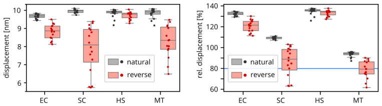
In atomistic simulations of four evolutionarily diverse ribosome exit tunnels, a model deca-alanine peptide pulled from the peptidyl transferase center toward the ribosomal surface consistently travels farther, more uniformly, and with a narrower spread of outcomes than the same peptide pulled in the reverse direction. Reverse translocation is markedly less successful in the yeast and mitochondrial tunnels, and the constriction site emerges as the main direction-dependent barrier. The authors interpret this as evidence that the tunnel's shape—narrow through the constriction site formed by uL4 and uL22, widening toward the exit—imposes an intrinsic bias that favors natural-direction transloca
Load-bearing premise
The load-bearing premise is that the asymmetry measured during fast, non-equilibrium pulling (peptide driven at 0.05 nm/ns over 200 ns) reflects the tunnel's intrinsic, rate-independent directionality rather than an artifact of the pulling protocol or of the asymmetric boundary conditions at the two tunnel ends.
Editorial extensions
If this is right
- Proteins such as mL45 and the N-terminal tail of NACβ, which insert into the tunnel in reverse orientation, must act against an intrinsic directional bias, explaining why they are associated with dedicated structural machinery.
- The directional preference is conserved across bacteria, yeast, human cytosolic ribosomes, and human mitochondrial ribosomes, suggesting it is a general architectural feature of the exit tunnel.
- The constriction site is the dominant barrier in both directions, but reverse translocation encounters it late and with a wide spread of outcomes, indicating kinetically distinct reverse routes rather than a single pathway.
- Because reverse trajectories that fail to reach the PTC are excluded from force averages, the true resistance to reverse translocation is likely even stronger than the reported force profiles suggest.
- Absolute pulling forces are inflated by the fast, non-equilibrium protocol; the authors expect the qualitative directional bias, but not the absolute force magnitudes, to survive at biological timescales.
Reading between the lines
- If the asymmetry is truly thermodynamic rather than kinetic, equilibrium free-energy calculations along the tunnel axis should reveal a higher barrier for reverse passage; no such equilibrium calculation is reported, so this is a testable prediction arising from the paper's claim.
- The reverse direction ends in a confined PTC pocket modeled without tRNA, while the forward direction ends in open bulk solvent; part of the apparent directionality could come from this boundary asymmetry, so a control with a symmetric open channel or a PTC containing tRNA would sharpen the intrinsic-bias claim.
- The result predicts that ribosomes relying heavily on reverse-occupancy factors—mitochondrial ribosomes in particular—would either need stronger insertion machinery or have somewhat less directional tunnels; comparing tunnel geometry against the abundance of such factors could test this.
- If reverse translocation is intrinsically obstructed, the transient reverse occupancy by NAC's sensing tail carries an energetic cost that could be measurable with existing arrest-peptide force sensors.
Signed reviews
Editorial analysis
A structured set of objections, weighed in public.
Referee Report
Summary. The paper uses constant-velocity force-probe molecular dynamics to pull a capped deca-alanine peptide through ribosomal exit-tunnel models from E. coli, S. cerevisiae, human cytosolic, and human mitochondrial ribosomes, comparing the natural (PTC-to-exit) direction with the reverse (exit-to-PTC) direction. The authors report that in all four tunnels the probed residue travels farther in the forward direction, that reverse runs fail more often (relative displacement < 80%) in the yeast and mitochondrial tunnels, and that force and solvation profiles differ between directions. They conclude that the tunnel architecture intrinsically favors natural-direction translocation and that reverse occupants such as mL45 and NAC require dedicated structural machinery to overcome this bias. The manuscript is clearly written, discloses several important limitations in the Conclusions, and presents raw per-replica data in boxplots, but the central claim is not yet supported because of an uncontrolled boundary asymmetry and the rate-dependence of the pulling protocol.
Significance. The question addressed—whether the ribosomal exit tunnel has an intrinsic directional bias—is interesting and timely, especially given recent structures of NAC and mL45 inserting into the tunnel in the reverse direction. The four-organism comparison is a strength, and the methods are described in sufficient detail to be reproduced. The paper also benefits from showing raw trajectory counts and from explicitly acknowledging that fast non-equilibrium pulling may affect the observed asymmetry. If the intrinsic-directionality claim could be established with appropriate controls, this would be a useful contribution. As it stands, however, the main conclusion goes beyond what the displacement observable can show, and the paper needs additional control simulations or a substantially softened claim.
major comments (3)
- [§3.1, Fig. 2; Methods §2.1] The displacement metric used to infer intrinsic directionality is confounded by asymmetric boundary conditions. In the forward direction the probe can pull the residue past the tunnel exit into open bulk water, so displacements can exceed the tunnel length. In the reverse direction the PTC end is a closed pocket—Methods §2.1 states explicitly that no tRNAs were retained at the PTC—so the residue's progress is capped at approximately the tunnel length. Even a perfectly symmetric channel would therefore show larger forward displacements and more replicas above any absolute threshold. No control is reported (e.g., a symmetric cylinder, an open/reservoir PTC, or an analysis truncated at the tunnel lumen) to separate this endpoint artifact from an intrinsic tunnel bias. Because the central claim of intrinsic directionality rests on this observable, this is a load-bearing issue.
- [§2.3 and §3.2] The pulled terminus is not the same chemical group in the two directions: forward runs pull the N-terminal acetyl group and reverse runs pull the C-terminal N-methylamide group. These caps differ in size, polarity, and hydrogen-bonding capacity, so the force and displacement asymmetries could reflect probe chemistry rather than tunnel architecture. The authors should either use the same pulled atom/group in both directions (e.g., attaching the spring to a backbone atom or using identical caps) or demonstrate that the difference between ACE and NME does not materially affect the conclusion.
- [Conclusions, final paragraph] The authors themselves state: 'part of the asymmetry we observe between forward and reverse translocation may reflect the kinetics of the pulling protocol rather than the underlying thermodynamics alone.' This is a direct caveat to the abstract's claim that the tunnel 'intrinsically favours' the natural direction. Intrinsic directionality implies a rate-independent or equilibrium property, but no umbrella-sampling/free-energy calculation or multi-loading-rate study is presented. The manuscript should either provide such evidence, or reframe the conclusion as a protocol-dependent observation (e.g., 'under the fast non-equilibrium pulling used here, reverse translocation is more hindered') and remove the 'intrinsic' language from the abstract and Conclusions.
minor comments (5)
- [§2.4] The 80% success threshold is introduced post hoc. Although it is applied symmetrically to both directions, the choice should be justified and a sensitivity analysis (e.g., 70% or 90%) reported to show that the main asymmetry does not hinge on this particular cutoff.
- [§3.1] No statistical tests accompany the displacement comparisons. The boxplots are informative, but a Mann-Whitney U test or similar per-tunnel comparison would strengthen the claim that the forward/reverse differences are not due to sampling noise, especially for EC and HS where the asymmetry appears modest.
- [Abstract/Introduction] The abstract and Introduction state that 'all known proteins translocate through the ribosomal exit tunnel' in the natural direction; given that the paper itself discusses reverse occupancy by mL45 and NAC, the phrasing should be refined to avoid an apparent contradiction.
- [General] There are minor language issues: 'has been proved reliable' should be 'has proven reliable'; 'targetting' should be 'targeting'; and Table 1 needs spacing in the organism abbreviations (e.g., 'E. coli').
- [§3.2] The statement that 'specific chemical interactions are expected minor' is too vague. Since deca-alanine has only methyl side chains, this is plausible, but the authors should specify what evidence or reasoning underlies this expectation.
Circularity Check
Endpoint boundary conditions make the displacement asymmetry a by-construction outcome; the 'intrinsic directionality' claim is partially circular.
-
self definitional
[Methods §2.1, §2.3, §2.4; Results §3.1]
"Methods §2.1: 'No tRNAs were retained at the PTC.' Methods §2.3: 'Only the Z Cartesian coordinate of the probed residue was biased... Each fpMD simulation lasted 200 ns, over which the force probe moved by 10 nm.' Methods §2.4: 'The total displacement of the peptide was defined as the difference between the final and initial positions of the probed residue.' Results §3.1: 'In every tunnel, translocation in the natural direction resulted in greater net displacement... indicating that the forward path is systematically less obstructed.'"
Forward runs start at the PTC and are pulled toward the open exit; once the peptide leaves the tunnel it can continue into bulk water, so its displacement is not bounded by the tunnel length. Reverse runs start at the exit and are pulled toward the PTC, which is a closed rRNA/protein wall because 'No tRNAs were retained at the PTC'; the maximum possible displacement is therefore the tunnel length. Even a perfectly symmetric channel with these boundary conditions would yield larger forward than reverse displacements. The paper uses this displacement difference as a 'direct readout' of intrinsic directionality, so the claimed architectural bias is partly manufactured by the metric's asymmetric endpoints rather than measured.
full rationale
The central claim rests on the displacement asymmetry in Fig. 2, which is not produced by fitting and is not a renamed known result; the 80% threshold is applied symmetrically. The self-citations (refs. 23, 28, 40) support methodology (force-field reliability, loading rate, conformational criterion), are published and externally falsifiable, and are not used as a uniqueness or ansatz argument; they therefore do not constitute load-bearing circularity. The paper's own Conclusions concede that 'part of the asymmetry we observe between forward and reverse translocation may reflect the kinetics of the pulling protocol rather than the underlying thermodynamics alone,' which further weakens the strong 'intrinsic' reading. The genuine circularity concern is the endpoint asymmetry: the forward direction opens into bulk solvent while the reverse direction terminates at a closed PTC, so the displacement metric itself encodes a directional advantage. A symmetric-channel control or a displacement analysis truncated at the tunnel lumen would be needed to separate intrinsic tunnel bias from boundary-condition bias. Because the observed failure rates and force profiles contain information beyond the pure boundary effect, the circularity is partial, not total.
Assumptions & free parameters
free parameters (4)
- Success threshold (relative displacement ≥ 80%) =
80% of tunnel length
- Tunnel wall selection radius =
1.8 nm nominal, increased to 2.2 nm where walls had holes
- Peptide initial-conformation selection criterion =
extended criterion from ref 28
- Pulling speed and probe stiffness =
0.05 nm/ns, 602.7 kJ mol−1 nm−2 (0.1 pN/ps loading rate)
assumptions (6)
- domain assumption AMBER force fields (ff10 for RNA, ff12SB for protein, Aduri parameters for modified RNA) accurately model peptide–rRNA–protein interactions in confined tunnels.
- domain assumption The four deposited cryo-EM structures represent the physiologically relevant tunnel conformations.
- domain assumption Constant-velocity pulling at 0.05 nm/ns (loading rate 0.1 pN/ps) yields directionality information transferable to in vivo translocation occurring on millisecond timescales.
- domain assumption Restrained tunnel cutouts (300 kJ mol−1 nm−2 harmonic restraints; no tRNA at the PTC) preserve the essential barrier physics of translocation.
- domain assumption The Z-coordinate displacement of the probed residue is a valid progress measure despite tunnel curvature and free XY motion.
- domain assumption Deca-alanine is a sufficient probe to test the general intrinsic directionality of the tunnel.
Cite this review
Pith. "Pith review of Reverse translocation of a nascent polypeptide through the ribosomal exit tunnels." pith.science (2026). https://pith.science/paper/LCBYDXZ5
@misc{pith2026260713195,
  author       = {Pith},
  title        = {Pith review of: Reverse translocation of a nascent polypeptide through the ribosomal exit tunnels},
  year         = {2026},
  howpublished = {\url{https://pith.science/paper/LCBYDXZ5}},
  note         = {Machine review of arXiv:2607.13195}
}
read the original abstract
Before they mature, all known proteins translocate through the ribosomal exit tunnel in a form of extended or partially folded nascent polypeptide. This translocation occurs in the natural direction from the peptidyl transferase centre buried deep in the large ribosomal subunit through a tunnel to the ribosomal surface. Some proteins, however, enter the ribosomal tunnel from outside and translocate in the reverse direction. In this work, we address a simple question: to what extent is the ribosomal tunnel intrinsically directional. To compare the forward and reverse peptide translocations, we performed biased molecular dynamics simulations and assessed the directionality of tunnels from four different organisms using a model poly-alanine decapeptide. Our simulations reveal that the tunnel architecture intrinsically favours the natural direction translocation from peptidyl transferase centre to the ribosomal surface. Consequently, proteins that occupy the tunnel in the reverse direction likely require dedicated structural machinery to overcome this intrinsic bias.
Figures


Figures from the paper (2 more)


Reference graph
Works this paper leans on
-
[1]
R.; Gerstein, M.; Steitz, T
Voss, N. R.; Gerstein, M.; Steitz, T. A.; Moore, P. B. The Geometry of the Ribosomal Polypeptide Exit Tunnel . J. Mol. Biol. 2006, 360, 893--906
2006
-
[2]
S.; Bhattacharya, N.; Cate, J
Dao Duc, K.; Batra, S. S.; Bhattacharya, N.; Cate, J. H. D.; Song, Y. S. Differences in the Path to Exit the Ribosome across the Three Domains of Life. Nucleic Acids Res. 2019, 47, 4198--4210
2019
-
[3]
E.; Nissen, P
Brodersen, D. E.; Nissen, P. The Social Life of Ribosomal Proteins. The FEBS Journal 2005, 272, 2098--2108
2005
-
[4]
N.; and Nierhaus , K
Wilson, D. N.; and Nierhaus , K. H. Ribosomal Proteins in the Spotlight . Critical Reviews in Biochemistry and Molecular Biology 2005, 40, 243--267
2005
-
[5]
A.; Rodnina, M
Samatova, E.; Komar, A. A.; Rodnina, M. V. How the Ribosome Shapes Cotranslational Protein Folding. Curr. Opin. Struc. Biol. 2024, 84, 102740
2024
-
[6]
H.; McGrath, H.; Nepomuceno, F
Kol \'a r , M. H.; McGrath, H.; Nepomuceno, F. C.; C ernekov \'a , M. Three Stages of Nascent Protein Translocation Through the Ribosome Exit Tunnel . WIREs RNA 2024, 15, e1873
2024
-
[7]
B.; Barrientos, A.; Battersby, B
Itoh, Y.; Andr \'e ll, J.; Choi, A.; Richter, U.; Maiti, P.; Best, R. B.; Barrientos, A.; Battersby, B. J.; Amunts, A. Mechanism of Membrane-Tethered Mitochondrial Protein Synthesis. Science 2021, 371, 846--849
2021
-
[8]
G.; Boehringer, D.; Filipovska, A.; Ban, N
Kummer, E.; Leibundgut, M.; Rackham, O.; Lee, R. G.; Boehringer, D.; Filipovska, A.; Ban, N. Unique Features of Mammalian Mitochondrial Translation Initiation Revealed by Cryo- EM . Nature 2018, 560, 263--267
2018
Show all 56 references
-
[9]
H.; Rabl, L.; Gamerdinger, M.; Goyal, V.; Khakzar, K
Lee, J. H.; Rabl, L.; Gamerdinger, M.; Goyal, V.; Khakzar, K. M.; Barbosa, N. M.; Abramovich, J.; Morales-Polanco , F.; K \"o hler, A.-K.; Samatova, E.; Rodnina, M. V.; Deuerling, E.; Frydman, J. NAC Controls Nascent Chain Fate through Tunnel Sensing and Chaperone Action. Natu...
2026
-
[10]
S.; Shan, S.-o.; Ban, N.; Deuerling, E
Jomaa, A.; Gamerdinger, M.; Hsieh, H.-H.; Wallisch, A.; Chandrasekaran, V.; Ulusoy, Z.; Scaiola, A.; Hegde, R. S.; Shan, S.-o.; Ban, N.; Deuerling, E. Mechanism of Signal Sequence Handover from NAC to SRP on Ribosomes during ER-protein Targeting. Science 2022, 375, 839--844
2022
-
[11]
A.; Wiedmann, M
Wiedmann, B.; Sakai, H.; Davis, T. A.; Wiedmann, M. A Protein Complex Required for Signal-Sequence-Specific Sorting and Translocation. Nature 1994, 370, 434--440
1994
-
[12]
G.; Sailer, C.; Schl \"o mer, R.; Sachs, N.; Jomaa, A.; Stengel, F.; Ban, N.; Deuerling, E
Gamerdinger, M.; Kobayashi, K.; Wallisch, A.; Kreft, S. G.; Sailer, C.; Schl \"o mer, R.; Sachs, N.; Jomaa, A.; Stengel, F.; Ban, N.; Deuerling, E. Early Scanning of Nascent Polypeptides inside the Ribosomal Tunnel by NAC . Mol. Cell 2019, 75, 996--1006.e8
2019
-
[13]
Time-Resolved Cryo-Electron Microscopy: Recent Progress
Frank, J. Time-Resolved Cryo-Electron Microscopy: Recent Progress. Journal of Structural Biology 2017, 200, 303--306
2017
-
[14]
P.; Aspinall, L.; Muench, S
Klebl, D. P.; Aspinall, L.; Muench, S. P. Time Resolved Applications for Cryo-EM ; Approaches, Challenges and Future Directions. Curr. Opin. Struc. Biol. 2023, 83, 102696
2023
-
[15]
V.; Gabrielli, S.; Kol \'a r , M
Bock, L. V.; Gabrielli, S.; Kol \'a r , M. H.; Grubm \"u ller, H. Simulation of Complex Biomolecular Systems : The Ribosome Challenge . Annual Review of Biophysics 2023, 52, 361--390
2023
-
[16]
T.; Hoang, T
Bui, P. T.; Hoang, T. X. Folding and Escape of Nascent Proteins at Ribosomal Exit Tunnel. J. Chem. Phys. 2016, 144, 095102
2016
-
[17]
T.; Hoang, T
Bui, P. T.; Hoang, T. X. Protein Escape at the Ribosomal Exit Tunnel: Effects of Native Interactions, Tunnel Length, and Macromolecular Crowding. J. Chem. Phys. 2018, 149, 045102
2018
-
[18]
T.; Hoang, T
Bui, P. T.; Hoang, T. X. Hydrophobic and Electrostatic Interactions Modulate Protein Escape at the Ribosomal Exit Tunnel. Biophys. J. 2021, 120, 4798--4808
2021
-
[19]
T.; Hoang, T
Bui, P. T.; Hoang, T. X. The Protein Escape Process at the Ribosomal Exit Tunnel Has Conserved Mechanisms across the Domains of Life. J. Chem. Phys. 2023, 158, 015102
2023
-
[20]
A.; Vu, Q
Nissley, D. A.; Vu, Q. V.; Trovato, F.; Ahmed, N.; Jiang, Y.; Li, M. S.; O'Brien, E. P. Electrostatic Interactions Govern Extreme Nascent Protein Ejection Times from Ribosomes and Can Delay Ribosome Recycling . J. Am. Chem. Soc. 2020, 142, 6103--6110
2020
-
[21]
P.; Stan, G.; Thirumalai, D.; Brooks, B
O'Brien, E. P.; Stan, G.; Thirumalai, D.; Brooks, B. R. Factors Governing Helix Formation in Peptides Confined to Carbon Nanotubes . Nano Letters 2008, 8, 3702--3708
2008
-
[22]
C.; von Heijne , G.; Mingarro, I
Ba \ n \'o -Polo , M.; Baeza-Delgado , C.; Tamborero, S.; Hazel, A.; Grau, B.; Nilsson, I.; Whitley, P.; Gumbart, J. C.; von Heijne , G.; Mingarro, I. Transmembrane but Not Soluble Helices Fold inside the Ribosome Tunnel. Nat. Commun. 2018, 9, 5246
2018
-
[23]
H.; Nagy, G.; Kunkel, J.; Vaiana, S
Kol \'a r , M. H.; Nagy, G.; Kunkel, J.; Vaiana, S. M.; Bock, L. V.; Grubm \"u ller, H. Folding of VemP into Translation-Arresting Secondary Structure Is Driven by the Ribosome Exit Tunnel. Nucleic Acids Res. 2022, 50, 2258--2269
2022
-
[24]
N.; Beckmann, R.; Schulten, K
Gumbart, J.; Schreiner, E.; Wilson, D. N.; Beckmann, R.; Schulten, K. Mechanisms of SecM-Mediated Stalling in the Ribosome . Biophys. J. 2012, 103, 331--341
2012
-
[25]
V.; Graf, M.; Innis, C
Arenz, S.; Bock, L. V.; Graf, M.; Innis, C. A.; Beckmann, R.; Grubm \"u ller, H.; Vaiana, A. C.; Wilson, D. N. A Combined Cryo- EM and Molecular Dynamics Approach Reveals the Mechanism of ErmBL-mediated Translation Arrest. Nat. Commun. 2016, 7, 12026
2016
-
[26]
Ligand Binding : Molecular Mechanics Calculation of the Streptavidin-Biotin Rupture Force
Grubm \"u ller, H.; Heymann, B.; Tavan, P. Ligand Binding : Molecular Mechanics Calculation of the Streptavidin-Biotin Rupture Force . Science 1996, 271, 997--999
1996
-
[27]
Force Spectroscopy of Single Biomolecules
Rief, M.; Grubm \"u ller, H. Force Spectroscopy of Single Biomolecules . ChemPhysChem 2002, 3, 255--261
2002
-
[28]
C.; Kol \'a r , M
Nepomuceno, F. C.; Kol \'a r , M. H. Sensitivity of Peptide Conformational Dynamics in Carbon Nanotubes to Directional Mechanical Forces. Phys. Chem. Chem. Phys. 2025, 27, 20050--20062
2025
-
[29]
L.; Ward, F
Watson, Z. L.; Ward, F. R.; M \'e heust, R.; Ad, O.; Schepartz, A.; Banfield, J. F.; Cate, J. H. Structure of the Bacterial Ribosome at 2 Resolution. eLife 2020, 9, e60482
2020
-
[30]
B.; Svidritskiy, E.; Susorov, D.; Lee, S.; Park, A.; Zvornicanin, S.; Demo, G.; Gao, F.-B.; Korostelev, A
Loveland, A. B.; Svidritskiy, E.; Susorov, D.; Lee, S.; Park, A.; Zvornicanin, S.; Demo, G.; Gao, F.-B.; Korostelev, A. A. Ribosome Inhibition by C9ORF72-ALS / FTD-associated Poly- PR and Poly- GR Proteins Revealed by Cryo- EM . Nat. Commun. 2022, 13, 2776
2022
-
[31]
C.; Pellegrino, S.; Jaako, P.; Warren, A
Faille, A.; Dent, K. C.; Pellegrino, S.; Jaako, P.; Warren, A. J. The Chemical Landscape of the Human Ribosome at 1.67 Resolution. 2023
2023
-
[32]
K.; Nobe, Y.; Izumikawa, K.; Aibara, S.; Andr \'e ll, J.; Whitford, P
Singh, V.; Itoh, Y.; Del'Olio, S.; Hassan, A.; Naschberger, A.; Flygaard, R. K.; Nobe, Y.; Izumikawa, K.; Aibara, S.; Andr \'e ll, J.; Whitford, P. C.; Barrientos, A.; Taoka, M.; Amunts, A. Mitoribosome Structure with Cofactors and Modifications Reveals Mechanism of Ligand Bin...
2024
-
[33]
Berendsen, H. J. C.; Grigera, J. R.; Straatsma, T. P. The Missing Term in Effective Pair Potentials. J. Phys. Chem. 1987, 91, 6269--6271
1987
-
[34]
S.; Cheatham, T
Joung, I. S.; Cheatham, T. E. Determination of Alkali and Halide Monovalent Ion Parameters for Use in Explicitly Solvated Biomolecular Simulations . J. Phys. Chem. B 2008, 112, 9020--9041
2008
-
[35]
D.; Cieplak, P.; Bayly, C
Cornell, W. D.; Cieplak, P.; Bayly, C. I.; Gould, I. R.; Merz, K. M.; Ferguson, D. M.; Spellmeyer, D. C.; Fox, T.; Caldwell, J. W.; Kollman, P. A. A Second Generation Force Field for the Simulation of Proteins , Nucleic Acids , and Organic Molecules . J. Am. Chem. Soc. 1995, 1...
1995
-
[36]
E.; Laughton, C
P \'e rez, A.; March \'a n, I.; Svozil, D.; Sponer, J.; Cheatham, T. E.; Laughton, C. A.; Orozco, M. Refinement of the AMBER Force Field for Nucleic Acids : Improving the Description of / Conformers . Biophys. J. 2007, 92, 3817--3829
2007
-
[37]
E.; Jure c ka, P
Zgarbov \'a , M.; Otyepka, M.; S poner, J.; Ml \'a dek, A.; Ban \'a s , P.; Cheatham, T. E.; Jure c ka, P. Refinement of the Cornell et al. Nucleic Acids Force Field Based on Reference Quantum Chemical Calculations of Glycosidic Torsion Profiles . J. Chem. Theory Comput. 2011,...
2011
-
[38]
A.; Martinez, C.; Kasavajhala, K.; Wickstrom, L.; Hauser, K
Maier, J. A.; Martinez, C.; Kasavajhala, K.; Wickstrom, L.; Hauser, K. E.; Simmerling, C. ff14SB : Improving the Accuracy of Protein Side Chain and Backbone Parameters from ff99SB . J. Chem. Theory Comput. 2015, 11, 3696--3713
2015
-
[39]
T.; Saro, P.; Taniga, H.; Schlegel, H
Aduri, R.; Psciuk, B. T.; Saro, P.; Taniga, H.; Schlegel, H. B.; SantaLucia, J. AMBER Force Field Parameters for the Naturally Occurring Modified Nucleosides in RNA . J. Chem. Theory Comput. 2007, 3, 1464--1475
2007
-
[40]
McGrath, H.; C ernekov \'a , M.; Kol \'a r , M. H. Binding of the Peptide Deformylase on the Ribosome Surface Modulates the Exit Tunnel Interior. Biophys. J. 2022, 121, 4443--4451
2022
-
[41]
V.; Graf, M.; Frister, J
Huter, P.; Arenz, S.; Bock, L. V.; Graf, M.; Frister, J. O.; Heuer, A.; Peil, L.; Starosta, A. L.; Wohlgemuth, I.; Peske, F.; Nov \'a c ek, J.; Berninghausen, O.; Grubm \"u ller, H.; Tenson, T.; Beckmann, R.; Rodnina, M. V.; Vaiana, A. C.; Wilson, D. N. Structural Basis for Po...
2017
-
[42]
C.; Sothiselvam, S.; Bock, L
Beckert, B.; Leroy, E. C.; Sothiselvam, S.; Bock, L. V.; Svetlov, M. S.; Graf, M.; Arenz, S.; Abdelshahid, M.; Seip, B.; Grubm \"u ller, H.; Mankin, A. S.; Innis, C. A.; V \'a zquez-Laslop , N.; Wilson, D. N. Structural and Mechanistic Basis for Translation Inhibition by Macro...
2021
-
[43]
J.; Murtola, T.; Schulz, R.; P \'a ll, S.; Smith, J
Abraham, M. J.; Murtola, T.; Schulz, R.; P \'a ll, S.; Smith, J. C.; Hess, B.; Lindahl, E. GROMACS : High Performance Molecular Simulations through Multi-Level Parallelism from Laptops to Supercomputers. SoftwareX 2015, 1--2, 19--25
2015
-
[44]
P- LINCS :\, A Parallel Linear Constraint Solver for Molecular Simulation
Hess, B. P- LINCS :\, A Parallel Linear Constraint Solver for Molecular Simulation . J. Chem. Theory Comput. 2008, 4, 116--122
2008
-
[45]
Canonical Sampling through Velocity Rescaling
Bussi, G.; Donadio, D.; Parrinello, M. Canonical Sampling through Velocity Rescaling. J. Chem. Phys. 2007, 126, 014101
2007
-
[46]
Path of Nascent Polypeptide in Exit Tunnel Revealed by Molecular Dynamics Simulation of Ribosome
Ishida, H.; Hayward, S. Path of Nascent Polypeptide in Exit Tunnel Revealed by Molecular Dynamics Simulation of Ribosome . Biophys. J. 2008, 95, 5962--5973
2008
-
[47]
M.; Snow, C
Petrone, P. M.; Snow, C. D.; Lucent, D.; Pande, V. S. Side-Chain Recognition and Gating in the Ribosome Exit Tunnel. Proc. Natl. Acad. Sci. U. S. A. 2008, 105, 16549--16554
2008
-
[48]
Early Nascent Polypeptide Dynamics Are Coupled to the Flexibility of the Ribosomal Tunnel Constriction
McGrath, H.; C ernekov \'a , M.; Kol \'a r , M. Early Nascent Polypeptide Dynamics Are Coupled to the Flexibility of the Ribosomal Tunnel Constriction. 2026
2026
-
[49]
Steered Molecular Dynamics and Mechanical Functions of Proteins
Isralewitz, B.; Gao, M.; Schulten, K. Steered Molecular Dynamics and Mechanical Functions of Proteins. Curr. Opin. Struc. Biol. 2001, 11, 224--230
2001
-
[50]
Heterogeneous and Rate-Dependent Streptavidin--Biotin Unbinding Revealed by High-Speed Force Spectroscopy and Atomistic Simulations
Rico, F.; Russek, A.; Gonz \'a lez, L.; Grubm \"u ller, H.; Scheuring, S. Heterogeneous and Rate-Dependent Streptavidin--Biotin Unbinding Revealed by High-Speed Force Spectroscopy and Atomistic Simulations. Proc. Natl. Acad. Sci. U. S. A. 2019, 116, 6594--6601
2019
-
[51]
H.; Kaiser, C
Goldman, D. H.; Kaiser, C. M.; Milin, A.; Righini, M.; Tinoco, I.; Bustamante, C. Mechanical Force Releases Nascent Chain--Mediated Ribosome Arrest in Vitro and in Vivo. Science 2015, 348, 457--460
2015
-
[52]
B.; Hedman, R.; Marino, J.; Wickles, S.; Bischoff, L.; Johansson, M.; M \"u ller-Lucks , A.; Trovato, F.; Puglisi, J
Nilsson, O. B.; Hedman, R.; Marino, J.; Wickles, S.; Bischoff, L.; Johansson, M.; M \"u ller-Lucks , A.; Trovato, F.; Puglisi, J. D.; O'Brien, E. P.; Beckmann, R.; von Heijne , G. Cotranslational Protein Folding inside the Ribosome Exit Tunnel . Cell Rep. 2015, 12, 1533--1540
2015
-
[53]
Geometric Differences in the Ribosome Exit Tunnel Impact the Escape of Small Nascent Proteins
Yu, S.; Srebnik, S.; Dao Duc, K. Geometric Differences in the Ribosome Exit Tunnel Impact the Escape of Small Nascent Proteins. Biophys. J. 2023, 122, 20--29
2023
-
[54]
D.; Aitken, C
Lucent, D.; Snow, C. D.; Aitken, C. E.; Pande, V. S. Non- Bulk-Like Solvent Behavior in the Ribosome Exit Tunnel . PLoS Comp. Biol. 2010, 6, e1000963
2010
-
[55]
The Polypeptide Tunnel Exit of the Mitochondrial Ribosome Is Tailored to Meet the Specific Requirements of the Organelle
Gruschke, S.; Ott, M. The Polypeptide Tunnel Exit of the Mitochondrial Ribosome Is Tailored to Meet the Specific Requirements of the Organelle. BioEssays 2010, 32, 1050--1057
2010
-
[56]
J.; Boehringer, D.; Leibundgut, M.; Bieri, P.; Leitner, A.; Schmitz, N.; Aebersold, R.; Ban, N
Greber, B. J.; Boehringer, D.; Leibundgut, M.; Bieri, P.; Leitner, A.; Schmitz, N.; Aebersold, R.; Ban, N. The Complete Structure of the Large Subunit of the Mammalian Mitochondrial Ribosome. Nature 2014, 515, 283--286 mcitethebibliography
2014
Reviewed August 2, 2026 · model on record in the stance chip above.
Discussion (0). Continue with ORCID to comment.