REVIEW 3 major objections 5 minor 60 references
Snap-and-tune: combining deep learning and test-time optimization for high-fidelity cardiovascular volumetric meshing
T0 review · 3 major / 5 minor · reviewed 2026-08-07 · deepseek-v4-flash
Pith's one-line read Adding a test-time tune step to deep-learned heart meshing improves accuracy and element quality without extra labels.
desk verdict Solid incremental advance in automated cardiac meshing, undermined only by thin statistical reporting and an unvalidated pseudo-labeler. read the letter →
The pith
A machine-rendered reading of the paper's core claim, the machinery that carries it, and where it could break.
The reading
What carries the argument
The key mechanism is the joint use of a template mesh, two neural networks, and a per-patient optimization. Both networks predict control-point displacements that deform the template; the snap network $h_\theta$ is trained with volumetric strain energy, while the pseudo-labeler $g_\phi$ is trained with only surface geometric regularizers (normal, Laplacian, edge length) and therefore fits high-curvature regions more freely. The tune phase re-initializes the template from the snap output, then optimizes new displacements $\tilde{U}$ at test time using a composite distance loss that combines chamfer distance to $g_\phi$'s pseudo-labels with a one-sided inter-part distance from processed neighboring-object surfaces to the mesh. Displacements are parameterized on a sparse control-point grid via b-spline interpolation and the scaling-and-squaring algorithm, keeping the deformation smooth and invertible.
What would settle it
Compute the standalone chamfer distance and thickness error of the pseudo-labeler $g_\phi$ against the ground-truth labels on the held-out test set. If $g_\phi$'s thickness error is substantially larger than the 0.016 mm reported for the full snap-and-tune mesh, that would indicate the tune step is improving element quality at the expense of spatial fidelity to the true anatomy, undercutting the paper's accuracy claim.
Extended reading notes
Core claim
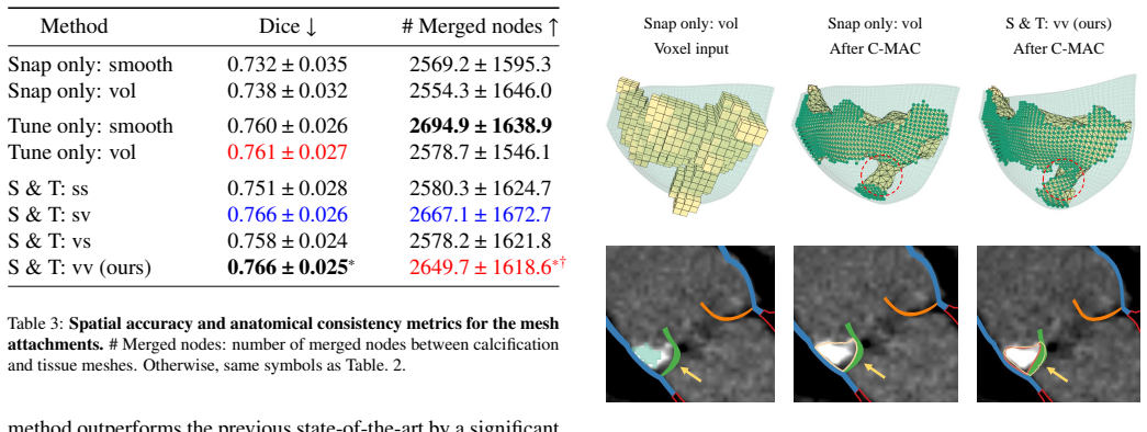
The central discovery is that the 'snap-and-tune' pipeline, with volumetric strain-energy regularization in both the network training and the test-time optimization, yields the best overall meshing performance. In head-to-head comparisons, the final method (S&T: vv) improves chamfer distance from 1.034 to 0.989 mm, thickness error from 0.051 to 0.016 mm, scaled Jacobian from 0.852 to 0.883, and skew from 0.356 to 0.299 relative to the previous state of the art (Snap only: vol), and it also improves the Dice score and number of merged nodes in attached calcification meshes. The paper attributes these gains to the tune phase's ability to overcome the rigidity of the learned mesh at high-curvature areas and to correct unrealistic gaps between neighboring structures.
Load-bearing premise
The tune phase treats the pseudo-labels produced by the surface-trained network $g_\phi$ as the correct target for the final mesh; if those pseudo-labels are systematically off from the true anatomy, the optimization will faithfully chase a biased target and could move the mesh away from the ground truth.
Editorial extensions
If this is right
- If the claimed improvements hold, clinicians can generate patient-specific volumetric meshes directly from CTA scans in about 38 seconds without manual mesh correction.
- The generated meshes can be imported directly into both engineering (Abaqus) and graphics (PhysX) simulation platforms, enabling valve opening and device deployment studies without further preprocessing.
- Because the tune phase adds no training labels, the strategy can be retrofitted to any existing DL-based volumetric mesh predictor to improve its output fidelity.
- The improved inter-part contact reduces common artifacts like bleeding between calcification and vessel wall, which matters for patient-specific simulations of calcific aortic stenosis.
- The demonstration that small mesh corrections alter valve opening shape and contact point distribution suggests mesh fidelity is a first-order factor in downstream simulation accuracy.
Reading between the lines
- The paper relies on $g_\phi$'s pseudo-labels as the tune-phase target but never reports $g_\phi$'s standalone accuracy against ground truth; if that learned target is biased, the tune step may optimize toward the wrong shape, so a direct evaluation of $g_\phi$'s chamfer distance would test this.
- The 38-second runtime is still an order of magnitude slower than the single forward pass (0.3 s); an obvious extension would be to learn the correction field itself, removing the test-time optimization while keeping its effect.
- The method is demonstrated on a single cardiac template and CTA dataset; generalizing to other anatomies or to congenital variants such as bicuspid aortic valves will require a way to change topology, since the template cannot.
- The tune phase's second loss, $\mathcal{D}_2$, is general to any pair of adjacent structures, so the same snap-and-tune recipe could plausibly be applied to cerebrovascular or orthopedic meshing where plaque-like contacts also matter.
Editorial analysis
A structured set of objections, weighed in public.
Referee Report
Summary. The paper proposes 'snap-and-tune' (S&T), a two-stage method for generating patient-specific cardiovascular volumetric meshes from cardiac CT. In the snap stage, a deep-learning template-deformation network (hθ) produces an initial volumetric mesh. In the tune stage, a per-subject test-time optimization refines that mesh using two loss-target pairs: (i) pseudo-labels from a second network gϕ trained with surface geometric regularizers instead of volumetric strain energy, and (ii) a one-sided distance term that pulls nearby calcification surfaces toward the mesh. Experiments on 35 test CT scans compare S&T variants against snap-only and tune-only baselines, reporting chamfer distance, Hausdorff distance, thickness error, scaled Jacobian, skew, attachment Dice, and merged-node counts. The central claim is that the final method (S&T: vv) significantly outperforms the previous state-of-the-art (Snap only: vol) in all metrics by a large margin, while requiring no additional training labels and remaining fully automated. The authors further demonstrate the simulation-readiness of their meshes by exporting them directly into Abaqus and PhysX and running solid-mechanics simulations.
Significance. If the claims are verified, the contribution is practically significant: it improves both spatial accuracy and element quality over a prior deep-learning pipeline without new training labels, it is fully automated at test time, and the generated meshes are directly usable in two established simulation platforms. The public code release, the inclusion of several clinically relevant metrics, and the downstream simulation demonstrations are concrete strengths. The evaluation spans 35 test subjects with multiple baselines, and the qualitative figures illustrate plausible improvements in high-curvature regions and in mesh attachment. However, two load-bearing aspects are not adequately supported in the current manuscript: the statistical basis for the headline 'significantly outperforms ... in all metrics by a large margin' claim is under-specified, and the quality of the pseudo-label target that drives the tune phase is never validated against ground truth. These issues are correctable and do not invalidate the overall approach, but they must be addressed before the central claims can be accepted.
major comments (3)
- [Section 4.4, Table 2] The headline claim that 'S&T: vv significantly outperforms the previous state-of-the-art method (Snap only: vol) in all metrics by a large margin' is not verifiable from the reported statistics. Table 2 reports only asterisk markers with 'p<0.05' and gives no test name (paired t-test, Wilcoxon signed-rank, or other), no statement that the comparisons are paired across the 35 test subjects, no confidence intervals or effect sizes, and no multiple-comparison correction across the five metrics and several contrasts. Some absolute differences are small (e.g., CD from 1.034±0.133 to 0.989±0.122 mm, HD from 4.510±1.767 to 4.410±1.690 mm), so the phrase 'large margin' needs quantitative support. Additionally, in the Thick err column, S&T: vv (0.016±0.001) is marked with † relative to 'Tune only: vol' (0.015±0.002); since S&T: vv has a larger (worse) mean, a two-sided test would indicate a significant worsening rather than an improvement, but the caption does not state the direction of the tests. Please report the full test specification, the direction of each significant contrast, corrected p-values or confidence intervals, and effect sizes such as paired Cohen's d.
- [Section 3.2, Eq. (9)] The tune phase optimizes the final mesh against pseudo-labels eY produced by gϕ, but the manuscript never reports the standalone accuracy of gϕ with respect to the ground-truth labels. This is load-bearing because gϕ is trained with surface geometric regularizers but no volumetric strain energy, so its pseudo-labels could be systematically biased away from the true anatomy; the tune step could then move the mesh away from ground truth while improving agreement with eY. Please report gϕ's own chamfer distance, Hausdorff distance, and thickness error against the ground-truth labels, and preferably provide a control experiment in which the tune objective uses the ground-truth surface (or a version with pseudo-labels replaced by ground-truth labels on the validation set) to confirm that the S&T: vv improvements over Snap only: vol are not an artifact of optimizing toward a biased target.
- [Section 4.5 and Section 3.2 (Alg. 1)] The manuscript does not specify what calcification segmentation is used in the tune phase and in the C-MAC attachment evaluation. Section 3.2 states that calcification 'can be done using simple thresholding and filtering operations as well as deep learning models,' but no threshold values, filter parameters, or model details are given. Section 4.5 evaluates attachment with C-MAC but never states whether the input voxelgrid is the human-annotated ground truth or an automatic prediction. If the same ground-truth segmentation is used both to drive the tune optimization and to compute attachment Dice and merged nodes, the attachment results in Table 3 would be partially circular; if an automatic segmentation was used, its accuracy should be reported to support the claim that the method is fully automated. Please clarify the exact inputs to the tune phase and to C-MAC, and report the accuracy of any automatic calcification segmentation.
minor comments (5)
- [Section 4.4] The heading reads 'we evaluated both the spacial accuracy and element quality'; 'spacial' should be 'spatial'.
- [Section 3.2] The text contains typos: 're-intialized' should be 're-initialized', and 'implemenations' should be 'implementations'.
- [Table 2 and Table 3 captions] The caption of Table 2 states 'Bold, blue, red: 1st, 2nd, 3rd best' but the significance markers are not defined in terms of the direction of the comparison. The caption of Table 3 says 'same symbols as Table. 2' but Table 3 uses only ∗ and †, not ‡, and the absence of ‡ is not explained.
- [Section 4.2] The default value of λuser is given as 1, but no sensitivity analysis is reported for this user-adjustable hyperparameter; a short robustness plot or a table showing the effect of varying λuser on CD and |Jac| would help users apply the method.
- [Figure 7 caption] The caption contains a typo: 'Dassault Systéms' should be 'Dassault Systèmes'.
Circularity Check
No significant circularity: the final mesh is evaluated against ground-truth labels, not the pseudo-labels optimized in the tune phase, so the headline improvement is not forced by construction.
full rationale
The derivation chain is self-contained against the ground-truth labels. The snap model hθ and pseudo-labeler gϕ are both trained on human-annotated surface meshes; the tune phase (Eq. 9) optimizes the final mesh toward gϕ's pseudo-labels, while the reported spatial accuracy and element-quality metrics (Table 2) are computed against the ground-truth labels and template mesh, not against the tune objective. Thus the main 'significantly outperforms' claim is an empirical comparison, not a tautology. The paper does rely on the authors' own prior pipeline ([18,19,20,58]) for the template, the strain-energy formulation, the chamfer loss, the SOTA anchor, and the C-MAC attachment evaluation, but each of those citations supplies a concrete algorithm or previously published result; the present comparison is measured on a held-out test set and includes several newly implemented baselines. The unvalidated standalone accuracy of gϕ and the missing details of the significance tests are correctness/verifiability concerns, not circularity. No equation in the paper defines a prediction as the fitted value of the same quantity, and no self-citation is used to forbid alternative explanations. The closest case, tune part 2 minimizing inter-part distance that later appears in the merged-node metric, is an objective/evaluation alignment rather than a definitional reduction, since the metric is computed through C-MAC on the same segmentation for all baselines.
Assumptions & free parameters
free parameters (3)
- Loss weighting hyperparameters λ0..λ5 and λuser =
{1, 10, 1, 10, 1, 0.01}; λuser default 1
- Training schedule hyperparameters =
8k epochs for hθ; 4k for gϕ; Adam lr 1e-4 networks, 1e-3 test-time
- Calcification segmentation threshold and filter parameters =
not specified
assumptions (5)
- domain assumption Template mesh M0 with fixed topology (LV myocardium, ascending aorta, aortic valve leaflets) is representative of all test patients and can be deformed to match them.
- ad hoc to paper Pseudo-labels from gϕ are sufficiently accurate targets for the tune phase.
- standard math Deformations are parameterized by b-spline control point displacements with scaling-and-squaring and are diffeomorphic.
- domain assumption Ground-truth surface labels and calcification segmentations are accurate enough for training and evaluation.
- ad hoc to paper Test-time calcification segmentation can be obtained automatically by thresholding/filtering with unspecified parameters.
Cite this review
Pith. "Pith review of Snap-and-tune: combining deep learning and test-time optimization for high-fidelity cardiovascular volumetric meshing." pith.science (2026). https://pith.science/paper/LWKIVEZ6
@misc{pith2026250608280,
  author       = {Pith},
  title        = {Pith review of: Snap-and-tune: combining deep learning and test-time optimization for high-fidelity cardiovascular volumetric meshing},
  year         = {2026},
  howpublished = {\url{https://pith.science/paper/LWKIVEZ6}},
  note         = {Machine review of arXiv:2506.08280}
}
read the original abstract
High-quality volumetric meshing from medical images is a key bottleneck for physics-based simulations in personalized medicine. For volumetric meshing of complex medical structures, recent studies have often utilized deep learning (DL)-based template deformation approaches to enable fast test-time generation with high spatial accuracy. However, these approaches still exhibit limitations, such as limited flexibility at high-curvature areas and unrealistic inter-part distances. In this study, we introduce a simple yet effective snap-and-tune strategy that sequentially applies DL and test-time optimization, which combines fast initial shape fitting with more detailed sample-specific mesh corrections. Our method provides significant improvements in both spatial accuracy and mesh quality, while being fully automated and requiring no additional training labels. Finally, we demonstrate the versatility and usefulness of our newly generated meshes via solid mechanics simulations in two different software platforms. Our code is available at https://github.com/danpak94/Deep-Cardiac-Volumetric-Mesh.
Figures


Figures from the paper (5 more)




Reference graph
Works this paper leans on
-
[1]
C. A. Taylor, C. Figueroa, Patient-specific modeling of cardiovascular mechanics, Annual review of biomedical engineering 11 (1) (2009) 109–134
work page 2009
-
[2]
Corral-Acero, F
J. Corral-Acero, F. Margara, M. Marciniak, C. Rodero, F. Loncaric, Y . Feng, A. Gilbert, J. F. Fernandes, H. A. Bukhari, A. Wajdan, M. V . Martinez, M. S. Santos, M. Shamohammdi, H. Luo, P. Westphal, P. Leeson, P. Di- Achille, V . Gurev, M. Mayr, L. Geris, P. Pathmanathan, T. Morrison, R. Cornelussen, F. Prinzen, T. Delhaas, A. Doltra, M. Sitges, E. J. Vi...
2020
-
[3]
C. Martin-Isla, V . M. Campello, C. Izquierdo, Z. Raisi- Estabragh, B. Baeßler, S. E. Petersen, K. Lekadir, Image- based cardiac diagnosis with machine learning: a review, Frontiers in cardiovascular medicine 7 (2020) 1
work page 2020
-
[4]
C. Chen, C. Qin, H. Qiu, G. Tarroni, J. Duan, W. Bai, D. Rueckert, Deep learning for cardiac image segmen- tation: a review, Frontiers in cardiovascular medicine 7 (2020) 25
work page 2020
-
[5]
M. Peirlinck, F. Sahli Costabal, J. Yao, J. M. Guccione, S. Tripathy, Y . Wang, D. Ozturk, P. Segars, T. M. Mor- rison, S. Levine, E. Kuhl, Precision medicine in human heart modeling: Perspectives, challenges, and opportu- nities, Biomechanics and Modeling in Mechanobiology 20 (3) (2021) 803–831. doi:10.1007/s10237-021-01421-z
-
[6]
F. Xu, E. L. Johnson, C. Wang, A. Jafari, C.-H. Yang, M. S. Sacks, A. Krishnamurthy, M.-C. Hsu, Computational in- vestigation of left ventricular hemodynamics following bioprosthetic aortic and mitral valve replacement, Mechan- ics Research Communications 112 (2021) 103604
work page 2021
-
[7]
A. Mohamed, C. Davatzikos, Finite element mesh gener- ation and remeshing from segmented medical images, in: 2004 2nd IEEE International Symposium on Biomedical Imaging: Nano to Macro (IEEE Cat No. 04EX821), IEEE, 2004, pp. 420–423
work page 2004
- [8]
Show all 60 references
-
[9]
Bathe, Finite element procedures, Klaus-Jurgen Bathe, 2006
K.-J. Bathe, Finite element procedures, Klaus-Jurgen Bathe, 2006
2006
-
[10]
Moccia, E
S. Moccia, E. De Momi, S. El Hadji, L. S. Mattos, Blood vessel segmentation algorithms—review of meth- ods, datasets and evaluation metrics, Computer methods and programs in biomedicine 158 (2018) 71–91
2018
-
[11]
Aoyama, L
G. Aoyama, L. Zhao, S. Zhao, X. Xue, Y . Zhong, H. Ya- mauchi, H. Tsukihara, E. Maeda, K. Ino, N. Tomii, S. Tak- agi, I. Sakuma, M. Ono, T. Sakaguchi, Automatic aortic valve cusps segmentation from ct images based on the cas- cading multiple deep neural networks, Journal of Im...
2022 doi
-
[12]
Zhuang, J
X. Zhuang, J. Shen, Multi-scale patch and multi-modality atlases for whole heart segmentation of mri, Medical image analysis 31 (2016) 77–87
2016
-
[13]
Antonelli, A
M. Antonelli, A. Reinke, S. Bakas, K. Farahani, A. Kopp- Schneider, B. A. Landman, G. Litjens, B. Menze, O. Ron- neberger, R. M. Summers, B. van Ginneken, M. Bilello, P. Bilic, P. F. Christ, R. K. G. Do, M. J. Gollub, S. H. Heckers, H. Huisman, W. R. Jarnagin, M. K. McHugo, S....
2022
-
[14]
Isensee, P
F. Isensee, P. F. Jaeger, S. A. Kohl, J. Petersen, K. H. Maier-Hein, nnu-net: a self-configuring method for deep learning-based biomedical image segmentation, Nature methods 18 (2) (2021) 203–211
2021
-
[15]
Wasserthal, H.-C
J. Wasserthal, H.-C. Breit, M. T. Meyer, M. Pradella, D. Hinck, A. W. Sauter, T. Heye, D. T. Boll, J. Cyriac, S. Yang, M. Bach, M. Segeroth, Totalsegmentator: Ro- bust segmentation of 104 anatomic structures in ct images, Radiology: Artificial Intelligence 5 (5) (2023) e230024...
2023 doi
-
[16]
B. A. Carabello, Aortic stenosis: from pressure overload to heart failure, Heart failure clinics 2 (4) (2006) 435–442
2006
-
[17]
C. M. Wojnarski, L. G. Svensson, E. E. Roselli, J. J. Idrees, A. M. Lowry, J. Ehrlinger, G. B. Pettersson, A. M. Gillinov, D. R. Johnston, E. G. Soltesz, J. L. Navia, D. F. Hammer, B. Griffin, M. Thamilarasan, V . Kalahasti, J. F. I. Sabik, E. H. Blackstone, B. W. Lytle, Aorti...
2015 doi
-
[18]
D. H. Pak, M. Liu, T. Kim, L. Liang, R. McKay, W. Sun, J. S. Duncan, Distortion energy for deep learning-based volumetric finite element mesh generation for aortic valves, in: Medical Image Computing and Computer Assisted 9 Intervention–MICCAI 2021: 24th International Confer- ...
2021
-
[19]
D. H. Pak, M. Liu, T. Kim, L. Liang, A. Caballero, J. Onofrey, S. S. Ahn, Y . Xu, R. McKay, W. Sun, R. Glea- son, J. S. Duncan, Patient-specific heart geometry mod- eling for solid biomechanics using deep learning, IEEE Transactions on Medical Imaging 43 (1) (2023) 203–215. do...
2023
-
[20]
D. H. Pak, M. Liu, T. Kim, C. Ozturk, R. McKay, E. T. Roche, R. Gleason, J. S. Duncan, Robust automated cal- cification meshing for personalized cardiovascular biome- chanics, NPJ Digital Medicine 7 (1) (2024) 213
2024
-
[21]
Ozturk, D
C. Ozturk, D. H. Pak, L. Rosalia, D. Goswami, M. E. Robakowski, R. McKay, C. T. Nguyen, J. S. Duncan, E. T. Roche, Ai-powered multimodal modeling of personalized hemodynamics in aortic stenosis, Advanced Science 12 (5) (2025) 2404755
2025
-
[22]
Schwarz, A
K. Schwarz, A. Sauer, M. Niemeyer, Y . Liao, A. Geiger, V oxgraf: Fast 3d-aware image synthesis with sparse voxel grids, Advances in Neural Information Processing Systems 35 (2022) 33999–34011
2022
-
[23]
C. Sun, M. Sun, H.-T. Chen, Direct voxel grid optimization: Super-fast convergence for radiance fields reconstruction, in: Proceedings of the IEEE/CVF conference on computer vision and pattern recognition, 2022, pp. 5459–5469
2022
-
[24]
C. R. Qi, H. Su, K. Mo, L. J. Guibas, Pointnet: Deep learn- ing on point sets for 3d classification and segmentation, in: Proceedings of the IEEE conference on computer vision and pattern recognition, 2017, pp. 652–660
2017
-
[25]
X. Yu, Y . Rao, Z. Wang, Z. Liu, J. Lu, J. Zhou, Pointr: Diverse point cloud completion with geometry-aware trans- formers, in: Proceedings of the IEEE/CVF international conference on computer vision, 2021, pp. 12498–12507
2021
-
[26]
Chibane, A
J. Chibane, A. Mir, G. Pons-Moll, Neural unsigned dis- tance fields for implicit function learning, in: Advances in Neural Information Processing Systems, V ol. 33, 2020, pp. 21638–21652
2020
-
[27]
Molaei, A
A. Molaei, A. Aminimehr, A. Tavakoli, A. Kazerouni, B. Azad, R. Azad, D. Merhof, Implicit neural representa- tion in medical imaging: A comparative survey, in: Pro- ceedings of the IEEE/CVF International Conference on Computer Vision, 2023, pp. 2381–2391
2023
-
[28]
Kerbl, G
B. Kerbl, G. Kopanas, T. Leimkühler, G. Drettakis, 3d gaussian splatting for real-time radiance field rendering., ACM Trans. Graph. 42 (4) (2023) 139–1
2023
-
[29]
J. J. Monaghan, Smoothed particle hydrodynamics and its diverse applications, Annual Review of Fluid Mechanics 44 (1) (2012) 323–346
2012
-
[30]
Raissi, P
M. Raissi, P. Perdikaris, G. E. Karniadakis, Physics- informed neural networks: A deep learning framework for solving forward and inverse problems involving nonlin- ear partial differential equations, Journal of Computational physics 378 (2019) 686–707
2019
-
[31]
V . Modi, N. Sharp, O. Perel, S. Sueda, D. I. Levin, Sim- plicits: Mesh-free, geometry-agnostic elastic simulation, ACM Transactions on Graphics (TOG) 43 (4) (2024) 1–11
2024
-
[32]
Yan, Y .-T
X. Yan, Y .-T. Jiang, C.-F. Li, R. R. Martin, S.-M. Hu, Multiphase sph simulation for interactive fluids and solids, ACM Transactions on Graphics (TOG) 35 (4) (2016) 1–11
2016
-
[33]
Sahin, M
T. Sahin, M. von Danwitz, A. Popp, Solving forward and inverse problems of contact mechanics using physics- informed neural networks, Advanced Modeling and Simu- lation in Engineering Sciences 11 (1) (2024) 11
2024
-
[34]
T. Kim, D. L. James, Skipping steps in deformable simula- tion with online model reduction, in: ACM SIGGRAPH Asia 2009 papers, 2009, pp. 1–9
2009
-
[35]
Fulton, V
L. Fulton, V . Modi, D. Duvenaud, D. I. Levin, A. Jacobson, Latent-space dynamics for reduced deformable simulation, in: Computer graphics forum, V ol. 38, Wiley Online Li- brary, 2019, pp. 379–391
2019
-
[36]
W. E. Lorensen, H. E. Cline, Marching cubes: A high resolution 3d surface construction algorithm, in: Seminal graphics: pioneering efforts that shaped the field, 1998, pp. 347–353
1998
-
[37]
G. M. Treece, R. W. Prager, A. H. Gee, Regularised march- ing tetrahedra: improved iso-surface extraction, Computers & Graphics 23 (4) (1999) 583–598
1999
-
[38]
Remelli, A
E. Remelli, A. Lukoianov, S. Richter, B. Guillard, T. Bagautdinov, P. Baque, P. Fua, Meshsdf: Differentiable iso-surface extraction, Advances in Neural Information Processing Systems 33 (2020) 22468–22478
2020
-
[39]
Chen, J.-c
L. Chen, J.-c. Xu, Optimal delaunay triangulations, Journal of Computational Mathematics (2004) 299–308
2004
-
[40]
Labelle, J
F. Labelle, J. R. Shewchuk, Isosurface stuffing: fast tetra- hedral meshes with good dihedral angles, in: ACM SIG- GRAPH 2007 papers, 2007, pp. 57–es
2007
-
[41]
Hang, Tetgen, a delaunay-based quality tetrahedral mesh generator, ACM Trans
S. Hang, Tetgen, a delaunay-based quality tetrahedral mesh generator, ACM Trans. Math. Softw 41 (2) (2015) 11
2015
-
[42]
Y . Hu, T. Schneider, B. Wang, D. Zorin, D. Panozzo, Fast tetrahedral meshing in the wild (2020)
2020
-
[43]
Fabri, S
A. Fabri, S. Pion, Cgal: The computational geometry algo- rithms library, in: Proceedings of the 17th ACM SIGSPA- TIAL international conference on advances in geographic information systems, 2009, pp. 538–539. 10
2009
-
[44]
Y . Liao, S. Donne, A. Geiger, Deep marching cubes: Learn- ing explicit surface representations, in: Proceedings of the IEEE conference on computer vision and pattern recogni- tion, 2018, pp. 2916–2925
2018
-
[45]
N. Wang, Y . Zhang, Z. Li, Y . Fu, W. Liu, Y .-G. Jiang, Pixel2mesh: Generating 3d mesh models from single rgb images, in: Proceedings of the European conference on computer vision (ECCV), 2018, pp. 52–67
2018
-
[46]
Gkioxari, J
G. Gkioxari, J. Malik, J. Johnson, Mesh r-cnn, in: Pro- ceedings of the IEEE/CVF international conference on computer vision, 2019, pp. 9785–9795
2019
-
[47]
H. Kato, Y . Ushiku, T. Harada, Neural 3d mesh renderer, in: Proceedings of the IEEE conference on computer vision and pattern recognition, 2018, pp. 3907–3916
2018
-
[48]
S. Liu, T. Li, W. Chen, H. Li, Soft rasterizer: A differ- entiable renderer for image-based 3d reasoning, in: Pro- ceedings of the IEEE/CVF international conference on computer vision, 2019, pp. 7708–7717
2019
-
[49]
Tewari, J
A. Tewari, J. Thies, B. Mildenhall, P. Srinivasan, E. Tretschk, Y . Wang, C. Lassner, V . Sitzmann, R. Martin- Brualla, S. Lombardi, T. Simon, C. Theobalt, M. Nießner, J. T. Barron, G. Wetzstein, M. Zollhöfer, V . Golyanik, Ad- vances in neural rendering, Computer Graphics For...
2022 doi
-
[50]
J. Gao, W. Chen, T. Xiang, A. Jacobson, M. McGuire, S. Fidler, Learning deformable tetrahedral meshes for 3d reconstruction, Advances in neural information processing systems 33 (2020) 9936–9947
2020
-
[51]
T. Shen, J. Gao, K. Yin, M.-Y . Liu, S. Fidler, Deep march- ing tetrahedra: a hybrid representation for high-resolution 3d shape synthesis, Advances in Neural Information Pro- cessing Systems 34 (2021) 6087–6101
2021
-
[52]
Grbic, T
S. Grbic, T. Mansi, R. Ionasec, I. V oigt, H. Houle, M. John, M. Schoebinger, N. Navab, D. Comaniciu, Image-based computational models for tavi planning: from ct images to implant deployment, in: International Conference on Medi- cal Image Computing and Computer-Assisted Inter...
2013
-
[53]
M. R. Labrosse, C. J. Beller, M. Boodhwani, C. Hudson, B. Sohmer, Subject-specific finite-element modeling of normal aortic valve biomechanics from 3d+ t tee images, Medical image analysis 20 (1) (2015) 162–172
2015
-
[54]
F. C. Ghesu, E. Krubasik, B. Georgescu, V . Singh, Y . Zheng, J. Hornegger, D. Comaniciu, Marginal space deep learning: efficient architecture for volumetric im- age parsing, IEEE transactions on medical imaging 35 (5) (2016) 1217–1228
2016
-
[55]
D. H. Pak, A. Caballero, W. Sun, J. S. Duncan, Efficient aortic valve multilabel segmentation using a spatial trans- former network, in: 2020 IEEE 17th international sym- posium on biomedical imaging (ISBI), IEEE, 2020, pp. 1738–1742
2020
-
[56]
F. Kong, S. C. Shadden, Learning whole heart mesh gener- ation from patient images for computational simulations, IEEE Transactions on Medical Imaging 42 (2) (2022) 533– 545
2022
-
[57]
T. Kim, D. Eberle, Dynamic deformables: implementation and production practicalities, in: ACM SIGGRAPH 2020 Courses, 2020, pp. 1–182
2020
-
[58]
D. H. Pak, M. Liu, S. S. Ahn, A. Caballero, J. A. Onofrey, L. Liang, W. Sun, J. S. Duncan, Weakly supervised deep learning for aortic valve finite element mesh generation from 3d ct images, in: Information Processing in Medi- cal Imaging: 27th International Conference, IPMI 20...
2021
-
[59]
Rueckert, L
D. Rueckert, L. I. Sonoda, C. Hayes, D. L. Hill, M. O. Leach, D. J. Hawkes, Nonrigid registration using free- form deformations: application to breast mr images, IEEE transactions on medical imaging 18 (8) (1999) 712–721
1999
-
[60]
A. V . Dalca, G. Balakrishnan, J. Guttag, M. R. Sabuncu, Unsupervised learning of probabilistic diffeomorphic reg- istration for images and surfaces, Medical image analysis 57 (2019) 226–236. 11
2019
Reviewed August 7, 2026 · model on record in the stance chip above.
Discussion (0). Sign in to comment.