REVIEW 3 major objections 5 minor 11 references
Motion correction of dynamic contrast enhanced MRI of the liver
T0 review · 3 major / 5 minor · reviewed 2026-08-14 · deepseek-v4-flash
Pith's one-line read Groupwise PCA-based registration aligns liver DCE-MRI time series better than pairwise registration and improves radiologists' alignment scores.
desk verdict A competent, honest application of an existing groupwise registration method to liver DCE-MRI, with a plausible quantitative comparison but a clinical-benefit claim that outruns the evidence. read the letter →
The pith
A machine-rendered reading of the paper's core claim, the machinery that carries it, and where it could break.
The reading
What carries the argument
The load-bearing mechanism is a true groupwise registration in which all volumes of the DCE-MRI series are transformed to a common space and the similarity of the whole series is scored at once, rather than each volume being registered to a reference volume. The dissimilarity metric is built from a principal component analysis of the correlation matrix of voxel intensities across time, with the cost $D_{\mathrm{PCA}} = \sum_{j=1}^{V} j\, \lambda_j$, where $\lambda_j$ are the eigenvalues of the correlation matrix; weighting the smaller eigenvalues more heavily makes the optimization put more emphasis on the low-variance directions that remain after contrast-related intensity change is accounted for. Because no reference image and no contrast-enhancement model is used, the method avoids the bias that pairwise reference-based registration and synthetic-image-based methods introduce. The transformation is a multi-resolution B-spline deformation, with the same sampling and interpolation settings used for the pairwise mutual-information baseline, so the comparison isolates the groupwise metric and common-space strategy.
What would settle it
Recompute the Dice overlap measures using independent lesion segmentations from two radiologists on a larger cohort; if the groupwise advantage over pairwise registration disappears or reverses, the central alignment claim fails. A reader-level test would be a blinded randomized comparison in which radiologists score original versus groupwise-registered series; the claim fails if alignment quality, reading time, and confidence are not better for the registered series.
Extended reading notes
Core claim
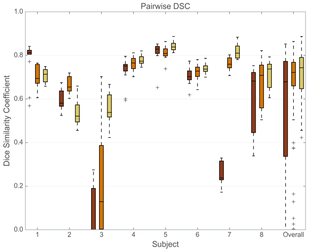
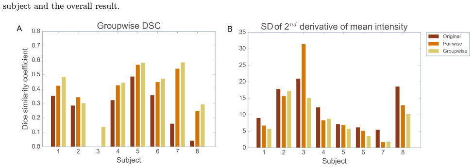
The central claim is that groupwise registration using a PCA-based dissimilarity metric achieves good and temporally smooth alignment of clinical DCE-MRI of the liver, and does so better than pairwise registration with a mutual information metric or no registration. In the eight-patient quantitative comparison, average overlap between the first-frame lesion mask and the other frames rose from 0.57 without registration to 0.67 with pairwise registration and 0.71 with groupwise registration; the stricter groupwise DSC rose from 0.25 to 0.37 and 0.41, and the standard deviation of the second derivative of mean lesion intensity over time fell from 12.2 to 11.1 and 8.5. The mean Jacobian determinant of the lesion deformation was close to 1 for both methods, with a smaller spread for groupwise registration, indicating stable lesion volumes and smoother spatial transformations. The paper also reports early clinical results in which radiologists scored average alignment quality at 2.1 before and 4 after registration, found the subtraction images diagnostically useful in all nine cases, estimated a time gain of almost a minute in four cases, and reported increased confidence in three cases. The authors note two exceptions: one series that was already well aligned in its first breath holds, and one where the groupwise method failed to align the first non-contrast image with the rest of the series.
Load-bearing premise
The quantitative comparison rests on the manual lesion outlines being accurate enough to serve as ground truth, yet in several images the lesion was hard to distinguish from the liver and the same observer's repeat outlines overlapped by only 81 percent (Dice).
Editorial extensions
If this is right
- Radiologists can rely on subtraction images from groupwise-registered liver DCE-MRI: in all nine clinical cases the subtraction images were judged good enough to use, and in two cases they were determinative for diagnosis.
- Lesion enhancement curves over time become smoother and less noisy, since the groupwise result shows the lowest standard deviation of the second derivative of mean lesion intensity.
- Lesion volume measurements over a DCE-MRI series are not systematically distorted by the transformation, because the mean Jacobian determinant in the lesion stayed close to 1.
- The registration approach does not depend on a contrast-enhancement model, so it is usable in short clinical acquisitions (16 volumes) where model fitting may be unreliable.
- For scans with little motion, registration adds no benefit, so the technique should be targeted at series where breath-hold inconsistency is actually present.
Reading between the lines
- A testable extension not reported here: because the PCA metric weights the smallest eigenvalues most heavily, the optimal eigenvalue weighting may need retuning when the number of time points drops well below 16, so the method should be stress-tested on shorter series.
- The one visible failure mode, where the first non-contrast frame was not aligned with the rest of the series, suggests a practical remedy worth testing: initialize the groupwise optimization with a pairwise alignment of the first frame, or evaluate alignment after re-anchoring to a later contrast-enhanced frame.
- The clinical gains reported are from an early study with nine patients; whether improved alignment changes diagnostic decisions in a larger, blinded reader study remains an open question.
Signed reviews
Editorial analysis
A structured set of objections, weighed in public.
Referee Report
Summary. The paper evaluates a groupwise PCA-based registration method, previously proposed by Huizinga et al. (2016), for motion correction of liver DCE-MRI. Using eight clinical data sets, the authors compare the groupwise method with a pairwise mutual-information registration and with no registration. Quantitative evaluation is based on manual lesion segmentations, computing pairwise DSC, groupwise DSC, temporal intensity smoothness, and Jacobian-determinant statistics. The paper additionally reports early results from an ongoing clinical evaluation in which three radiologists scored groupwise-registered images from nine patients. The central claim is that groupwise registration provides better alignment than pairwise registration or no registration, and that radiologists benefit from the registered images and subtraction images.
Significance. If the conclusion were established, the paper would provide a useful clinical validation of a model-free groupwise registration approach for abdominal DCE-MRI, an application where contrast dynamics make pairwise reference-based registration difficult. Strengths of the design include the use of evaluation metrics that are independent of the PCA registration objective, the comparison against a conventional pairwise baseline, and the inclusion of a clinical reader study. The main weakness is that the quantitative evidence is based on only eight patients, with small performance differences, no statistical significance testing, and a segmentation ground truth whose repeatability (intra-observer DSC 0.81) is low enough to cover the reported gains. The early clinical evaluation is qualitatively favorable but is presented as ongoing and without formal analysis.
major comments (3)
- [Section 3.1, Figures 2 and 3] The central quantitative claim that groupwise registration is superior is not statistically supported. The reported between-method differences are small (mean DSC 0.71 vs. 0.67; groupwise DSC 0.41 vs. 0.37; temporal smoothness SD 8.5 vs. 11.1) and are computed from only eight patients. No confidence intervals, paired significance tests, or effect-size measures are provided. Given the small sample, the authors should add appropriate paired statistical tests or bootstrap confidence intervals, and should state whether the observed differences are significant at any prespecified level.
- [Section 2.3.1 and Section 2.1] The DSC and groupwise DSC metrics are computed from manual lesion segmentations whose intra-observer Dice coefficient is only 0.81, and the text acknowledges that in some images the lesion was hard to distinguish from liver parenchyma. With reported between-method DSC differences of about 0.04, the segmentation reproducibility margin is an order of magnitude larger than the method advantage, so the comparison may be dominated by annotation variability rather than by registration quality. The authors should provide a sensitivity analysis (e.g., perturbing segmentations or using segmentation uncertainty) or otherwise demonstrate that the reported gains exceed the segmentation noise.
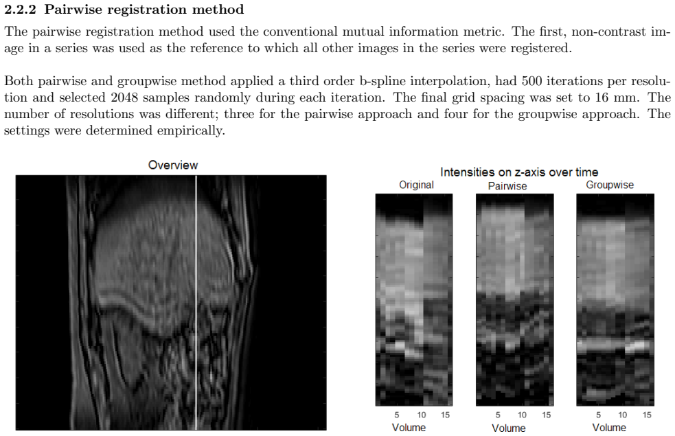
- [Section 2.2.2 and Section 2.2.1] The pairwise and groupwise registration settings are not matched: the pairwise method uses three resolutions and the groupwise method uses four, and the text states that the settings were determined empirically. This confound prevents a clean attribution of the observed improvement to the groupwise metric rather than to favorable tuning. The authors should justify the choice of different settings or report experiments showing that the advantage of groupwise registration is robust across reasonable variations of resolutions, grid spacing, and iteration counts.
minor comments (5)
- [Abstract and Section 2.2.1] The text uses the phrase 'principle component analysis' instead of 'principal component analysis' in the abstract and in the method description.
- [Section 2.2.1, equation for C] The notation in the correlation matrix formula is unclear: the matrix S is described as 'a diagonal matrix with the column-wise standard deviations of M', but the formula includes S−1 without defining the inverse operation. If S is diagonal, the inverse is clear, but this should be stated explicitly to avoid confusion.
- [Section 2.3.1, groupwise DSC definition] The formula '16 × S1 ⋂ S2... ⋂ S16' followed by the denominator 'S1+S2...+S16' is ambiguous; it should be made explicit that the numerator is the size of the intersection of all sixteen masks and the denominator is the sum of the individual mask volumes.
- [Section 3, figure callouts] The text refers to 'Figure 4' twice for two different purposes: once for the Jacobian determinant boxplot and later for the radiologist quality scores. The figure numbers should be corrected so that each figure is cited unambiguously.
- [Section 3.2] The sentence 'All the original images had a score of 2 or 3, except in one case a score of 5 was assigned' is ambiguous about whether 'one case' refers to one patient, one image series, or one radiologist's score; this should be clarified.
Circularity Check
No significant circularity: the paper is an empirical validation study whose outcome measures are independent of the registration objective.
full rationale
The paper is an application/validation study rather than a derivation, so there is no chain in which a prediction reduces to its input. The groupwise PCA-based registration method is taken from Huizinga et al. (2016) and is not derived here; the current authors do not overlap with that method's authors. The central comparison is empirical: groupwise PCA registration versus pairwise mutual-information registration and no registration, assessed with DSC and groupwise DSC computed from manual lesion segmentations, temporal smoothness (SD of the second derivative of the mean intensity in the lesion), Jacobian determinant statistics, and radiologist scoring. None of these evaluation quantities is used as a fitting target or defined in terms of the PCA dissimilarity metric D_PCA = sum_j j*lambda_j. The registration settings are admittedly empirical ('The settings were determined empirically'), and the manual segmentations are acknowledged to be difficult in some images ('the lesion was hard to distinguish from the liver parenchyma', intra-observer DSC 0.81); these are validity and statistical-strength concerns, not circularity. The only self-citation is the Elastix toolbox paper [9], on which Pluim is a coauthor, but it is cited purely as the software implementation and is not load-bearing for the claimed result. No equation is equivalent to its input by construction, no fitted parameter is renamed as a prediction, and no uniqueness or ansatz is imported via self-citation. The reported gains are small and the sample size is limited, but that is an evidence-quality issue, not a circular-reasoning issue.
Assumptions & free parameters
free parameters (1)
- Registration hyperparameters (grid spacing, iterations, samples, resolutions) =
16 mm, 500 iterations, 2048 samples, 3-4 resolutions
assumptions (4)
- domain assumption PCA on the correlation matrix of image intensities separates contrast enhancement from patient motion in DCE-MRI time series.
- domain assumption The manual lesion segmentations, with an intra-observer DSC of 0.81, are accurate enough to serve as ground truth for the alignment metrics.
- standard math B-spline interpolation and the Elastix registration framework are appropriate for 3D DCE-MRI data.
- domain assumption The first non-contrast image is a valid reference for pairwise registration and for propagating lesion masks.
Cite this review
Pith. "Pith review of Motion correction of dynamic contrast enhanced MRI of the liver." pith.science (2026). https://pith.science/paper/MWIRZ6WY
@misc{pith2026190808254,
  author       = {Pith},
  title        = {Pith review of: Motion correction of dynamic contrast enhanced MRI of the liver},
  year         = {2026},
  howpublished = {\url{https://pith.science/paper/MWIRZ6WY}},
  note         = {Machine review of arXiv:1908.08254}
}
read the original abstract
Motion correction of dynamic contrast enhanced magnetic resonance images (DCE-MRI) is a challenging task, due to changes in image appearance. In this study a groupwise registration, using a principle component analysis (PCA) based metric,1 is evaluated for clinical DCE MRI of the liver. The groupwise registration transforms the images to a common space, rather than to a reference volume as conventional pairwise methods do, and computes the similarity metric on all volumes simultaneously. This groupwise registration method is compared to a pairwise approach using a mutual information metric. Clinical DCE MRI of the abdomen of eight patients were included. Per patient one lesion in the liver was manually segmented in all temporal images (N=16). The registered images were compared for accuracy, spatial and temporal smoothness after transformation, and lesion volume change. Compared to a pairwise method or no registration, groupwise registration provided better alignment. In our recently started clinical study groupwise registered clinical DCE MRI of the abdomen of nine patients were scored by three radiologists. Groupwise registration increased the assessed quality of alignment. The gain in reading time for the radiologist was estimated to vary from no difference to almost a minute. A slight increase in reader confidence was also observed. Registration had no added value for images with little motion. In conclusion, the groupwise registration of DCE MR images results in better alignment than achieved by pairwise registration, which is beneficial for clinical assessment.
Figures
Figures from the paper (2 more)


Reference graph
Works this paper leans on
-
[1]
Huizinga, W., Poot, D. H. J., Guyader, J.-M., Klaassen, R., Coolen, B. F., van Kranenburg, M., van Geuns, R. J. M., Uitterdijk, A., Polfliet, M., Vandemeulebroucke, J., Leemans, A., Niessen, W. J., and Klein, S., ``Pca-based groupwise image registration for quantitative mri,'' Med. Image Anal. 29 , 65--78 (2016)
work page 2016
-
[2]
Choyke, P. L., Dwyer, A. J., and Knopp, M. V., ``Functional tumor imaging with dynamic contrast- enhanced magnetic resonance imaging,'' J. Magn. Reson. Imaging 17 , 509--520 (2003)
work page 2003
-
[3]
Wollny, G., Kellman, P., Santos, A., and Ledesma-Carbayo, M. J., ``Automatic motion compensation of free breathing acquired myocardial perfusion data by using independent component analysis,'' Med. Image Anal. 16 , 1015--1028 (2012)
work page 2012
-
[4]
Buonaccorsi, G. A., O’Connor, J. P. B., Caunce, A., Roberts, C., Cheung, S., Watson, Y., Davies, K., Hope, L., Jackson, A., Jayson, G. C., and Parker, G. J. M., ``Tracer kinetic model-driven registration for dynamic contrast-enhanced mri time-series data,'' Magn. Reson. Med. 58 , 1010--1019 (2007)
work page 2007
-
[5]
Hayton, P., Brady, M., Tarassenko, L., and Moore, N., ``Analysis of dynamic mr breast images using a model of contrast enhancement,'' Med. Image Anal. 1 , 207--224 (1997)
work page 1997
-
[6]
Bhushan, M., Schnabel, J. A., Risser, L., Heinrich, M. P., Brady, J. M., and Jenkinson, M., ``Motion correction and parameter estimation in dce mri sequences : Application to colorectal cancer,'' Lect. Notes Comput. Sci. MICCAI 2011 , 476--483 (2011)
work page 2011
-
[7]
Melbourne, A., Atkinson, D., White, M., Collins, D., Leach, M., and Hawkes, D., ``Registration of dynamic contrast-enhanced mri using a progressive principal component registration (ppcr),'' Phys. Med. Biol. 52 , 5147--5156 (2007)
work page 2007
-
[8]
Hamy, V., Dikaios, N., Punwani, S., Melbourne, A., Latifoltojar, A., Makanyanga, J., Chouhan, M., Helbren, E., Menys, A., Taylor, S., and Atkinson, D., ``Respiratory motion correction in dynamic mri using robust data decomposition registration - application to dce-mri,'' Med. Image Anal. 18 , 301--313 (2014)
work page 2014
Show all 11 references
-
[9]
A., and Pluim, J
Klein, S., Staring, M., Murphy, K., Viergever, M. A., and Pluim, J. P. W., ``elastix: A toolbox for intensity-based medical image registration,'' IEEE Trans. Med. Imaging 29 , 196--205 (2010)
2010
-
[10]
K., Wilson, G
Sundarakumar, D. K., Wilson, G. J., Osman, S. F., Zaidi, S. F., and Maki, J. H., ``Evaluation of image registration in subtracted 3d dynamic contrast-enhanced mri of treated hepatocellular carcinoma,'' Am. J. Roentgenol. 204 , 287--296 (2015)
2015
-
[11]
write newline
" write newline "" before.all 'output.state := FUNCTION blank.sep after.quote 'output.state := FUNCTION fin.entry output.state after.quoted.block = 'skip 'add.period if write newline FUNCTION new.block output.state before.all = 'skip output.state after.quote = after.quoted.blo...
Reviewed August 14, 2026 · model on record in the stance chip above.
Discussion (0). Continue with ORCID to comment.