REVIEW 3 major objections 6 minor 14 references
Multi-step Cascaded Networks for Brain Tumor Segmentation
T0 review · 3 major / 6 minor · reviewed 2026-08-14 · deepseek-v4-flash
Pith's one-line read Cascaded 3D U-nets hit 0.886 whole-tumor Dice on BraTS 2019.
desk verdict A credible BraTS 2019 cascade method with real validation scores, but its central advantage claim rests on no baseline at all. read the letter →
The pith
A machine-rendered reading of the paper's core claim, the machinery that carries it, and where it could break.
The reading
What carries the argument
The central mechanism is the multi-step cascade with mask multiplication. In each finer step, the previous step's predicted binary mask is multiplied voxel-wise into the chosen MRI modality before it enters the next 3D U-net, so the network concentrates on the remaining candidate region; this acts as a spatial-attention prior consistent with the nested containment hierarchy of whole tumor, tumor core, and enhancing tumor. The auxiliary deep-supervision outputs in the expanding pathway propagate gradients to earlier layers, and focal loss reweights easy versus hard voxels to counter the strong background-foreground imbalance.
What would settle it
Replace the predicted whole-tumor mask with the ground-truth whole-tumor mask during inference on the validation set. If the tumor-core and enhancing-tumor Dice scores improve substantially, the cascade's accuracy is bottlenecked by first-step recall; if they stay the same, error propagation from the first step is not the limiting factor. The paper itself reports no ablation of this kind.
Extended reading notes
Core claim
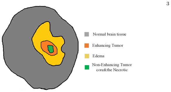
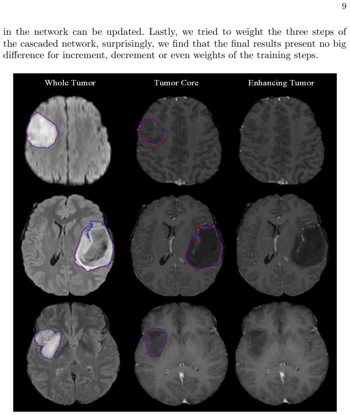
The central claim is that converting the multi-class tumor segmentation problem into three linked binary problems, ordered by the anatomical hierarchy, improves accuracy over a single network that must separate all structures at once. Each cascade step is a 3D U-net with three auxiliary deep-supervision outputs; the loss is focal loss. The chosen modalities mirror the manual annotation protocol: Flair and T1ce for whole tumor, T1ce alone for tumor core and enhancing tumor. The authors report mean Dice of 0.886 for whole tumor, 0.813 for tumor core, and 0.771 for enhancing tumor on the BraTS 2019 validation data, and note that the most common failure is predicting the whole tumor as tumor core, especially for LGG cases.
Load-bearing premise
The whole pipeline assumes the first step's whole-tumor mask contains the true tumor core and enhancing tumor; if that first mask misses any tumor voxels, the two finer steps never get a chance to find them.
Editorial extensions
If this is right
- The cascade turns one hard multi-class segmentation problem into three simpler binary problems, which should generalize better when labeled training data are limited.
- Because each step sees only the voxels inside the previous mask, the final enhancing-tumor prediction is constrained to lie inside the predicted tumor core, enforcing anatomical consistency by construction.
- End-to-end training outperformed a curriculum in which the steps were trained one at a time, suggesting joint parameter updates help the cascade fit the training data.
- Weighting the three cascade steps up or down changed final results little, so the method is insensitive to that hyperparameter.
- The observed confusion of whole tumor for tumor core points to inter-volume intensity variation as a remaining source of error.
Reading between the lines
- An implication the paper leaves untested: the pipeline's ceiling is set by the recall of the first-step whole-tumor mask; any voxel missed there is unrecoverable by the tumor-core and enhancing-tumor steps. A direct test would be to feed ground-truth whole-tumor masks into the later steps and measure how much the tumor-core and enhancing-tumor Dice improve.
- The hard mask multiplication could be replaced by a soft probability map from the previous step, which would let gradients flow across steps and might allow the finer networks to correct small whole-tumor misses; the paper does not explore this.
- The same coarse-to-fine cascade with per-step modality selection could transfer to other nested lesion structures, such as pancreas regions or prostate zones, wherever a containment hierarchy is known a priori.
- Because the authors observe worse variance on tumor core than enhancing tumor and attribute it to LGG cases, a stratified analysis by tumor grade, or a histogram-equalization preprocessing step, is a concrete next experiment suggested by their own discussion.
Signed reviews
Editorial analysis
A structured set of objections, weighed in public.
Referee Report
Summary. The paper proposes a multi-step cascaded 3D U-Net for glioma substructure segmentation on BraTS 2019. The pipeline first segments the whole tumor (WT) from Flair and T1ce, then segments the tumor core (TC) and enhancing tumor (ET) from T1ce with the previous mask multiplied into the input as prior information; all steps are trained end-to-end with deep supervision and focal loss. On the official BraTS 2019 validation platform the authors report mean Dice coefficients of 0.886 for WT, 0.813 for TC, and 0.771 for ET, with code publicly available. The paper also describes N4 bias-field correction, random cropping, data augmentation, and a curriculum-learning comparison that reportedly showed no benefit over end-to-end training.
Significance. If the reported scores are accurate, the paper describes a competitive automatic glioma segmentation pipeline on a standard benchmark; the use of the official evaluation platform and the release of code are strengths. However, the paper's central causal claims—that the cascade itself is more effective than a one-step network and that per-step modality selection is beneficial—are not backed by any ablation in the manuscript. The descriptive results are plausible, but the attribution of the scores to the cascade and modality choices is an unsupported step that can be remedied with targeted experiments.
major comments (3)
- [Section 2.1, Figure 2, and Conclusion] The conclusion states that the multi-step network 'has proved to be more effective than the one-step counterpart,' but the manuscript reports no one-step baseline or three-independent-networks baseline trained with the same architecture and data. Because the TC and ET steps multiply the previous WT mask into the input, any WT false negative (validation WT sensitivity is 0.921 in Table 1) removes those voxels from the reach of the later steps. The manuscript should report an ablation that either (a) feeds oracle ground-truth WT masks into steps 2 and 3 to measure the ceiling imposed by the WT step, or (b) trains a single multi-class network and/or three independent binary networks under identical conditions. Without such an experiment, the TC/ET Dice cannot be attributed to the cascade mechanism rather than to the WT step's recall.
- [Section 2.1 and Conclusion] The conclusion also claims that choosing specific modalities for each step 'improves our result a lot compared to the method to use all modalities,' but no experiment comparing modality subsets is reported anywhere in the paper. This is a load-bearing claim for the method's design; an ablation using all four modalities in each cascade step, or in the one-step baseline, is needed before the claim can be accepted.
- [Section 3.3 and Table 1] No analysis is provided of the spatial overlap between WT false negatives and true TC/ET voxels. Since TC and ET are strict subsets of WT, even a modest WT sensitivity loss could disproportionately remove enhancing-tumor voxels, and this is exactly the failure mode that the paper needs to rule out. I recommend quantifying the fraction of true TC/ET voxels that fall inside WT false-negative regions on the validation set, and relating that quantity to the observed TC/ET Dice.
minor comments (6)
- [Section 3.2] The text 'adding guassion blur' should read 'adding Gaussian blur.'
- [Section 3.3] The word 'mothed' should be 'method.'
- [Conclusion] The word 'mamual' should be 'manual.'
- [Section 2.2] Please define how the three auxiliary outputs are combined with the main output in the total loss, including the weighting coefficients, and provide the values of alpha_t and gamma used in Eq. (1).
- [Introduction and References] The phrase 'Inspired by Havaei and iek' should name Cicek et al.; the intended citation to reference [4] is otherwise unclear.
- [Figure 3] The architectural details are not fully specified: the number of feature maps, downsampling factors, and the exact locations where auxiliary outputs are attached should be stated to make the network reproducible.
Circularity Check
No circularity: the central claims are external validation scores on a fixed benchmark; the cascade's coarse-to-fine masking is the method under test, not an input-dependent derivation.
full rationale
The paper reports an empirical segmentation method evaluated on the BraTS 2019 benchmark with online platform metrics. There is no derivation chain in which an output is defined in terms of fitted inputs, no parameter fitted to a subset and then renamed as a prediction, and no load-bearing self-citation. The multi-step cascade uses each previous mask as an additional input to the next step; this is the proposed architecture under evaluation, not a circular argument. The absence of an oracle-mask ablation is a legitimate experimental limitation concerning whether the cascade causes the observed performance, but it does not make the reported Dice scores circular. Self-citations in the introduction are background references, not justifications for the paper's conclusions. Therefore no specific circular step can be exhibited, and the appropriate score is 0.
Assumptions & free parameters
free parameters (8)
- initial learning rate =
0.001, decayed to 0.0005
- random crop size =
96 x 96 x 96
- batch size =
1
- number of training epochs =
50
- focal loss alpha_t =
not reported
- focal loss gamma =
not reported
- data augmentation settings =
not reported
- 3D U-net architecture dimensions =
not specified in text
assumptions (4)
- domain assumption BraTS 2019 ground truth segmentations are accurate and follow a consistent annotation protocol
- domain assumption The hierarchical containment of tumor substructures is a valid prior for segmentation
- domain assumption Preprocessed multimodal MRI with N4 correction and z-score normalization is sufficient input for the network
- ad hoc to paper Mask multiplication propagates only useful prior information into the next cascade step
Cite this review
Pith. "Pith review of Multi-step Cascaded Networks for Brain Tumor Segmentation." pith.science (2026). https://pith.science/paper/NCMYR45U
@misc{pith2026190805887,
  author       = {Pith},
  title        = {Pith review of: Multi-step Cascaded Networks for Brain Tumor Segmentation},
  year         = {2026},
  howpublished = {\url{https://pith.science/paper/NCMYR45U}},
  note         = {Machine review of arXiv:1908.05887}
}
read the original abstract
Automatic brain tumor segmentation method plays an extremely important role in the whole process of brain tumor diagnosis and treatment. In this paper, we propose a multi-step cascaded network which takes the hierarchical topology of the brain tumor substructures into consideration and segments the substructures from coarse to fine .During segmentation, the result of the former step is utilized as the prior information for the next step to guide the finer segmentation process. The whole network is trained in an end-to-end fashion. Besides, to alleviate the gradient vanishing issue and reduce overfitting, we added several auxiliary outputs as a kind of deep supervision for each step and introduced several data augmentation strategies, respectively, which proved to be quite efficient for brain tumor segmentation. Lastly, focal loss is utilized to solve the problem of remarkably imbalance of the tumor regions and background. Our model is tested on the BraTS 2019 validation dataset, the preliminary results of mean dice coefficients are 0.886, 0.813, 0.771 for the whole tumor, tumor core and enhancing tumor respectively. Code is available at https://github.com/JohnleeHIT/Brats2019
Figures


Figures from the paper (3 more)


Reference graph
Works this paper leans on
-
[1]
Proceedings MICCAI-BRATS (2014) 36–39
Zikic, D., Ioannou, Y., Brown, M., Criminisi, A.: Segmentation of brain tumor tissues with convolutional neural networks. Proceedings MICCAI-BRATS (2014) 36–39
work page 2014
-
[2]
Zhao, L., Jia, K.: Deep feature learning with discrimination mechanism for brain tumor segmentation and diagnosis. In: 2015 International Conference on Intelligent Information Hiding and Multimedia Signal Processing (IIH-MSP), IEEE (2015) 306–309
work page 2015
-
[3]
Medical image analysis 35 (2017) 18–31
Havaei, M., Davy, A., Warde-Farley, D., Biard, A., Courville, A., Bengio, Y., Pal, C., Jodoin, P.M., Larochelle, H.: Brain tumor segmentation with deep neural networks. Medical image analysis 35 (2017) 18–31
2017
-
[4]
C ¸ i¸ cek,¨O., Abdulkadir, A., Lienkamp, S.S., Brox, T., Ronneberger, O.: 3d u- net: learning dense volumetric segmentation from sparse annotation. In: Interna- tional conference on medical image computing and computer-assisted intervention, Springer (2016) 424–432
work page 2016
-
[5]
Medical image analysis 36 (2017) 61–78
Kamnitsas, K., Ledig, C., Newcombe, V.F., Simpson, J.P., Kane, A.D., Menon, D.K., Rueckert, D., Glocker, B.: Efficient multi-scale 3d cnn with fully connected crf for accurate brain lesion segmentation. Medical image analysis 36 (2017) 61–78
work page 2017
-
[6]
Pattern Recognition 88 (2019) 90–100
Chen, S., Ding, C., Liu, M.: Dual-force convolutional neural networks for accurate brain tumor segmentation. Pattern Recognition 88 (2019) 90–100
work page 2019
-
[7]
IEEE transactions on medical imaging 37(8) (2018) 1943–1954
Ma, C., Luo, G., Wang, K.: Concatenated and connected random forests with mul- tiscale patch driven active contour model for automated brain tumor segmentation of mr images. IEEE transactions on medical imaging 37(8) (2018) 1943–1954
work page 2018
-
[8]
IEEE transactions on medical imaging 34(10) (2014) 1993–2024
Menze, B.H., Jakab, A., Bauer, S., Kalpathy-Cramer, J., Farahani, K., Kirby, J., Burren, Y., Porz, N., Slotboom, J., Wiest, R., et al.: The multimodal brain tumor image segmentation benchmark (brats). IEEE transactions on medical imaging 34(10) (2014) 1993–2024
work page 2014
Show all 14 references
-
[9]
In: Proceedings of the IEEE international conference on computer vision
Lin, T.Y., Goyal, P., Girshick, R., He, K., Doll´ ar, P.: Focal loss for dense object detection. In: Proceedings of the IEEE international conference on computer vision. (2017) 2980–2988
2017
-
[10]
Scientific data 4 (2017) 170117 11
Bakas, S., Akbari, H., Sotiras, A., Bilello, M., Rozycki, M., Kirby, J.S., Freymann, J.B., Farahani, K., Davatzikos, C.: Advancing the cancer genome atlas glioma mri collections with expert segmentation labels and radiomic features. Scientific data 4 (2017) 170117 11
2017
-
[11]
arXiv preprint arXiv:1811.02629 (2018)
Bakas, S., Reyes, M., Jakab, A., Bauer, S., Rempfler, M., Crimi, A., Shinohara, R.T., Berger, C., Ha, S.M., Rozycki, M., et al.: Identifying the best machine learning algorithms for brain tumor segmentation, progression assessment, and overall survival prediction in the brats c...
2018 arXiv
-
[12]
The Cancer Imaging Archive 286 (2017)
Bakas, S., Akbari, H., Sotiras, A., Bilello, M., Rozycki, M., Kirby, J., Freymann, J., Farahani, K., Davatzikos, C.: Segmentation labels and radiomic features for the pre-operative scans of the tcga-lgg collection. The Cancer Imaging Archive 286 (2017)
2017
-
[13]
the cancer imaging archive (2017)
Bakas, S., Akbari, H., Sotiras, A., Bilello, M., Rozycki, M., Kirby, J., Freymann, J., Farahani, K., Davatzikos, C.: Segmentation labels and radiomic features for the pre-operative scans of the tcga-gbm collection. the cancer imaging archive (2017). (2017)
2017
-
[14]
IEEE transactions on medical imaging 29(6) (2010) 1310
Tustison, N.J., Avants, B.B., Cook, P.A., Zheng, Y., Egan, A., Yushkevich, P.A., Gee, J.C.: N4itk: improved n3 bias correction. IEEE transactions on medical imaging 29(6) (2010) 1310
2010
Reviewed August 14, 2026 · model on record in the stance chip above.
Discussion (0). Continue with ORCID to comment.