REVIEW 3 major objections 4 minor 29 references
Automated retinal vessel segmentation based on morphological preprocessing and 2D-Gabor wavelets
T0 review · 3 major / 4 minor · reviewed 2026-08-14 · deepseek-v4-flash
Pith's one-line read Retinal vessel segmentation without training labels reaches 94.32% accuracy on DRIVE.
desk verdict The pipeline is a plausible configuration of standard tools, but the abstract's superiority claim is contradicted by the paper's own Table 2 and should be revised before publication. read the letter →
The pith
A machine-rendered reading of the paper's core claim, the machinery that carries it, and where it could break.
The reading
What carries the argument
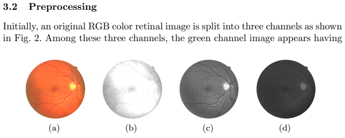
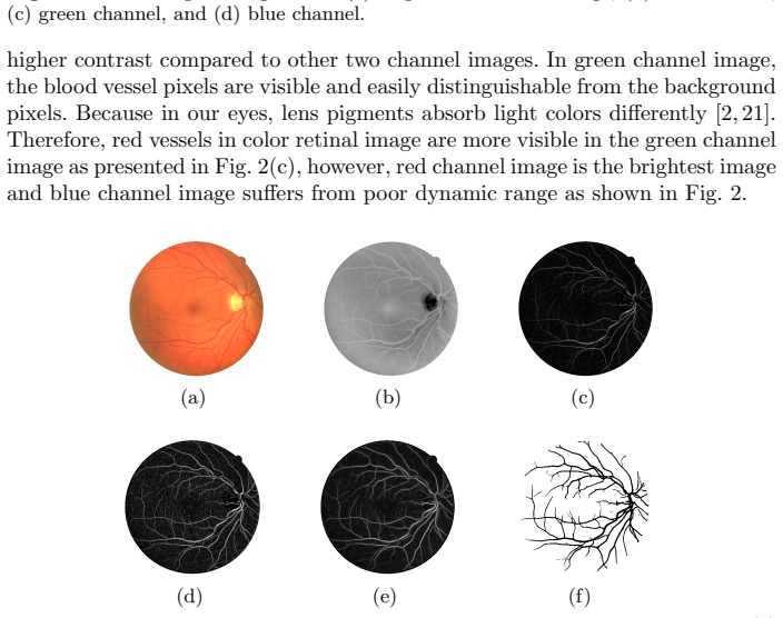
The load-bearing mechanism is the ordering of three complementary operators. White top-hat transformation with a circular structuring element 11 pixels in diameter extracts image details smaller than that width, which by the paper's estimate covers all blood vessels (1–10 pixels) while rejecting larger structures such as optic disc, fovea, and exudates. CLAHE then raises the local contrast of those remaining vessel pixels. A bank of 2D-Gabor wavelets—orientations sampled every 20° from 0° to 180° and radial frequencies from 0.7 to 1.5, with bandwidth constant $K=2.2$—represents thick and thin vessels at different angles; taking the maximum wavelet response per pixel and applying Otsu's global threshold produces the final binary vessel map.
What would settle it
Tune nothing: run the published pipeline with the stated parameters on the STARE and CHASE DB1 databases and compare against their manual ground truths; if accuracy falls materially below the DRIVE figure, or if the structuring element must be re-tuned per database to recover performance, the claim that the algorithm generalizes in its fixed form is refuted.
Extended reading notes
Core claim
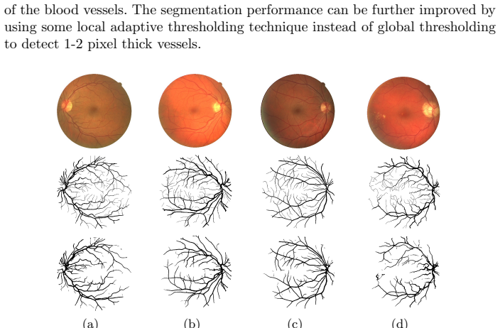
The central claim is that adding a top-hat morphological transform before CLAHE and Gabor filtering is the decisive improvement: it removes large non-vessel structures from the inverted green channel before contrast enhancement, so the subsequent filter bank responds mainly to vessel pixels. The paper reports that on the DRIVE test set this pipeline yields an average accuracy of 94.32%, sensitivity of 75.03%, specificity of 97.12%, area under the ROC curve of 0.9524, and kappa agreement of 0.7374, with a small accuracy spread ($\sigma=0.0049$). It further claims that these results compare favorably with established supervised and unsupervised algorithms in accuracy, sensitivity, and kappa agreement, and that the method handles a pathological image containing exudates better than a comparable level-set/region-growing approach.
Load-bearing premise
The reported results rest on the assumption that the fixed filter settings (11-pixel structuring element, Gabor frequency range, and bandwidth constant) were chosen without peeking at the DRIVE test labels and will carry over to other fundus images unchanged.
Editorial extensions
If this is right
- A training-free pipeline can reach accuracy within about two percentage points of the strongest supervised classifiers on DRIVE, so unsupervised filtering approaches remain a practical baseline for retinal screening.
- Because the structuring element diameter is tied to the maximum vessel width (1–10 pixels on DRIVE), applying the same algorithm to other databases requires only rescaling that one parameter to the new image resolution, a transfer step the paper itself notes.
- The small standard deviation of accuracy (0.0049) across the 20 DRIVE test images suggests the method performs consistently on images with different pigmentation, illumination, and pathology.
- The authors' suggestion that local adaptive thresholding could replace Otsu implies that thin-vessel detection (1–2 pixel vessels) is the main remaining bottleneck, and a thresholding upgrade is a natural next step.
Reading between the lines
- An independent rerun on STARE or CHASE with the same fixed parameters would separate genuine generalizability from DRIVE-specific tuning; the paper does not provide that test.
- The top-hat-plus-Gabor response could also serve as input features to a lightweight supervised classifier, potentially combining the preprocessing's robustness with learned thresholds.
- Swapping Otsu for local adaptive thresholding should preferentially recover 1–2 pixel thin vessels, at the risk of extra false positives around lesions.
Signed reviews
Editorial analysis
A structured set of objections, weighed in public.
Referee Report
Summary. This paper presents an unsupervised retinal vessel segmentation method. The pipeline is: take the green channel of the retinal image, invert it and apply the fundus mask, apply a white top-hat transform with a circular structuring element of diameter 11, apply CLAHE, filter with a bank of multiscale 2D-Gabor wavelets (radial frequencies 0.7 to 1.3 at intervals of 0.2, orientations 0 to 180 degrees at 20 degree intervals, K=2.2), take the maximum response per pixel, and threshold globally with Otsu. The method is evaluated on the DRIVE test set (20 images) using accuracy, sensitivity, specificity, area under the ROC curve, and kappa agreement. Reported averages are Acc=0.9432 (std=0.0049), Se=0.7503, Sp=0.9717, Az=0.9524, and kappa=0.7374. The authors claim that their algorithm produces better performance than major algorithms with respect to accuracy, sensitivity, and kappa agreement.
Significance. The paper is an empirical benchmark study. Its main potential value is that the proposed method is fully unsupervised, requires no classifier training, and is evaluated on the public DRIVE test set with standard metrics and detailed per-image results in Table 1. If the performance claims are supported, the method would be a useful simple baseline for retinal vessel segmentation. However, the strength of the paper depends on the validity of the superiority claim and on the robustness of the reported metrics to parameter selection; both currently require additional evidence. The paper does not provide code, and the comparison table is assembled from previously published reports, so the comparison is only as reliable as those sources.
major comments (3)
- [Abstract and Section 4 (Table 2)] The statement that the algorithm 'produces better performance concerning the accuracy, sensitivity, and kappa agreement' is not supported by the reported numbers. The proposed accuracy (0.9432) is lower than at least seven rows in Table 2: Soares et al. (0.9466), Staal et al. (0.9441), Ricci et al. (0.9595), Lahiri et al. (0.9530), Zhao et al. (0.9477), the 2nd observer (0.9473), and Miri et al. (0.9458). The sensitivity (0.7503) is also lower than the 2nd observer (0.7760) and Gou et al. (0.7526). If the comparison set is meant to be a subset of 'major algorithms', that subset is not defined. Please specify the comparison set and provide paired statistical tests (e.g., per-image McNemar or bootstrap tests) for the differences, or revise the claimed superiority.
- [Section 3.3] The parameter choices are described as selected by 'few experiments on retinal images and Gabor wavelet filters'; the structuring element diameter is set to 11 pixels based on the stated DRIVE vessel width range of 1-10 pixels. Because the evaluation is performed on the DRIVE test set and no separate validation split is described, the reported accuracy may be optimistically biased by test-set-informed parameter selection. Please describe a validation protocol (for example, tuning on the DRIVE training set and then evaluating on the test set) and evaluate on external databases such as STARE or CHASE DB1 to support the generalization claim. The final section lists testing on those databases as future work, but such testing is necessary to support the current claims.
- [Section 4 (ROC description)] The sentence defining the ROC computation states 'false positive rate (XR = 1−Sp) and true positive rate (ZR = Sp)', which equates the true positive rate with specificity. This is inconsistent with Eq. (6), where sensitivity is Se = Z/(Z+Y), and with standard ROC analysis, where TPR = Se. If this definition was used to compute the reported Az = 0.9524, the value is not a valid area under the ROC curve. Please correct the notation and confirm whether all reported Az values were computed with TPR = Se and FPR = 1−Sp.
minor comments (4)
- [Section 5 and Figure captions] The conclusion text is duplicated several times, and the figure captions repeat the same 'Fig. 7' caption and also label the same material as 'Fig. 5'. The manuscript should be cleaned up so that each section and figure appears exactly once with a unique caption.
- [Section 4 (ROC description)] The symbols XR and ZR are used without definition; please define them clearly as the false positive rate and the true positive rate, respectively, and ensure they are used consistently with Eq. (6).
- [Equation (3)] In Eq. (3), the notation ψθ(a,x,y,x0,y0) uses the scale parameter a both as an argument of the function and inside the argument list, which is confusing. Please use distinct symbols for the scale parameter and the spatial coordinates.
- [Throughout] There are several typographical errors, including 'Lee at el.' instead of 'Lee et al.', 'we have been proposed' instead of 'we have proposed', and inconsistent use of 'Tw' versus 'Twav' for the wavelet transform. A careful proofreading pass is needed.
Circularity Check
No circularity: the segmentation output is evaluated against external DRIVE ground truth, not derived from it.
full rationale
This paper is an empirical benchmark evaluation, not a derivation. The processing chain is (1) green-channel extraction and inversion, (2) white top-hat transform (Eq. 1), (3) CLAHE, (4) multiscale 2D-Gabor wavelet responses with maximum selection (Eqs. 2-5), and (5) Otsu thresholding. None of these equations takes the ground-truth labels, accuracy, sensitivity, specificity, kappa, or AUC as an input. The reported metrics (Eqs. 6-7) are computed by comparing the final binary segmentation against the external DRIVE gold standard, so the claimed accuracy of 94.32% and kappa of 0.7374 are measured outcomes, not quantities that the method was fitted to reproduce. The structural-element diameter is set from the reported vessel-width range of 1-10 pixels, and the Gabor parameter K=2.2 is said to be 'selected by performing few experiments on retinal images and Gabor wavelet filters'; this is informal tuning and a possible generalization risk, but it is not circular because the tuned parameters do not encode the benchmark scores. The paper contains no self-citations that carry the argument, no uniqueness theorem imported from the authors' own prior work, and no renaming of a known result as a new derivation. The main substantive concern raised elsewhere is that Table 2 appears to contradict the abstract's claim of superior accuracy, since several listed methods (e.g., Ricci et al. 0.9595, Zhao et al. 0.9477, the 2nd observer 0.9473) have higher accuracy than the presented 0.9432. That is a correctness and evidence-quality problem, not a circularity problem, because the comparison is still external and the failure does not arise from the method silently reusing its own target. Accordingly, no circular step can be identified from the paper's equations, citations, or evaluation protocol.
Assumptions & free parameters
free parameters (5)
- Structuring element diameter =
11 pixels
- Gabor bandwidth K =
2.2
- Radial frequency grid =
0.7 to 1.5 in steps of 0.2
- Orientation grid =
0 to 180 degrees in steps of 20 degrees
- CLAHE settings (clip limit, tile size) =
not reported
assumptions (4)
- domain assumption The green channel provides the highest contrast for vessel pixels.
- domain assumption All non-vessel structures in DRIVE images are larger than an 11-pixel disk.
- domain assumption Otsu thresholding yields a meaningful vessel/background partition of the Gabor response.
- domain assumption The first observer's manual segmentation (set A) is a valid ground truth.
Cite this review
Pith. "Pith review of Automated retinal vessel segmentation based on morphological preprocessing and 2D-Gabor wavelets." pith.science (2026). https://pith.science/paper/NKUM7QYR
@misc{pith2026190804123,
  author       = {Pith},
  title        = {Pith review of: Automated retinal vessel segmentation based on morphological preprocessing and 2D-Gabor wavelets},
  year         = {2026},
  howpublished = {\url{https://pith.science/paper/NKUM7QYR}},
  note         = {Machine review of arXiv:1908.04123}
}
read the original abstract
Automated segmentation of vascular map in retinal images endeavors a potential benefit in diagnostic procedure of different ocular diseases. In this paper, we suggest a new unsupervised retinal blood vessel segmentation approach using top-hat transformation, contrast-limited adaptive histogram equalization (CLAHE), and 2-D Gabor wavelet filters. Initially, retinal image is preprocessed using top-hat morphological transformation followed by CLAHE to enhance only the blood vessel pixels in the presence of exudates, optic disc, and fovea. Then, multiscale 2-D Gabor wavelet filters are applied on preprocessed image for better representation of thick and thin blood vessels located at different orientations. The efficacy of the presented algorithm is assessed on publicly available DRIVE database with manually labeled images. On DRIVE database, we achieve an average accuracy of 94.32% with a small standard deviation of 0.004. In comparison with major algorithms, our algorithm produces better performance concerning the accuracy, sensitivity, and kappa agreement.
Figures

Figures from the paper (2 more)

Reference graph
Works this paper leans on
-
[1]
IEEE Transactions on medical Imaging 25(9), 1214–1222 (2006)
Soares, J.V., Leandro, J.J., Cesar, R.M., Jelinek, H.F., Cree, M.J.: Retinal vessel segmentation using the 2-D Gabor wavelet and supervised classification. IEEE Transactions on medical Imaging 25(9), 1214–1222 (2006)
work page 2006
-
[2]
Pattern Recognition 47(7), 2437–2446 (2014)
Zhao, Y.Q., Wang, X.H., Wang, X.F., Shih, F.Y.: Retinal vessels segmentation based on level set and region growing. Pattern Recognition 47(7), 2437–2446 (2014)
work page 2014
-
[3]
IEEE Transactions on medical imaging 8(3), 263–269 (1989)
Chaudhuri, S., Chatterjee, S., Katz, N., Nelson, M., Goldbaum, M.: Detection of blood vessels in retinal images using two-dimensional matched filters. IEEE Transactions on medical imaging 8(3), 263–269 (1989)
work page 1989
-
[4]
IEEE Transactions on Medical imaging 19(3), 203–210 (2000)
Hoover, A., Kouznetsova, V., Goldbaum, M.: Locating blood vessels in retinal im- ages by piecewise threshold probing of a matched filter response. IEEE Transactions on Medical imaging 19(3), 203–210 (2000)
work page 2000
-
[5]
Computers in biology and medicine 40(4), 438–445 (2010)
Zhang, B., Zhang, L., Zhang, L., Karray, F.: Retinal vessel extraction by matched filter with first-order derivative of Gaussian. Computers in biology and medicine 40(4), 438–445 (2010)
work page 2010
-
[6]
IEEE transactions on medical imaging 35(11), 2369–2380 (2016)
Liskowski, P., Krawiec, K.: Segmenting retinal blood vessels with deep neural net- works. IEEE transactions on medical imaging 35(11), 2369–2380 (2016)
work page 2016
-
[7]
Machine Vision and Applications 29(4), 655–666 (2018)
Gou, D., Wei, Y., Fu, H., Yan, N.: Retinal vessel extraction using dynamic multi- scale matched filtering and dynamic threshold processing based on histogram fit- ting. Machine Vision and Applications 29(4), 655–666 (2018)
work page 2018
-
[8]
Computer methods and programs in biomedicine 108(1), 407–433 (2012)
Fraz, M.M., Remagnino, P., Hoppe, A., Uyyanonvara, B., Rudnicka, A.R., Owen, C.G., Barman, S.A.: Blood vessel segmentation methodologies in retinal images–a survey. Computer methods and programs in biomedicine 108(1), 407–433 (2012)
work page 2012
Show all 29 references
-
[9]
IEEE transactions on medical imaging 23(4), 501–509 (2004)
Staal, J., Abr` amoff, M.D., Niemeijer, M., Viergever, M.A., Van Ginneken, B.: Ridge-based vessel segmentation in color images of the retina. IEEE transactions on medical imaging 23(4), 501–509 (2004)
2004
-
[10]
Applied Soft Computing 22, 94– 100 (2014)
Franklin, S.W., Rajan, S.E.: Retinal vessel segmentation employing ANN technique by Gabor and moment invariants-based features. Applied Soft Computing 22, 94– 100 (2014)
2014
-
[11]
Medical image analysis 19(1), 46–57 (2015)
Azzopardi, G., Strisciuglio, N., Vento, M., Petkov, N.: Trainable COSFIRE filters for vessel delineation with application to retinal images. Medical image analysis 19(1), 46–57 (2015)
2015
-
[12]
IEEE transactions on medical imaging 26(10), 1357– 1365 (2007)
Ricci, E., Perfetti, R.: Retinal blood vessel segmentation using line operators and support vector classification. IEEE transactions on medical imaging 26(10), 1357– 1365 (2007)
2007
-
[13]
Computer- ized Medical Imaging and Graphics 55, 68–77 (2017)
Zhu, C., Zou, B., Zhao, R., Cui, J., Duan, X., Chen, Z., Liang, Y.: Retinal vessel segmentation in colour fundus images using extreme learning machine. Computer- ized Medical Imaging and Graphics 55, 68–77 (2017)
2017
-
[14]
arXiv preprint arXiv:1707.02022 (2017)
Sadek, I., Elawady, M., Shabayek, A.E.R.: Automatic classification of bright retinal lesions via deep network features. arXiv preprint arXiv:1707.02022 (2017)
2017 arXiv
-
[15]
Journal of medical systems 38(8), 85 (2014)
Rahebi, J., Hardala¸ c, F.: Retinal blood vessel segmentation with neural network by using gray-level co-occurrence matrix-based features. Journal of medical systems 38(8), 85 (2014)
2014
-
[16]
Expert Systems with Applications 39(9), 7600–7610 (2012)
Li, Q., You, J., Zhang, D.: Vessel segmentation and width estimation in retinal images using multiscale production of matched filter responses. Expert Systems with Applications 39(9), 7600–7610 (2012)
2012
-
[17]
IEEE transactions on medical imag- ing 25(12), 1531–1546 (2006)
Sofka, M., Stewart, C.V.: Retinal vessel centerline extraction using multiscale matched filters, confidence and edge measures. IEEE transactions on medical imag- ing 25(12), 1531–1546 (2006)
2006
-
[18]
IEEE Transactions on Biomedical Engineering 58(5), 1183–1192 (2011)
Miri, M.S., Mahloojifar, A.: Retinal image analysis using curvelet transform and multistructure elements morphology by reconstruction. IEEE Transactions on Biomedical Engineering 58(5), 1183–1192 (2011)
2011
-
[19]
Procedia Computer Science 65, 612–622 (2015)
Hassan, G., El-Bendary, N., Hassanien, A.E., Fahmy, A., Snasel, V., et al.: Retinal blood vessel segmentation approach based on mathematical morphology. Procedia Computer Science 65, 612–622 (2015)
2015
-
[20]
IEEE Transactions on Biomedical Engineering 62(7), 1738–1749 (2015)
Roychowdhury, S., Koozekanani, D.D., Parhi, K.K.: Iterative vessel segmentation of fundus images. IEEE Transactions on Biomedical Engineering 62(7), 1738–1749 (2015)
2015
-
[21]
Medical image analysis 11(6), 555–566 (2007)
Walter, T., Massin, P., Erginay, A., Ordonez, R., Jeulin, C., Klein, J.C.: Automatic detection of microaneurysms in color fundus images. Medical image analysis 11(6), 555–566 (2007)
2007
-
[22]
Dougherty, E.R., Lotufo, R.A.: Hands-on morphological image processing, vol. 59. SPIE press (2003)
2003
-
[23]
Biomedical Signal Processing and Con- trol 8(1), 71–80 (2013)
Fathi, A., Naghsh-Nilchi, A.R.: Automatic wavelet-based retinal blood vessels seg- mentation and vessel diameter estimation. Biomedical Signal Processing and Con- trol 8(1), 71–80 (2013)
2013
-
[24]
IEEE Transactions on pattern analysis and machine intelligence 18(10), 959–971 (1996)
Lee, T.S.: Image representation using 2D Gabor wavelets. IEEE Transactions on pattern analysis and machine intelligence 18(10), 959–971 (1996)
1996
-
[25]
IEEE Transactions on acoustics, speech, and signal processing 36(7), 1169–1179 (1988)
Daugman, J.G.: Complete discrete 2D Gabor transforms by neural networks for im- age analysis and compression. IEEE Transactions on acoustics, speech, and signal processing 36(7), 1169–1179 (1988)
1988
-
[26]
In: Engineering in Medicine and Biology Society (EMBC), 2016 IEEE 38th Annual International Conference of the
Lahiri, A., Roy, A.G., Sheet, D., Biswas, P.K.: Deep neural ensemble for retinal vessel segmentation in fundus images towards achieving label-free angiography. In: Engineering in Medicine and Biology Society (EMBC), 2016 IEEE 38th Annual International Conference of the. pp. 13...
2016
-
[27]
IEEE transactions on image processing 10(7), 1010–1019 (2001)
Zana, F., Klein, J.C.: Segmentation of vessel-like patterns using mathematical mor- phology and curvature evaluation. IEEE transactions on image processing 10(7), 1010–1019 (2001)
2001
-
[28]
Medical image analysis 11(1), 47–61 (2007)
Martinez-Perez, M.E., Hughes, A.D., Thom, S.A., Bharath, A.A., Parker, K.H.: Segmentation of blood vessels from red-free and fluorescein retinal images. Medical image analysis 11(1), 47–61 (2007)
2007
-
[29]
In: Medical Imaging 2004: Image Processing
Niemeijer, M., Staal, J., van Ginneken, B., Loog, M., Abramoff, M.D.: Comparative study of retinal vessel segmentation methods on a new publicly available database. In: Medical Imaging 2004: Image Processing. vol. 5370, pp. 648–657. International Society for Optics and Photonics (2004)
2004
Reviewed August 14, 2026 · model on record in the stance chip above.
Discussion (0). Continue with ORCID to comment.