REVIEW 3 major objections 4 minor 46 references
Patient Pose Assessment Using a CT-Based Framework for Synthetic Data Generation
T0 review · 3 major / 4 minor · reviewed 2026-08-07 · deepseek-v4-flash
Pith's one-line read A CT-based framework that generates synthetic depth-image/radiograph pairs from existing scans can train pose-assessment networks that transfer to real patients, improving accuracy by up to 11 percentage points.
desk verdict Plausible, well-engineered pipeline for synthetic depth/radiograph pairs from CT, but the headline 11 pp transfer gain is built on two subjects and seed-level statistics. read the letter →
The pith
A machine-rendered reading of the paper's core claim, the machinery that carries it, and where it could break.
The reading
What carries the argument
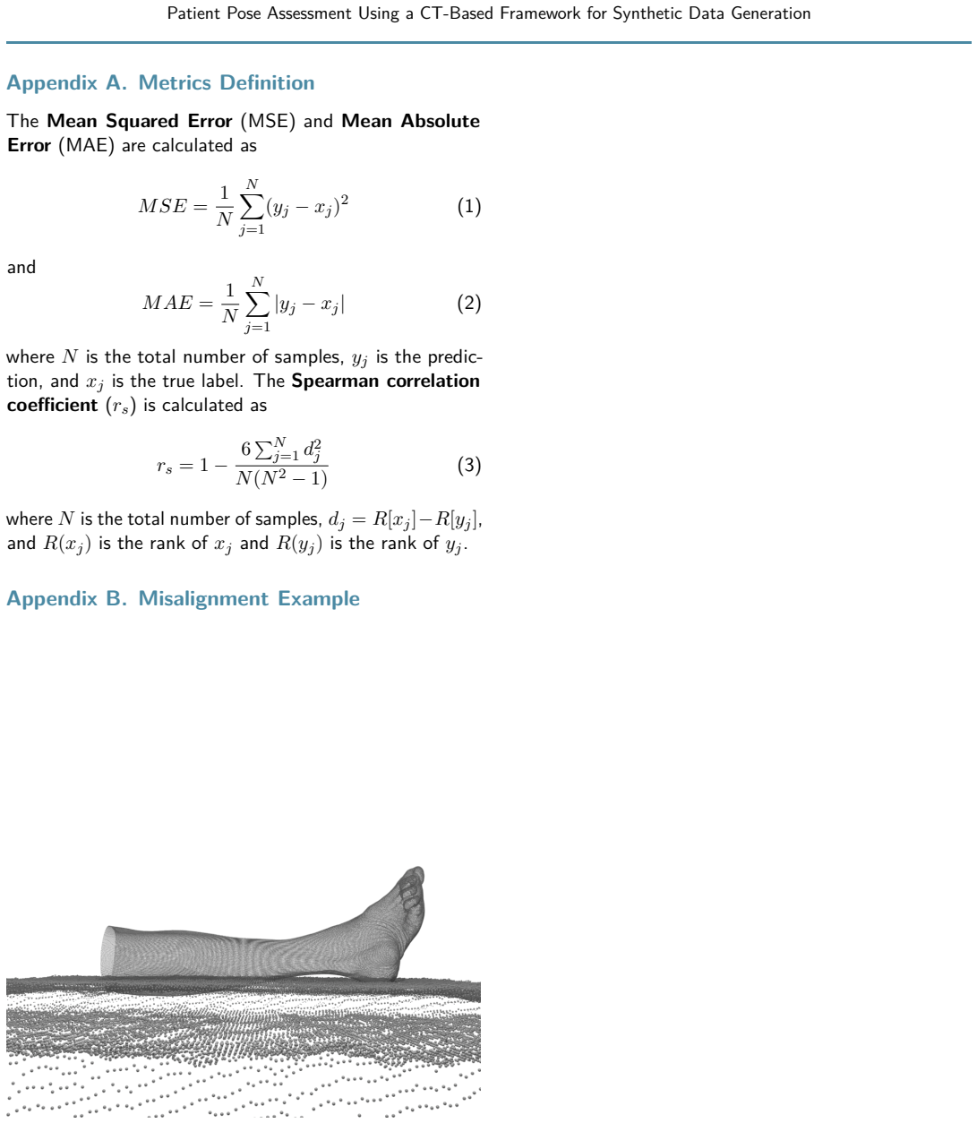
The load-bearing object is the data-generation pipeline that creates paired depth images and radiographs from a single CT. Surface extraction via Marching Cubes at -500 Hounsfield units yields a patient point cloud; normal-vector shifts and Gaussian bumps simulate body-shape variation; scene composition aligns the point cloud onto a recorded X-ray table so depth images look like real time-of-flight camera frames; and a Monte Carlo simulation using voxel material and mass-density maps renders the paired radiograph, with expert radiologist labels for joint-space visibility defining ground truth. These paired synthetic images are then used for task-specific pretraining of two EfficientNet-B0 regressors, one per camera view, with camera-view-specific weight initialization.
What would settle it
Measure inter-rater agreement between expert quality ratings of synthetic radiographs and real radiographs of the same anatomical pose; near-chance agreement, or a fine-tuning experiment in which synthetic pretraining performs worse than from-scratch training, would falsify the transfer claim.
Extended reading notes
Core claim
The central claim is that a CT-based synthetic data-generation framework can substitute for scarce real depth-image/radiograph pairs in training pose-assessment networks. From each CT, the framework extracts the body surface as a point cloud, augments it to vary body shape and simulate edema-like bumps, places it on a recorded X-ray-room table, rotates it through 181 medial poses per foot, renders depth images from two time-of-flight camera views, and produces a matching synthetic radiograph by Monte Carlo X-ray transport with material-specific voxels. Radiologists label each synthetic radiograph's diagnostic quality from 1 to 3, and this label is attached to all corresponding depth images. Pretraining two EfficientNet-B0 regressors on these synthetic images, then fine-tuning on real data, improves accuracy on the two-preparation dataset to 90.45 percent versus 79.25 percent from scratch and 77.37 percent with ImageNet pretraining; the same recipe improves pose ranking on a living-subject dataset with surrogate labels. The paper reads this as evidence that synthetic CT-derived labels are usable proxies for real radiographic quality in pose assessment.
Load-bearing premise
The load-bearing premise is that expert quality labels assigned to synthetic radiographs are valid for real patient poses; if the synthetic joint-space appearance differs systematically from real radiographs, the learned transfer could be wrong.
Editorial extensions
If this is right
- Pretraining on synthetic CT-derived depth images improves downstream pose assessment on real data by up to 11 percentage points over training from scratch, and by more than ImageNet pretraining.
- Depth images alone can predict radiograph diagnostic quality, reaching 90.45 percent accuracy compared with the 93 percent reported for assessing real radiographs directly.
- Retrospective CT scans can be turned into large training sets with deliberately non-diagnostic poses, avoiding the ethical and regulatory problem of radiographing subjects without indication.
- Synthetic-pretrained features transfer to a weakly labeled living-subject dataset, improving pose margin accuracy by about 8 percentage points over from-scratch training.
Reading between the lines
- The paper's own suggestion that quality labels could be assigned automatically, for example with an existing radiograph-quality network, points to a fully automated loop: CT in, labeled depth-image training set out; the reported experiments do not test whether an automated labeler would preserve the 11-point gain.
- Because the largest gain comes from camera-view-specific pretraining and task-specific size augmentation, the mechanism may be domain-adapted feature learning rather than radiograph realism; an ablation that swaps Monte Carlo radiographs for cheaper ray-traced projections would separate photorealism from label-transfer value.
- The real-world clinical dataset uses surrogate positioning labels rather than radiographic ground truth, so the actual clinical benefit of fewer retakes and less radiation remains untested until a prospective study measures retake rates.
Editorial analysis
A structured set of objections, weighed in public.
Referee Report
Summary. The paper presents a framework that generates synthetic depth-image/radiograph pairs from CT scans, using Marching Cubes surface extraction, point-cloud augmentation, scene composition, and Monte Carlo radiograph simulation, to create a pretraining dataset of 3,077 upper-ankle poses from 10 CTs. The authors evaluate the synthetic data by training EfficientNet-B0 regressors and then transfer to two real datasets: a two-subject anatomical preparations dataset with radiograph-derived quality labels and a weakly labeled 18-subject clinical dataset. The central claim is that synthetic pretraining improves real-data pose assessment by up to 11 percentage points in Accuracy.
Significance. The framework addresses a real data-acquisition bottleneck in medical imaging, and the public release of the synthetic and real-world clinical datasets is a useful contribution. If the transfer results are reliable, the approach could reduce radiation exposure by supporting pose feedback before radiograph acquisition. The paper's strengths include a physically based radiograph synthesis pipeline that avoids deep-learning-based label leakage, a clear experimental setup with multiple pretraining variants, and an honest discussion of the weak labels in the clinical dataset. The main uncertainty lies in the statistical support for the headline effect size and in the validation of synthetic-radiograph labels.
major comments (3)
- [6.1, Table 2] The statistical analysis in Section 6.1 does not support the headline 'up to 11 pp' claim. The permutation test is applied to MAE only, whereas the abstract's 'up to 11 percentage points' refers to Accuracy (Table 2: 79.25% vs. 90.45%). Furthermore, the test treats the 10 random seeds as independent observations for a fixed dataset of two anatomical preparations. With only two subjects and reciprocal folds, the effective number of subject-level observations is two, so a seed-level test cannot establish that the improvement generalizes to new subjects. Please report the per-fold (per-preparation) Accuracy values and perform an analysis that respects the subject-level design, or revise the claim to reflect the strength of the evidence.
- [5.2, Table 2] The 'camera view specific without augmentation' configuration (90.45% Accuracy) is selected from nine pretraining conditions in Table 2, but no multiple-comparison correction or pre-registered selection procedure is described. Because the best configuration is identified on the same two-subject dataset on which it is then evaluated, the reported gain is at risk of selection bias. Moreover, this exact configuration (without augmentation) is not among the configurations tested on the independent real-world clinical dataset in Table 3, so the confirmation there does not directly validate the best Table 2 result. Please acknowledge this limitation, report all configurations with per-subject results, or validate the selected configuration on the clinical dataset.
- [3.4] The synthetic radiographs are the sole source of labels for the synthetic pretraining data, but the manuscript reports only that 'expert radiologists have validated that the visual quality is suitable' (Section 3.4). No quantitative comparison between synthetic and real radiograph labels, no inter-rater agreement (e.g., Cohen's kappa or ICC), and no analysis of whether joint-space visibility is rendered equivalently in synthetic and real images is provided. If synthetic radiographs obscure or exaggerate joint space differently from real radiographs, the pretraining labels would be systematically biased. Please add a label-consistency experiment or explicitly restrict the claim to the specific rendering validated by the visual inspection.
minor comments (4)
- [Table 2] The grouping of the augmentation columns under 'unified camera view' and 'camera view specific' is visually ambiguous because the header line does not clearly separate the two strategies from the 'wo/ aug' column; please adjust the formatting for readability.
- [5.2, Experiment 2b] The text says 'subject-level three-fold cross-validation' but does not specify how the left and right feet of the same subject are assigned to folds; since both feet of a subject are captured, please clarify whether they are kept together in the same fold.
- [5.4, Pose Delta] The notation uses N both for the number of samples in Accuracy and for the number of subject-foot pairs in Pose Delta; please use distinct symbols to avoid confusion.
- [6.1] The one-sided paired permutation test is described as exact, but the p-values are reported only as p=0.0049 and p=0.0020; please state the number of permutations used or confirm that an exact enumeration over the 2^10 possible sign flips was performed.
Circularity Check
No significant circularity: the synthetic-pretraining claim is evaluated on independent real data, and the noted limitations concern evidence strength rather than definitional dependence.
full rationale
The derivation chain is not circular at any load-bearing point. The synthetic training signal is produced by the framework (MC radiograph simulation plus depth projection) and labeled by radiologists (Section 4), while the claimed transfer gain is measured on real data: the anatomical preparations dataset with radiologist-assigned labels and the real-world clinical dataset with weak labels, compared against from-scratch and ImageNet initialization (Tables 2 and 3). No fitted parameter is renamed as a prediction; the 11 pp figure is a direct accuracy comparison on held-out real preparations. The self-citations to Laufer et al. 2024 (preparations dataset and two-view architecture) and Laufer et al. 2026 (camera positioning) are supporting infrastructure, not a derivation that assumes the conclusion. The limitation noted in Section 7 (small, single-site datasets; weak labels) and the seeds-as-observations statistical choice (Section 6.1) weaken evidential force but do not make any prediction equal to its input by construction. Therefore no circularity is identified.
Assumptions & free parameters
free parameters (6)
- MCA threshold =
-500 HU
- rotation range and step =
90 degrees in 0.5 degree steps
- augmentation shift magnitude =
not specified
- bump parameters =
2-3 bumps, Gaussian size and position manual
- simulated X-ray paths =
2e10
- non-linear value mapping =
unspecified
assumptions (6)
- domain assumption Surface extracted from CT at -500 HU is sufficient to render realistic depth images of the target anatomy.
- domain assumption Diagnostic quality of an ankle radiograph is primarily determined by pose, particularly rotational alignment around the longitudinal axis, and is visible in the joint space.
- domain assumption Synthetic radiographs generated by MCGPU with PENELOPE materials are visually and diagnostically equivalent enough to real radiographs for labeling.
- domain assumption A plane exists under every X-ray examination that can be matched to place the anatomy realistically.
- domain assumption Anatomy-specific definitions of rotation axis and plane generalize to other anatomies.
- domain assumption Radiologist ratings on a 1 to 3 scale with 0.5 increments provide reliable labels.
Cite this review
Pith. "Pith review of Patient Pose Assessment Using a CT-Based Framework for Synthetic Data Generation." pith.science (2026). https://pith.science/paper/NV3N6SVW
@misc{pith2026260806126,
  author       = {Pith},
  title        = {Pith review of: Patient Pose Assessment Using a CT-Based Framework for Synthetic Data Generation},
  year         = {2026},
  howpublished = {\url{https://pith.science/paper/NV3N6SVW}},
  note         = {Machine review of arXiv:2608.06126}
}
read the original abstract
An adequate diagnostic quality of radiographs is essential for reliable diagnoses and treatment planning. The patient's pose during radiography is one of the most important factors determining the diagnostic quality. Since patient positioning is difficult and not standardized, an automated AI-based approach using depth images to automatically assess the patient's pose before the radiograph has been taken would be helpful. Due to regulatory hurdles, however, it is difficult in practice to acquire the required depth images and corresponding radiographs. In this paper, we present a framework that can generate such training data synthetically from Computed Tomography scans. We further show that by pretraining on our generated synthetic dataset consisting of 3077 image pairs of upper ankle joints, the pose assessment of real upper ankle joints can be improved by up to 11 percentage points.
Figures


Figures from the paper (4 more)


Reference graph
Works this paper leans on
-
[1]
Laufer, Manuel and Haas, Julius and Mairh \"o fer, Dominik and Sieren, Malte and Gerdes, Hauke and Reis, Fabio Leal dos and Bischof, Arpad and K \"a ster, Thomas and Barth, Erhardt and Barkhausen, J \"o rg and Martinetz, Thomas. Evaluation of Time-of-flight Camera Positioning for AI-based Patient Pose Assessment in Radiography. Bildverarbeitung f \"u r di...
work page 2026
-
[2]
Russakoff, D.B. and Rohlfing, T. and Mori, K. and Rueckert, D. and Ho, A. and Adler, J.R. and Maurer, C.R. , year =. Fast Generation of Digitally Reconstructed Radiographs Using Attenuation Fields with Application to. IEEE Transactions on Medical Imaging , volume =. doi:10.1109/TMI.2005.856749 , urldate =
-
[3]
Badal, Andreu and Badano, Aldo , year =. Accelerating. Medical Physics , volume =. doi:10.1118/1.3231824 , urldate =
-
[4]
Saiti, E. and Theoharis, T. , title =. 2022 , issue_date =. doi:10.1016/j.cag.2022.06.012 , journal =
-
[5]
Rybakov, Oleksiy and Bier, Bastian and Maier, Jennifer and Unberath, Mathias and Maier, Andreas , booktitle =
-
[6]
Segars, W. P. and Sturgeon, G. and Mendonca, S. and Grimes, Jason and Tsui, B. M. W. , title =. Medical Physics , volume =. doi:https://doi.org/10.1118/1.3480985 , url =. https://aapm.onlinelibrary.wiley.com/doi/pdf/10.1118/1.3480985 , abstract =
-
[7]
Olya Grove, Khairan Rajab and Les A. Piegl , title =. Computer-Aided Design and Applications , volume =. 2010 , publisher =. doi:10.1080/16864360.2010.10738807 , URL =
-
[8]
Unberath, Mathias and Zaech, Jan-Nico and Lee, Sing Chun and Bier, Bastian and Fotouhi, Javad and Armand, Mehran and Navab, Nassir , editor =. Medical. 2018 , pages =. doi:10.1007/978-3-030-00937-3_12 , abstract =
Show all 46 references
-
[9]
, year =
Chougule, Vikas and Mulay, Arati and Ahuja, B. , year =
-
[10]
Advancing Medical Imaging: A Comparative Exploration of Generative Adversarial Networks for Chest X-ray Synthesis , year=
Keerthi, R and Kiran, Kuval and Kiran, Ss and Likitha, P , booktitle=. Advancing Medical Imaging: A Comparative Exploration of Generative Adversarial Networks for Chest X-ray Synthesis , year=
-
[11]
, journal=
Liu, Jian and Lin, Tim H. , journal=. A Framework for the Synthesis of X-Ray Security Inspection Images Based on Generative Adversarial Networks , year=
-
[12]
Chapter 50 - Fast Simulation of Radiographic Images Using a Monte Carlo x-Ray Transport Algorithm Implemented in
Badal, Andreu and Badano., Aldo , editor =. Chapter 50 - Fast Simulation of Radiographic Images Using a Monte Carlo x-Ray Transport Algorithm Implemented in. 2011 , series =. doi:10.1016/B978-0-12-384988-5.00050-4 , abstract =
2011 doi
-
[13]
Workshop Proceedings , volume=
PENELOPE-2006: A Code System for Monte Carlo Simulation of Electron and Photon Transport , author=. Workshop Proceedings , volume=
2006
-
[14]
and Cline, Harvey E
Lorensen, William E. and Cline, Harvey E. , title =. SIGGRAPH Comput. Graph. , month = aug, pages =. 1987 , issue_date =. doi:10.1145/37402.37422 , abstract =
1987
-
[15]
Efficient Implementation of Marching Cubes' Cases with Topological Guarantees , volume =
Lewiner, Thomas and Lopes, Hélio and Vieira, Antonio and Tavares, Geovan , year =. Efficient Implementation of Marching Cubes' Cases with Topological Guarantees , volume =. Journal of Graphics Tools , doi =
-
[16]
A density-based algorithm for discovering clusters in large spatial databases with noise , year =
Ester, Martin and Kriegel, Hans-Peter and Sander, J\". A density-based algorithm for discovering clusters in large spatial databases with noise , year =. Proceedings of the Second International Conference on Knowledge Discovery and Data Mining , pages =
-
[17]
arXiv:1801.09847 , year =
Qian-Yi Zhou and Jaesik Park and Vladlen Koltun , title =. arXiv:1801.09847 , year =
-
[18]
Medical Imaging 2024: Image Processing , editor =
Manuel Laufer and Dominik Mairh. Medical Imaging 2024: Image Processing , editor =. 2024 , doi =
2024
-
[19]
doi:10.5281/zenodo.3828935 , license =
Falcon, William and. doi:10.5281/zenodo.3828935 , license =
-
[20]
and Alghamdi, Sami A
Alashban, Yazeed and Shubayr, Nasser and Alghamdi, Abdulrahman A. and Alghamdi, Sami A. and Boughattas, Sami , year =. An Assessment of Image Reject Rates for Digital Radiography in. Journal of Radiation Research and Applied Sciences , volume =. doi:10.1016/j.jrras.2022.01.023...
-
[21]
Reject Rate Analysis in Digital Radiography: An
Atkinson, Samantha and Neep, Michael and Starkey, Deborah , year =. Reject Rate Analysis in Digital Radiography: An. Journal of Medical Radiation Sciences , volume =. doi:10.1002/jmrs.343 , urldate =
-
[22]
Robust Chest X-Ray Quality Assessment Using Convolutional Neural Networks and Atlas Regularization , booktitle =
von Berg, Jens and Kr. Robust Chest X-Ray Quality Assessment Using Convolutional Neural Networks and Atlas Regularization , booktitle =. 2020 , month = mar, volume =. doi:10.1117/12.2549541 , urldate =
2020 doi
-
[23]
Deng, Jia and Dong, Wei and Socher, Richard and Li, Li-Jia and Li, Kai and. 2009. 2009 , month = jun, pages =. doi:10.1109/CVPR.2009.5206848 , abstract =
2009
-
[24]
2012 , month = dec, journal =
Positioning Errors and Quality Assessment in Panoramic Radiography , author =. 2012 , month = dec, journal =. doi:10.5624/isd.2012.42.4.207 , urldate =
2012 doi
-
[25]
, year =
Hansen, Lasse and Siebert, Marlin and Diesel, Jasper and Heinrich, Mattias P. , year =. Fusing Information from Multiple. International Journal of Computer Assisted Radiology and Surgery , volume =. doi:10.1007/s11548-019-02044-7 , urldate =
-
[26]
2015 , month = oct, journal =
Image Rejects in General Direct Digital Radiography , author =. 2015 , month = oct, journal =. doi:10.1177/2058460115604339 , urldate =
2015 doi
- [27]
-
[28]
Kadkhodamohammadi, Abdolrahim and Gangi, Afshin and. A. 2017. 2017 , month = mar, pages =. doi:10.1109/WACV.2017.47 , abstract =
2017 doi
- [29]
-
[30]
The Assessment of Image Quality and Diagnostic Value in
Kjelle, Elin and Chilanga, Catherine , year =. The Assessment of Image Quality and Diagnostic Value in. Insights into Imaging , volume =. doi:10.1186/s13244-022-01169-9 , urldate =
-
[31]
and Schanche, A
Kjelle, E. and Schanche, A. K. and Hafskjold, L. , year =. To Keep or Reject, That Is the Question -. Radiography , volume =. doi:10.1016/j.radi.2020.06.020 , urldate =
2020 doi
-
[32]
Estimation of the Ankle-Joint Space Visibility in x-Ray Images Using Convolutional Neural Networks , booktitle =
K. Estimation of the Ankle-Joint Space Visibility in x-Ray Images Using Convolutional Neural Networks , booktitle =. 2023 , month = apr, volume =. doi:10.1117/12.2651757 , urldate =
2023 doi
-
[33]
Kr. Medical. 2022 , month = apr, volume =. doi:10.1117/12.2611734 , urldate =
2022 doi
-
[34]
and Reiser, Ingrid and Liu, Lili and Kinsey, Tiffany and S
Little, Kevin J. and Reiser, Ingrid and Liu, Lili and Kinsey, Tiffany and S. Unified. 2017 , month = feb, journal =. doi:10.1016/j.jacr.2016.07.011 , urldate =
2017 doi
-
[35]
Mairh. An. Proceedings of the. 2021 , month = aug, pages =
2021
-
[36]
doi:10.1016/j.jvcir.2018.07.010 , urldate =
2018 , month = aug, journal =. doi:10.1016/j.jvcir.2018.07.010 , urldate =
2018 doi
-
[37]
2022 , month = nov, journal =
Automated Quality Assessment of Chest Radiographs Based on Deep Learning and Linear Regression Cascade Algorithms , author =. 2022 , month = nov, journal =. doi:10.1007/s00330-022-08771-x , urldate =
2022 doi
-
[38]
Proceedings of the 33rd
Paszke, Adam and Gross, Sam and Massa, Francisco and Lerer, Adam and Bradbury, James and Chanan, Gregory and Killeen, Trevor and Lin, Zeming and Gimelshein, Natalia and Antiga, Luca and Desmaison, Alban and K. Proceedings of the 33rd. 2019 , month = dec, number =
2019
-
[39]
2012 , journal =
Fast Time-of-Flight Camera Based Surface Registration for Radiotherapy Patient Positioning , author =. 2012 , journal =. doi:10.1118/1.3664006 , urldate =
2012 doi
-
[40]
2021 , month = jan, journal =
Multi-View Classification with Convolutional Neural Networks , author =. 2021 , month = jan, journal =. doi:10.1371/journal.pone.0245230 , urldate =
2021 doi
-
[41]
Dropout:
Srivastava, Nitish and Hinton, Geoffrey and Krizhevsky, Alex and Sutskever, Ilya and Salakhutdinov, Ruslan , year =. Dropout:. Journal of Machine Learning Research , volume =
- [42]
-
[43]
2021 , journal =
Digital Radiography Reject Analysis of Examinations with Multiple Rejects: An. 2021 , journal =. doi:10.1002/jmrs.468 , urldate =
2021 doi
-
[44]
Interactive Teaching Environment for Diagnostic Radiography with Real-Time
Sujar, Aaron and Kelly, Graham and Garc. Interactive Teaching Environment for Diagnostic Radiography with Real-Time. 2022 , month = jan, journal =. doi:10.1007/s11548-021-02499-7 , urldate =
2022 doi
-
[45]
Development and
Takamoto, Hiroki and Nishine, Hiroki and Sato, Shohei and Sun, Guanghao and Watanabe, Sadao and Seokjin, Kim and Asai, Masahito and Mineshita, Masamichi and Matsui, Takemi , year =. Development and. Frontiers in Physiology , volume =
-
[46]
International
Tan, Mingxing and Le, Quoc , year =. International
Reviewed August 7, 2026 · model on record in the stance chip above.
Discussion (0). Sign in to comment.