MorVess: Morphology-Aware Pulmonary Vessel Segmentation Network
Pith reviewed 2026-06-26 00:44 UTC · model grok-4.3
The pith
MorVess jointly predicts vessel masks with distance and thickness maps to improve pulmonary vessel segmentation in CT scans.
A machine-rendered reading of the paper's core claim, the machinery that carries it, and where it could break.
Core claim
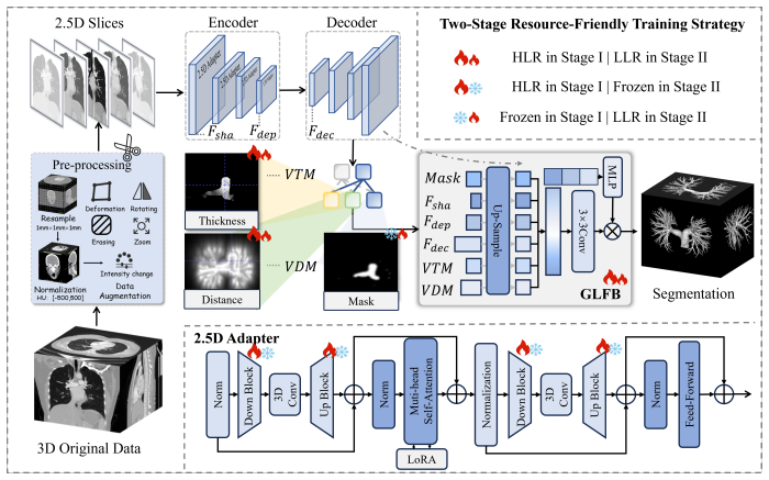
MorVess is a morphology-aware segmentation framework that integrates differentiable geometric priors with large-scale foundation model adaptation. It jointly predicts vessel masks, distance maps, and thickness maps to supply explicit supervision for vascular boundaries, centerline consistency, and smooth diameter transitions. A lightweight 2.5D adapter bridges 3D spatial context and 2D SAM representations while a global-local fusion block aggregates multi-level semantics and geometric cues. On two challenging pulmonary CT benchmarks the method yields superior Dice, clDice, and HD95 scores and substantially improves small-vessel recovery and global connectivity.
What carries the argument
Joint prediction of vessel masks, distance maps, and thickness maps that supplies explicit supervision for boundaries and topology, together with a 2.5D adapter and global-local fusion block.
If this is right
- Small branches become recoverable because distance and thickness supervision enforce centerline and diameter consistency.
- Global connectivity improves because the geometric maps reduce fragmentation under voxel-wise loss alone.
- The 2.5D adapter allows pretrained 2D foundation models to handle 3D tubular structures without full 3D retraining.
- Quantitative vessel analysis gains reliability from the explicit diameter and boundary predictions.
Where Pith is reading between the lines
- The same joint-map supervision could transfer to segmentation of other tubular anatomy such as coronary or cerebral vessels.
- Clinical workflows that rely on vessel diameter measurements might obtain more consistent results without additional post-processing steps.
- The framework suggests a route for embedding geometric priors into other foundation-model adaptations in medical imaging.
Load-bearing premise
That jointly predicting distance and thickness maps will supply effective explicit supervision for vascular boundaries and topology without the auxiliary predictions introducing errors.
What would settle it
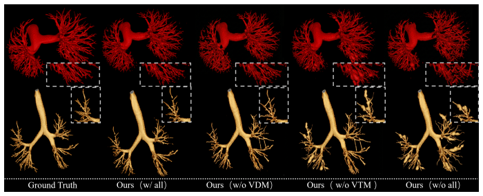
An ablation experiment on the same two CT benchmarks in which removing the distance-map and thickness-map prediction heads produces no drop or an increase in clDice and HD95 scores.
Figures
read the original abstract
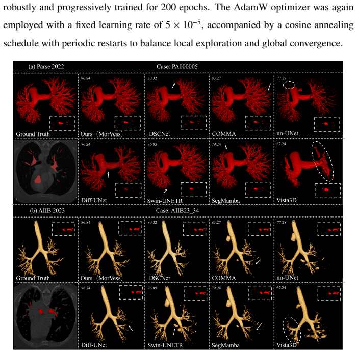
Accurate pulmonary vessel segmentation remains challenging due to the sparse, tortuous, and multi-scale nature of vascular structures, where small branches are easily lost and topology integrity is difficult to preserve under voxel-wise supervision. Existing deep segmentation models primarily optimize binary masks, lacking explicit geometric constraints, thus struggling to recover continuous tubular morphology and fine vascular connectivity. In this study, we introduce MorVess, a morphology-aware segmentation framework that integrates differentiable geometric priors with large-scale foundation model adaptation to achieve fine-grained vascular parsing. MorVess jointly predicts vessel masks, distance maps, and thickness maps, providing explicit supervision for vascular boundaries, centerline consistency, and smooth diameter transitions. A lightweight 2.5D adapter bridges 3D spatial context and 2D SAM representations, while a global-local fusion block aggregates multi-level semantics and geometric cues for high-fidelity topology reconstruction. Across two challenging pulmonary CT benchmarks, MorVess delivers superior Dice, clDice, and HD95 scores, substantially improving small-vessel recovery and global connectivity. These results demonstrate that embedding geometric intelligence into pretrained vision models offers a principled and scalable pathway toward precise vessel analysis and clinically reliable structural quantification. Our source code is available at https://github.com/MaoFuyou/MorVess.
Editorial analysis
A structured set of objections, weighed in public.
Referee Report
Summary. The paper proposes MorVess, a morphology-aware framework for pulmonary vessel segmentation in CT that jointly predicts binary masks, distance maps, and thickness maps to enforce geometric constraints on boundaries and topology. It incorporates a lightweight 2.5D adapter to bridge 3D context with 2D SAM representations and a global-local fusion block to aggregate multi-level semantics and geometric cues. The central claim is that this yields superior Dice, clDice, and HD95 scores on two challenging pulmonary CT benchmarks, with particular gains in small-vessel recovery and global connectivity.
Significance. If the empirical claims are substantiated, the approach of embedding explicit geometric supervision via auxiliary distance and thickness heads into a SAM-adapted architecture could meaningfully advance topology-preserving segmentation for sparse, multi-scale tubular structures in medical imaging. The open-source code release is a positive factor for reproducibility.
major comments (3)
- [Abstract] Abstract: The central claim of superior Dice, clDice, and HD95 performance (with substantial gains in small-vessel recovery) is asserted without any numerical values, baseline comparisons, ablation studies, or error analysis, which prevents assessment of whether the data and method support the claims.
- [Method] Method (joint prediction of auxiliary maps): No separate quantitative metrics (e.g., MAE or correlation) are reported for the distance and thickness map heads, nor is there an ablation removing these heads; this leaves open whether the auxiliary predictions reinforce or compete with the primary mask loss and whether they are used at inference.
- [Experiments] Experiments: The absence of any reported results, tables, or figures quantifying the claimed improvements on the two benchmarks means the load-bearing assertion of better small-vessel recovery and connectivity cannot be evaluated.
Simulated Author's Rebuttal
We thank the referee for the constructive feedback. The comments correctly identify that the current manuscript version lacks explicit numerical results, ablations, and auxiliary-task metrics in the sections highlighted. We will revise the manuscript to incorporate these elements, thereby strengthening the empirical support for our claims.
read point-by-point responses
-
Referee: [Abstract] Abstract: The central claim of superior Dice, clDice, and HD95 performance (with substantial gains in small-vessel recovery) is asserted without any numerical values, baseline comparisons, ablation studies, or error analysis, which prevents assessment of whether the data and method support the claims.
Authors: We agree that the abstract should contain concrete numerical evidence. In the revised manuscript we will insert the key quantitative results (Dice, clDice, HD95) together with the main baseline comparisons and a brief reference to the ablation findings that demonstrate the contribution of the morphology-aware components. revision: yes
-
Referee: [Method] Method (joint prediction of auxiliary maps): No separate quantitative metrics (e.g., MAE or correlation) are reported for the distance and thickness map heads, nor is there an ablation removing these heads; this leaves open whether the auxiliary predictions reinforce or compete with the primary mask loss and whether they are used at inference.
Authors: We will add MAE and Pearson correlation values for both auxiliary heads on the validation sets. We will also include an ablation that removes the distance and thickness heads while keeping all other components fixed. The auxiliary maps are used exclusively during training to provide geometric supervision; at inference only the binary mask is output. This clarification and the new quantitative results will be inserted into the Method section. revision: yes
-
Referee: [Experiments] Experiments: The absence of any reported results, tables, or figures quantifying the claimed improvements on the two benchmarks means the load-bearing assertion of better small-vessel recovery and connectivity cannot be evaluated.
Authors: We will insert the full quantitative tables (including per-method Dice, clDice, HD95, and small-vessel-specific metrics) and the corresponding figures for both benchmarks. These tables will also report the ablation results mentioned above so that the contribution of each design choice can be directly assessed. revision: yes
Circularity Check
No circularity detected; standard supervised multi-task learning
full rationale
The paper describes a standard deep segmentation architecture (MorVess) that jointly optimizes a primary mask loss together with auxiliary distance-map and thickness-map heads on labeled CT data. No equation or claim reduces a reported performance metric to a fitted parameter by construction, no uniqueness theorem is imported from self-citation, and no ansatz is smuggled through prior work. The geometric priors are supplied by explicit ground-truth maps during training, which is an ordinary multi-task supervision pattern whose outputs are independently measurable and falsifiable on the same benchmarks. The derivation chain therefore remains self-contained against external data rather than internally tautological.
Axiom & Free-Parameter Ledger
free parameters (1)
- multi-task loss weighting
axioms (1)
- domain assumption Geometric maps (distance and thickness) can be accurately regressed from image features and provide useful supervision signals
Reference graph
Works this paper leans on
-
[1]
Moccia, E
S. Moccia, E. De Momi, S. El Hadji, L. S. Mattos, Blood vessel segmenta- tion algorithms—review of methods, datasets and evaluation metrics, Computer methods and programs in biomedicine 158 (2018) 71–91
2018
-
[2]
S. S. Murugaraj, K. Vadivelu, P. T. Sambandam, B. S. Kumar, Lung vessel seg- mentation and abnormality classification based on hybrid mobile-lenet using ct image, Biomedical Signal Processing and Control 100 (2025) 107072
2025
-
[3]
X. Deng, J. Luo, P. Huang, P. He, J. Li, Y . Liu, H. Xiao, P. Feng, Mcranet: Mtsl- based connectivity region attention network for pd-l1 status segmentation in h&e stained images, Computers in Biology and Medicine 184 (2025) 109357
2025
-
[4]
Y . Chen, B. Zou, Z. Guo, Y . Huang, Y . Huang, F. Qin, Q. Li, C. Wang, Scunet++: Swin-unet and cnn bottleneck hybrid architecture with multi-fusion dense skip connection for pulmonary embolism ct image segmentation, in: Proceedings of the IEEE/CVF winter conference on applications of computer vision, 2024, pp. 7759–7767
2024
-
[5]
J. Li, Y . Huang, X. Ye, H. Yang, Topology-joint curvilinear segmentation network using confidence-based bezier topological representation, Engineering Applica- tions of Artificial Intelligence 143 (2025) 110045
2025
-
[6]
Lin, W.-C
K.-C. Lin, W.-C. Ko, Y .-D. Tsai, C.-Y . Chang, Y .-H. Yang, Y .-S. Huang, Y .- C. Chang, Hemorrhage risk prediction after computed tomography-guided lung 30 biopsy: Clinical parameters and quantitative pulmonary vascular analysis, Jour- nal of the Formosan Medical Association 124 (2025) 79–86
2025
-
[7]
X. Bai, M. Liu, Y . Chen, H. Yang, Q. Tian, Chest-omdl: Organ-specific multi- disease detection and localization in chest computed tomography using weakly supervised deep learning from free-text radiology report, in: Medical Imaging with Deep Learning, 2025
2025
-
[8]
Z. Fu, Z. Fu, C. Lu, J. Yan, J. Fei, H. Han, Robust implementation of fore- ground extraction and vessel segmentation for x-ray coronary angiography image sequence, Pattern Recognition 145 (2024) 109926
2024
-
[9]
D. Song, W. Huang, J. Liu, M. J. Islam, H. Yang, S. Wang, H. Zheng, S. Wang, Optimized vessel segmentation: A structure-agnostic approach with small vessel enhancement and morphological correction, IEEE Transactions on Image Pro- cessing 34 (2025) 7168–7179. doi:10.1109/TIP.2025.3530318
-
[10]
X. Liu, H. Shen, W. Zhong, W. Xiong, Z. Chen, Dsdc-net: Semi-supervised superficial octa vessel segmentation for false positive reduction, Pattern Recog- nition 165 (2025) 111592
2025
-
[11]
J. Chen, M. Liang, C. Peng, J. Zhang, S. Huo, Improving maritime data: A ma- chine learning-based model for missing vessel trajectories reconstruction, IEEE Transactions on Vehicular Technology 74 (2025) 9565–9578. doi:10.1109/TVT. 2025.3539077
work page doi:10.1109/tvt 2025
-
[12]
Zhang, J
X. Zhang, J. Zhang, L. Ma, P. Xue, Y . Hu, D. Wu, Y . Zhan, J. Feng, D. Shen, Pro- gressive deep segmentation of coronary artery via hierarchical topology learning, in: International conference on medical image computing and computer-assisted intervention, Springer, 2022, pp. 391–400
2022
-
[13]
Y . Wu, S. Qi, M. Wang, S. Zhao, H. Pang, J. Xu, L. Bai, H. Ren, Transformer- based 3d u-net for pulmonary vessel segmentation and artery-vein separation from ct images, Medical & Biological Engineering & Computing 61 (2023) 2649–2663. 31
2023
-
[14]
Y . Qi, Y . He, X. Qi, Y . Zhang, G. Yang, Dynamic snake convolution based on topological geometric constraints for tubular structure segmentation, in: Pro- ceedings of the IEEE/CVF international conference on computer vision, 2023, pp. 6070–6079
2023
-
[15]
Z. Xing, T. Ye, Y . Yang, G. Liu, L. Zhu, Segmamba: Long-range sequential mod- eling mamba for 3d medical image segmentation, in: International conference on medical image computing and computer-assisted intervention, Springer, 2024, pp. 578–588
2024
-
[16]
H. Wang, Y . Chen, W. Chen, H. Xu, H. Zhao, B. Sheng, H. Fu, G. Yang, L. Zhu, Serp-mamba: Advancing high-resolution retinal vessel segmentation with selec- tive state-space model, IEEE Transactions on Medical Imaging 44 (2025) 4811–
2025
-
[17]
doi:10.1109/TMI.2025.3533374
-
[18]
Yagis, S
E. Yagis, S. Aslani, Y . Jain, Y . Zhou, S. Rahmani, J. Brunet, A. Bellier, C. Wer- lein, M. Ackermann, D. Jonigk, et al., Deep learning for 3d vascular segmentation in hierarchical phase contrast tomography: a case study on kidney, Scientific Re- ports 14 (2024) 27258
2024
-
[19]
AlMohimeed, M
I. AlMohimeed, M. Y . Sikkandar, A. Mohanarathinam, V . S. Parvathy, M. K. Ishak, F. K. Karim, S. M. Mostafa, Sandpiper optimization algorithm with region growing based robust retinal blood vessel segmentation approach, IEEE Access 12 (2024) 28612–28620
2024
-
[20]
Vigneshwaran, S
P. Vigneshwaran, S. Velmurugan, Detection of intracranial aneurysms by using 3-dimensional multiscale vessel enhanced filtering, in: 2024 Intelligent Systems and Machine Learning Conference (ISML), IEEE, 2024, pp. 395–401
2024
-
[21]
X. Chen, J. Jiang, X. Zhang, Automatic 3d coronary artery segmentation based on local region active contour model, Journal of Thoracic Disease 16 (2024) 2563
2024
-
[22]
T. Lv, G. Yang, Y . Zhang, J. Yang, Y . Chen, H. Shu, L. Luo, Vessel segmentation using centerline constrained level set method, Multimedia Tools and Applications 78 (2019) 17051–17075. 32
2019
-
[23]
G. Shi, H. Zhang, J. Tian, COMMA: coordinate-aware modulated mamba net- work for 3d dispersed vessel segmentation, IEEE Transactions on Image Process- ing (2026)
2026
-
[24]
L. Xia, H. Zhang, Y . Wu, R. Song, Y . Ma, L. Mou, J. Liu, Y . Xie, M. Ma, Y . Zhao, 3d vessel-like structure segmentation in medical images by an edge-reinforced network, Medical Image Analysis 82 (2022) 102581
2022
-
[25]
X. Luo, L. Peng, Z. Ke, J. Lin, Z. Yu, Pa-net: A hybrid architecture for retinal vessel segmentation, Pattern Recognition 161 (2025) 111254
2025
-
[26]
Bertels, T
J. Bertels, T. Eelbode, M. Berman, D. Vandermeulen, F. Maes, R. Bisschops, M. B. Blaschko, Optimizing the dice score and jaccard index for medical image segmentation: Theory and practice, in: International conference on medical im- age computing and computer-assisted intervention, Springer, 2019, pp. 92–100
2019
-
[27]
Y . Wang, Y . Chen, S. Jiang, W. Yu, M. Liu, B. Wu, J. Zong, F. Qin, C. Wang, Q. Tian, Smart: Style-modulated robust test-time adaptation for cross-domain brain tumor segmentation in mri, 2025.arXiv:2509.17925
arXiv 2025
-
[28]
Huang, X
Y . Huang, X. Yang, L. Liu, H. Zhou, A. Chang, X. Zhou, R. Chen, J. Yu, J. Chen, C. Chen, et al., Segment anything model for medical images?, Medical Image Analysis 92 (2024) 103061
2024
-
[29]
S. He, R. Bao, J. Li, J. Stout, A. Bjornerud, P. E. Grant, Y . Ou, Computer-vision benchmark segment-anything model (sam) in medical images: Accuracy in 12 datasets, arXiv preprint arXiv:2304.09324 (2023)
arXiv 2023
- [30]
-
[31]
J. Wu, Z. Wang, M. Hong, W. Ji, H. Fu, Y . Xu, M. Xu, Y . Jin, Medical sam adapter: Adapting segment anything model for medical image segmentation, Medical image analysis 102 (2025) 103547. 33
2025
-
[32]
H. Wang, S. Guo, J. Ye, Z. Deng, J. Cheng, T. Li, J. Chen, Y . Su, Z. Huang, Y . Shen, B. Fu, S. Zhang, J. He, Sam-med3d: A vision foundation model for general-purpose segmentation on volumetric medical images, IEEE Trans- actions on Neural Networks and Learning Systems 36 (2025) 17599–17612. doi:10.1109/TNNLS.2025.3586694
-
[33]
N. Ravi, V . Gabeur, Y .-T. Hu, R. Hu, C. Ryali, T. Ma, H. Khedr, R. Rädle, C. Rol- land, L. Gustafson, E. Mintun, J. Pan, K. V . Alwala, N. Carion, C.-Y . Wu, R. Gir- shick, P. Dollar, C. Feichtenhofer, SAM 2: Segment anything in images and videos, in: International Conference on Learning Representations, 2025
2025
-
[34]
S. Shit, J. C. Paetzold, A. Sekuboyina, I. Ezhov, A. Unger, A. Zhylka, J. P. Pluim, U. Bauer, B. H. Menze, clDice-a novel topology-preserving loss function for tubular structure segmentation, in: Proceedings of the IEEE/CVF conference on computer vision and pattern recognition, 2021, pp. 16560–16569
2021
-
[35]
P. Shi, J. Hu, Y . Yang, Z. Gao, W. Liu, T. Ma, Centerline boundary dice loss for vascular segmentation, in: International Conference on Medical Image Comput- ing and Computer-Assisted Intervention, Springer, 2024, pp. 46–56
2024
- [36]
- [37]
-
[38]
Y . Nan, X. Xing, S. Wang, Z. Tang, F. N. Felder, S. Zhang, R. E. Ledda, X. Ding, R. Yu, W. Liu, et al., Hunting imaging biomarkers in pulmonary fibrosis: bench- marks of the aiib23 challenge, Medical Image Analysis 97 (2024) 103253
2024
-
[39]
Y . Chu, G. Luo, L. Zhou, S. Cao, G. Ma, X. Meng, J. Zhou, C. Yang, D. Xie, D. Mu, et al., Deep learning-driven pulmonary artery and vein segmentation 34 reveals demography-associated vasculature anatomical differences, Nature Com- munications 16 (2025) 2262
2025
-
[40]
Zhang, Y
M. Zhang, Y . Wu, H. Zhang, Y . Qin, H. Zheng, W. Tang, C. Arnold, C. Pei, P. Yu, Y . Nan, et al., Multi-site, multi-domain airway tree modeling, Medical Image Analysis 90 (2023) 102957
2023
-
[41]
Y . He, P. Guo, Y . Tang, A. Myronenko, V . Nath, Z. Xu, D. Yang, C. Zhao, B. Si- mon, M. Belue, et al., Vista3d: Versatile imaging segmentation and annotation model for 3d computed tomography, in: Proceedings of the IEEE/CVF Confer- ence on Computer Vision and Pattern Recognition (CVPR), 2025, pp. 20863– 20873
2025
-
[42]
Liang, F
B. Liang, F. Peng, D. Luo, Q. Zeng, H. Wen, B. Zheng, Z. Zou, L. An, H. Wen, X. Wen, et al., Automatic segmentation of 15 critical anatomical labels and mea- surements of cardiac axis and cardiothoracic ratio in fetal four chambers using nnu-netv2, BMC Medical Informatics and Decision Making 24 (2024) 128
2024
-
[43]
Hatamizadeh, V
A. Hatamizadeh, V . Nath, Y . Tang, D. Yang, H. R. Roth, D. Xu, Swin unetr: Swin transformers for semantic segmentation of brain tumors in mri images, in: International MICCAI brainlesion workshop, Springer, 2021, pp. 272–284
2021
-
[44]
Z. Xing, L. Wan, H. Fu, G. Yang, Y . Yang, L. Yu, B. Lei, L. Zhu, Diff-unet: A diffusion embedded network for robust 3d medical image segmentation, Medical Image Analysis 105 (2025) 103654. doi:10.1016/j.media.2025.103654
-
[45]
K. Hu, Y . Zhu, T. Zhou, Y . Zhang, C. Cao, F. Xiao, X. Gao, Dsc-net: A novel interactive two-stream network by combining transformer and cnn for ultrasound image segmentation, IEEE Transactions on Instrumentation and Measurement 72 (2023) 1–12
2023
-
[46]
K. N. Telöken, I. D. Ghisleni, F. S. F. Zinani, D. P. Wermuth, Computational hemodynamic analysis of idealized coronary arteries with cylindrical and conical stents, Computer Methods and Programs in Biomedicine 278 (2026) 109285. doi:https://doi.org/10.1016/j.cmpb.2026.109285. 35
-
[47]
Spagnolo, S
P. Spagnolo, S. A. Guler, N. Chaudhuri, Z. Udwadia, L. Sesé, B. Kaul, J. I. En- ghelmayer, C. Valenzuela, A. Malhotra, C. J. Ryerson, Y . H. Khor, T. J. Corte, V . Cottin, Global epidemiology and burden of interstitial lung disease, The Lancet Respiratory Medicine 13 (2025) 739–755. doi:https://doi.org/10. 1016/S2213-2600(25)00129-8. 36
2025
discussion (0)
Sign in with ORCID, Apple, or X to comment. Anyone can read and Pith papers without signing in.