REVIEW 3 major objections 4 minor 1 cited by
Spiral waves speed up cell cycle oscillations in the frog cytoplasm
T0 review · 3 major / 4 minor · reviewed 2026-08-11 · deepseek-v4-flash
Pith's one-line read The paper reports spiral waves in frog egg extract that shorten the cell-cycle period nearly twofold, and argues the speed-up is a generic property of excitable media with strong time-scale separation.
desk verdict New observation and honest modeling, but the twofold speedup rests on three droplets and an assumed Cdk1 reporter link. read the letter →
The pith
A machine-rendered reading of the paper's core claim, the machinery that carries it, and where it could break.
The reading
What carries the argument
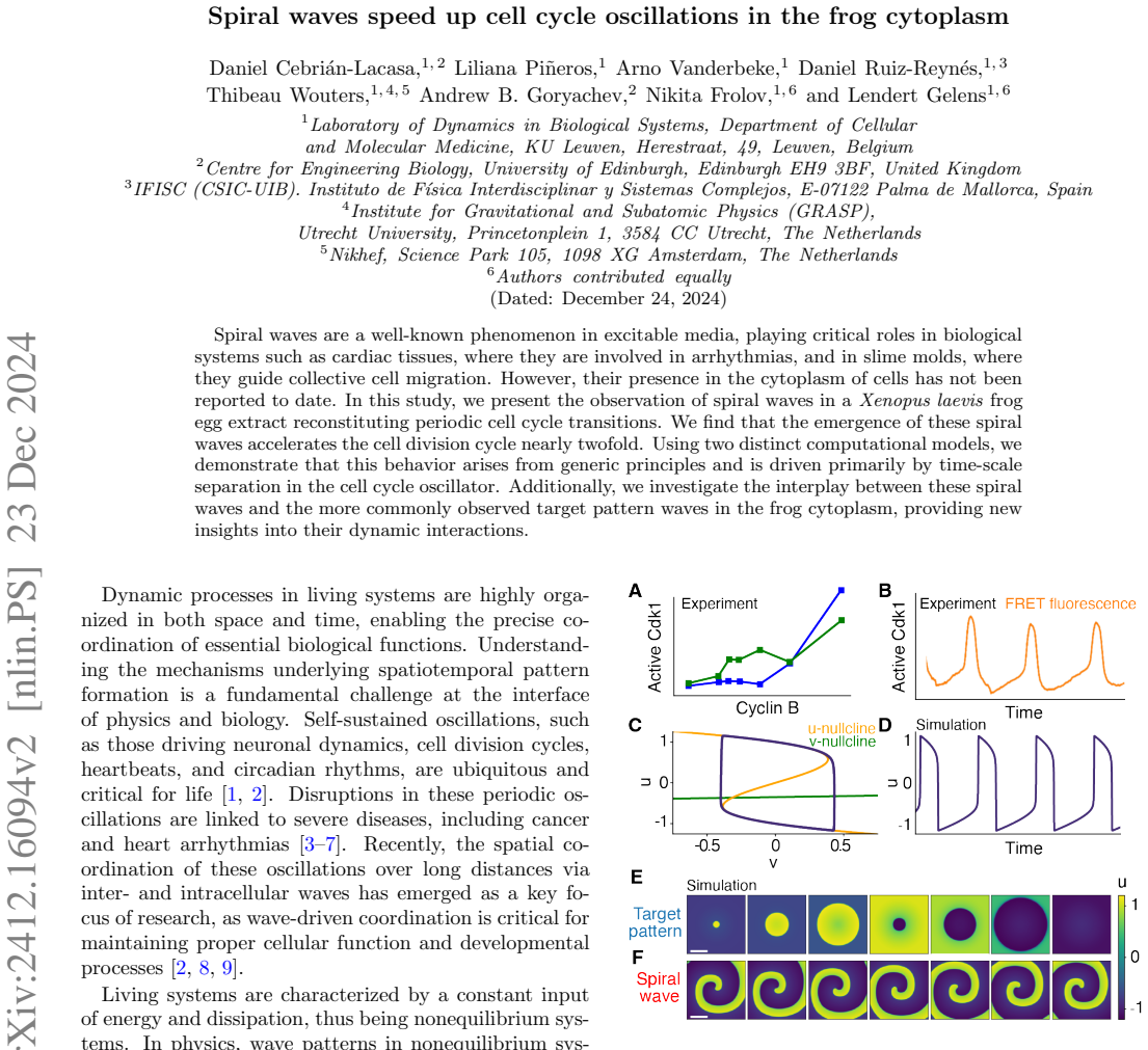
The central mechanism is the time-scale separation in the cell-cycle oscillator, captured by the two-variable FitzHugh-Nagumo model $\partial_t u = D\Delta u - u^3 + u - v$, $\partial_t v = D\Delta v + \varepsilon(u - b v + a)$, where $u$ is a fast activator and $v$ a slow recovery variable. With strong separation ($\varepsilon$ small), the medium is excitable and spiral waves rotate quickly: the angular velocity $\omega_s$ grows as $\varepsilon$ shrinks, so the spiral period $2\pi/\omega_s$ falls below the natural oscillation period $T_M$. Target patterns, by contrast, have a period bounded by the pacemaker and the medium and do not shorten. Singular perturbation theory gives the wave-speed scalings ($\sqrt{D/\varepsilon}$ for small $\varepsilon$), and a Hill-function fit to the simulated period reduction estimates $\bar\varepsilon_{CC}\approx0.008$ for the cell-cycle model and $\bar\varepsilon_{FHN}\approx0.004$ for FitzHugh-Nagumo, placing the frog cytoplasm in the regime where spirals accelerate the clock.
What would settle it
Image a Cdk1 activity biosensor together with the tubulin or nuclear reporter in the same droplets and compare the Cdk1 period in spiral, target-pattern, and wave-free regions: if the Cdk1 oscillation period is not shortened wherever a spiral rotates, the claim that spiral waves accelerate the cell cycle is falsified. A complementary check in simulation is to increase $\varepsilon$ beyond the fitted range; the period reduction should vanish as spirals become phase waves and their period approaches the medium's natural period.
Extended reading notes
Core claim
The paper's central claim is that spiral waves occur in the cytoplasm of frog egg extract and that, wherever a spiral rotates, the cell-cycle period shortens by up to about a factor of two. In large oil-encapsulated droplets, the authors observe both target patterns and spiral waves in fluorescent tubulin and in a nuclear marker; spirals arise when droplets merge or when a wave bends around an air bubble, matching the standard creation mechanisms for spirals in excitable media. Period measurements from five large droplets show that spiral-wave droplets oscillate faster than target-pattern or wave-free droplets, and the paper interprets both reporter readouts as proxies for Cdk1 activity waves. To show the effect is generic, the authors simulate the FitzHugh-Nagumo model and a cell-cycle model across the time-scale separation parameter $\varepsilon$; both models produce the same period reduction, with the spiral's rotation period dropping below the medium's natural period when $\varepsilon$ is small. The paper concludes that spiral-wave acceleration of the cell cycle is a robust property of excitable media with strong time-scale separation, not a special feature of frog cytoplasm.
Load-bearing premise
The load-bearing premise is that the fluorescent tubulin and nuclear waves seen in the droplets faithfully report underlying Cdk1 activity waves, so a shorter spiral period means a shorter cell cycle; the paper states this as a hypothesis and does not directly measure Cdk1 activity in the spiral droplets.
Editorial extensions
If this is right
- If the central claim is correct, a spatially extended cytoplasm can run its cell-cycle clock faster than the same biochemistry in a small, wave-free droplet; the spiral wave itself acts as a pacemaker that shortens division time by up to about twofold.
- The period reduction should be strongest in large systems with strong time-scale separation; small droplets that cannot host spirals should keep the ordinary oscillation period.
- Target patterns and spirals compete by fastest periodicity: a faster spiral should entrain the medium and win over a slower pacemaker-driven target pattern.
- Because spirals form when droplets merge or when waves bend around obstacles, these manipulations should provide a controlled experimental route to accelerating the cell cycle in extract.
- Target patterns that appear to adopt the spiral period can be explained by two nearby spiral tips merging into target-like waves, so no intrinsic resetting of the pacemaker is needed.
Reading between the lines
- Inference: if spiral-wave acceleration is generic in excitable media, other biological oscillators with strong time-scale separation — for example cardiac or neuronal tissue — may show the same period-shortening effect, not just frog cytoplasm.
- Inference: in an intact embryo, a spiral wave would locally speed up cleavage cycles and could desynchronize divisions across the embryo; the paper provides a concrete spatial mechanism for division timing varying within one cytoplasm.
- Inference: a decisive follow-up experiment is to co-image a Cdk1 FRET sensor with the tubulin or nuclear reporter in spiral droplets; if the Cdk1 period does not shorten while the reporters do, the acceleration claim would need to be revised.
- Inference: the fitted time-scale separations come from only five large droplets; measuring wave speed and period across many droplets would sharpen the estimate and test whether the reported twofold acceleration is quantitatively reproducible.
Signed reviews
Editorial analysis
A structured set of objections, weighed in public.
Referee Report
Summary. The paper reports the experimental observation of spiral waves in Xenopus laevis egg extract droplets using fluorescent tubulin and NLS-GFP reporters, alongside target patterns. It claims that spiral waves reduce the cell-cycle oscillation period by up to nearly twofold, and supports this with numerical simulations of the FitzHugh-Nagumo model and a cell-cycle model, attributing the period reduction to strong time-scale separation. The paper also studies how spiral waves and target patterns compete, proposing a double-tip mechanism for apparent exceptions. The central scientific claim is that cytoplasmic spiral waves can act as a spatial mechanism that accelerates the cell-cycle clock.
Significance. If the central claim holds, this is a novel and significant observation: spiral-wave dynamics in the cytoplasm have not been reported before, and a spatial wave pattern that shortens the cell-cycle period would be an important addition to the understanding of mitotic wave coordination in early Xenopus embryos. The numerical part is a strength: the authors use two distinct models, show that the period reduction is robust across parameter sets, and provide analytical wave-speed approximations for the target-pattern case, all of which make the modeling credible. However, the experimental evidence for the twofold speedup is currently very thin, and the identification of the observed tubulin/NLS-GFP waves as Cdk1 activity waves is explicitly stated as a hypothesis rather than directly demonstrated. The significance of the paper therefore hinges on additional experimental validation.
major comments (3)
- [Experimental results, Fig. 2F] The central claim of a nearly twofold period reduction is supported by only three spiral-wave droplets and two target-pattern droplets, with no error bars, no confidence intervals, and no statistical test reported. The normalization procedure uses the median of no-wave droplets, but with n=3 and n=2 the aggregated ratio is not a reliable measure of the effect size. Additional replicates and a proper inferential comparison (e.g., a mixed-effects model or permutation test) are required to establish the magnitude of the claimed speedup.
- [Experimental results, 'We hypothesize that underlying Cdk1 activity waves drive...'] The paper does not directly measure Cdk1 activity in the spiral droplets; the tubulin and NLS-GFP signals are downstream readouts (microtubule polymerization and nuclear envelope breakdown). The authors state the link as a hypothesis and provide supporting evidence from synchronization with sperm chromatin and matching wave speeds with earlier Cdk1-FRET experiments, but this does not exclude a cytoskeletal or reporter-driven origin of the spiral pattern. A direct Cdk1 activity sensor (e.g., Cdk1 FRET) imaged in the same spiral droplets is needed to establish that the measured period is genuinely the cell-cycle period. Without this, the abstract's assertion that spiral waves 'accelerate the cell division cycle' is not fully supported.
- [Numerical results, Fig. 3A and text following] The estimate of the time-scale separation in the extract, ε_CC ≈ 0.008 and ε_FHN ≈ 0.004, is obtained by fitting the model response curves to the experimental period reduction. This is a fit, not a prediction, and the two models yield different values. The conclusion that time-scale separation is the primary driver in the extract therefore depends on the model choice and on the very small experimental sample used for the fit. An independent estimate of ε from biochemical parameters or from a direct measurement of wave speed versus period would make the mechanistic claim more robust.
minor comments (4)
- [Data availability] The data availability statement lists repositories as '[Upcoming]' with no links or accession numbers; the final version should provide the actual deposited code and data, since the experimental and numerical reproducibility are important for the paper's claims.
- [Fig. 2F and text] The notation 'PN W' in the figure axis is confusing; it appears to mean normalized period, but it is not defined in the caption or text. Please use a clear symbol such as T̂ and define it.
- [Fig. 4A and 'Exceptional parameters'] The description of the exceptional parameters '(a, bP, bM) = (0,0,1)' is too terse; please specify which model is used, the meaning of bP and bM, and why these parameters are exceptional.
- [General] The paper switches between 'spiral waves speed up cell cycle oscillations' and 'reduce the cell cycle period'; both are fine, but please ensure that 'accelerate' is consistently defined as a decrease in period, not an increase in speed, to avoid ambiguity.
Circularity Check
No circularity found: the experimental observation is independent of the models, and the fitted time-scale-separation parameter is a transparent estimate rather than a disguised prediction.
full rationale
The paper's central claim, that spiral waves in frog egg extract droplets accelerate cell-cycle oscillations nearly twofold, rests on direct fluorescence time-lapse measurements (Fig. 2), not on the computational models. The models are used to test whether such a period reduction is a generic property of excitable media; the simulated period reduction as a function of the time-scale-separation parameter epsilon is a model output, not an input imposed by the data. The subsequent Hill-function fit, where the authors write "Fitting the Hill function to these response curves, we estimated the time-scale separation in the cytoplasmic extract," is transparent parameter estimation used to interpret the experiment, and the paper does not present the simulated twofold reduction as an independent forward prediction. No uniqueness theorem or load-bearing claim is imported from the authors' prior work: the FitzHugh-Nagumo equations are stated explicitly, the second cell-cycle model is a standard published model, and the wave-speed scalings are derived from singular perturbation theory. The identification of tubulin/NLS-GFP waves with Cdk1 activity is explicitly offered as a hypothesis and is supported by synchronization with nuclear envelope dynamics and by consistency with earlier Cdk1 wave-speed measurements; even if that identification were questioned, it would be an evidence gap rather than a circular derivation. Self-citations occur but are not load-bearing, and the central experimental result is independent of them. Accordingly, no step of the derivation is equivalent to its inputs by construction.
Assumptions & free parameters
free parameters (2)
- Time-scale separation epsilon (FHN model) =
0.004 (fitted)
- Time-scale separation epsilon (cell cycle model) =
0.008 (fitted)
assumptions (4)
- domain assumption Reporter waves reflect Cdk1 activity
- domain assumption FHN model is a valid model of the cell cycle oscillator
- domain assumption The cell cycle model of Parra-Rivas et al. [30] is applicable
- domain assumption Time-scale separation in the extract is small enough for the spiral speedup regime
Cite this review
Pith. "Pith review of Spiral waves speed up cell cycle oscillations in the frog cytoplasm." pith.science (2026). https://pith.science/paper/OY4YECDB
@misc{pith2026241216094,
  author       = {Pith},
  title        = {Pith review of: Spiral waves speed up cell cycle oscillations in the frog cytoplasm},
  year         = {2026},
  howpublished = {\url{https://pith.science/paper/OY4YECDB}},
  note         = {Machine review of arXiv:2412.16094}
}
read the original abstract
Spiral waves are a well-known phenomenon in excitable media, playing critical roles in biological systems such as cardiac tissues, where they are involved in arrhythmias, and in slime molds, where they guide collective cell migration. However, their presence in the cytoplasm of cells has not been reported to date. In this study, we present the observation of spiral waves in a Xenopus laevis frog egg extract reconstituting periodic cell cycle transitions. We find that the emergence of these spiral waves accelerates the cell division cycle nearly twofold. Using two distinct computational models, we demonstrate that this behavior arises from generic principles and is driven primarily by time-scale separation in the cell cycle oscillator. Additionally, we investigate the interplay between these spiral waves and the more commonly observed target pattern waves in the frog cytoplasm, providing new insights into their dynamic interactions.
Figures


Figures from the paper (1 more)

Forward citations
Cited by 1 Pith paper
-
Data-driven discovery of dynamical models in biology
A review benchmarking regression, network, and decomposition methods on the Oregonator model under the Koopman operator framework, with illustrative experiments on simulated data.
Reference graph
Works this paper leans on
-
[1]
B. Nov´ ak and J. J. Tyson, Design principles of biochemi- cal oscillators, Nature Reviews Molecular Cell Biology 9, 981 (2008)
work page 2008
-
[2]
C. Beta and K. Kruse, Intracellular oscillations and waves, Annual Review of Condensed Matter Physics 8, 239 (2017)
work page 2017
-
[3]
A. M. Pertsov, J. M. Davidenko, R. Salomonsz, W. T. Baxter, and J. Jalife, Spiral waves of excitation underlie reentrant activity in isolated cardiac muscle., Circulation Research 72, 631 (1993)
work page 1993
-
[4]
J. M. Davidenko, R. Salomonsz, A. M. Pertsov, W. T. Baxter, and J. Jalife, Effects of pacing on stationary reen- trant activity: theoretical and experimental study, Cir- culation research 77, 1166 (1995)
work page 1995
-
[5]
R. R. Aliev and A. V. Panfilov, A simple two-variable model of cardiac excitation, Chaos, Solitons & Fractals 7, 293 (1996)
work page 1996
-
[6]
M. P. Nash and A. V. Panfilov, Electromechanical model of excitable tissue to study reentrant cardiac arrhyth- mias, Progress in Biophysics and Molecular Biology 85, 501 (2004)
work page 2004
-
[7]
A. Panfilov, H. Dierckx, and V. Volpert, Reaction– diffusion waves in cardiovascular diseases, Physica D: Nonlinear Phenomena 399, 1 (2019)
work page 2019
- [8]
Show all 47 references
-
[9]
V. E. Deneke and S. Di Talia, Chemical waves in cell and developmental biology, Journal of Cell Biology 217, 1193 (2018)
2018
-
[10]
P. C. Fife, Pattern formation in reacting and diffusing systems, The Journal of Chemical Physics64, 554 (1976)
1976
-
[11]
J. P. Keener, Waves in excitable media, SIAM Journal on Applied Mathematics 39, 528 (1980)
1980
-
[12]
J. J. Tyson and J. P. Keener, Singular perturbation the- ory of traveling waves in excitable media (a review), Physica D: Nonlinear Phenomena 32, 327 (1988)
1988
-
[13]
Meron, Pattern formation in excitable media, Physics Reports 218, 1 (1992)
E. Meron, Pattern formation in excitable media, Physics Reports 218, 1 (1992)
1992
-
[14]
Rombouts and L
J. Rombouts and L. Gelens, Synchronizing an oscil- latory medium: The speed of pacemaker-generated 6 waves, Physical Review Research2, 10.1103/PhysRevRe- search.2.043038 (2020)
2020 doi
-
[15]
J. P. Keener and J. J. Tyson, Spiral waves in the Belousov-Zhabotinskii reaction, Physica D: Nonlinear Phenomena 21, 307 (1986)
1986
-
[16]
Cebri´ an-Lacasa, P
D. Cebri´ an-Lacasa, P. Parra-Rivas, D. Ruiz-Reyn´ es, and L. Gelens, Six decades of the FitzHugh–Nagumo model: A guide through its spatio-temporal dynamics and influ- ence across disciplines, Physics Reports 1096, 1 (2024)
2024
-
[17]
J. J. Tyson, K. A. Alexander, V. Manoranjan, and J. Murray, Spiral waves of cyclic AMP in a model of slime mold aggregation, Physica D: Nonlinear Phenomena 34, 193 (1989)
1989
-
[18]
Pla de Re- cuperacio, Transformaci´ o i Resiliencia
and on the cell cortex [44, 45]. The observation of spiral waves in the cytoplasmic ex- tract opens new avenues for investigating their functional roles in embryonic development. Unlike target patterns, spiral waves can shorten the cell cycle period, potentially altering early...
-
[19]
Lechleiter, S
J. Lechleiter, S. Girard, E. Peralta, and D. Clapham, Spi- ral calcium wave propagation and annihilation in Xeno- pus laevis oocytes, Science 252, 123 (1991)
1991
-
[20]
Siegert and C
F. Siegert and C. J. Weijer, Spiral and concentric waves organize multicellular dictyostelium mounds, Current Bi- ology 5, 937 (1995)
1995
-
[21]
H. Z. Ford, A. Manhart, and J. R. Chubb, Controlling periodic long-range signalling to drive a morphogenetic transition, eLife , e83796 (2023)
2023
-
[22]
J. R. Pomerening, E. D. Sontag, and J. E. Ferrell Jr, Building a cell cycle oscillator: hysteresis and bistabil- ity in the activation of Cdc2, Nature cell biology 5, 346 (2003)
2003
-
[23]
Maryu and Q
G. Maryu and Q. Yang, Nuclear-cytoplasmic com- partmentalization of Cyclin B1-Cdk1 promotes ro- bust timing of mitotic events, Cell Reports 41, 10.1016/j.celrep.2022.111870 (2022)
2022
-
[24]
J. B. Chang and J. E. Ferrell Jr, Mitotic trigger waves and the spatial coordination of the Xenopus cell cycle, Nature 500, 603 (2013)
2013
-
[25]
O. Puls, D. Ruiz-Reyn´ es, F. Tavella, M. Jin, Y. Kim, L. Gelens, and Q. Yang, Mitotic waves in frog egg ex- tracts: Transition from phase waves to trigger waves, bioRxiv 10.1101/2024.01.18.576267 (2024)
2024 doi
-
[26]
R. A. Fontanilla and R. Nuccitelli, Characterization of the sperm-induced calcium wave in Xenopus eggs using confocal microscopy, Biophysical journal75, 2079 (1998)
1998
-
[27]
K. Hara, P. Tydeman, and M. Kirschner, A cytoplas- mic clock with the same period as the division cycle in Xenopus eggs., Proceedings of the National Academy of Sciences 77, 462 (1980)
1980
-
[28]
F. E. Nolet, A. Vandervelde, A. Vanderbeke, L. Pi˜ neros, J. B. Chang, and L. Gelens, Nuclei determine the spa- tial origin of mitotic waves, eLife 9, 10.7554/eLife.52868 (2020)
2020 doi
-
[29]
Afanzar, G
O. Afanzar, G. K. Buss, T. Stearns, and J. Ferrell, James E, The nucleus serves as the pacemaker for the cell cycle, eLife 9, e59989 (2020)
2020
-
[30]
Novak and J
B. Novak and J. J. Tyson, Numerical analysis of a com- prehensive model of M-phase control in Xenopus oocyte extracts and intact embryos, Journal of Cell Science 106, 1153 (1993)
1993
-
[31]
Parra-Rivas, D
P. Parra-Rivas, D. Ruiz-Reyn´ es, and L. Gelens, Cell cycle oscillations driven by two interlinked bistable switches, Molecular Biology of the Cell 34, ar56 (2023)
2023
-
[32]
FitzHugh, Impulses and physiological states in theo- retical models of nerve membrane, Biophysical Journal 1, 445 (1961)
R. FitzHugh, Impulses and physiological states in theo- retical models of nerve membrane, Biophysical Journal 1, 445 (1961)
1961
-
[33]
Nagumo, S
J. Nagumo, S. Arimoto, and S. Yoshizawa, An active pulse transmission line simulating nerve axon, Proceed- ings of the IRE 50, 2061 (1962)
1962
-
[34]
Pi˜ neros, N
L. Pi˜ neros, N. Frolov, D. Ruiz-Reyn´ es, A. Van Eynde, G. Cavin-Meza, R. Heald, and L. Gelens, The nuclear-cytoplasmic ratio controls the cell cycle pe- riod in compartmentalized frog egg extract, bioRxiv 10.1101/2024.07.28.605512 (2024)
2024 doi
-
[35]
G. A. Anderson, L. Gelens, J. C. Baker, and J. E. Fer- rell, Desynchronizing embryonic cell division waves re- veals the robustness of Xenopus laevis development, Cell reports 21, 37 (2017)
2017
-
[36]
Johmura, N.-K
Y. Johmura, N.-K. Soung, J.-E. Park, L.-R. Yu, M. Zhou, J. K. Bang, B.-Y. Kim, T. D. Veenstra, R. L. Erikson, and K. S. Lee, Regulation of microtubule-based micro- tubule nucleation by mammalian polo-like kinase 1, Pro- ceedings of the National Academy of Sciences 108, 11446 (2011)
2011
-
[37]
Ishihara, K
K. Ishihara, K. S. Korolev, and T. J. Mitchison, Physical basis of large microtubule aster growth, eLife 5, e19145 (2016)
2016
-
[38]
Rinaldin, A
M. Rinaldin, A. Kickuth, B. Dalton, Y. Xu, S. Di Talia, and J. Brugu´ es, Robust cytoplasmic partitioning by solving an intrinsic cytoskeletal instability, bioRxiv 10.1101/2024.03.12.584684 (2024)
2024 doi
-
[39]
V. S. Zykov, Spiral wave initiation in excitable me- dia, Philosophical Transactions of the Royal Society A: Mathematical, Physical and Engineering Sciences 376, 20170379 (2018)
2018
-
[40]
G. Bub, A. Shrier, and L. Glass, Spiral wave generation in heterogeneous excitable media, Phys. Rev. Lett. 88, 058101 (2002)
2002
-
[41]
J. A. Sepulchre and A. Babloyantz, Motions of spiral waves in oscillatory media and in the presence of obsta- cles, Phys. Rev. E 48, 187 (1993)
1993
-
[42]
Agladze, J
K. Agladze, J. P. Keener, S. C. M¨ uller, and A. Panfilov, Rotating spiral waves created by geometry, Science 264, 1746 (1994)
1994
-
[43]
Rombouts and L
J. Rombouts and L. Gelens, Analytical approximations for the speed of pacemaker-generated waves, Phys. Rev. E 104, 014220 (2021)
2021
-
[44]
K. J. Lee, Wave pattern selection in an excitable system, Phys. Rev. Lett. 79, 2907 (1997)
1997
-
[45]
W. M. Bement, M. Leda, A. M. Moe, A. M. Kita, M. E. Larson, A. E. Golding, C. Pfeuti, K.-C. Su, A. L. Miller, A. B. Goryachev, et al., Activator–inhibitor coupling be- tween Rho signalling and actin assembly makes the cell cortex an excitable medium, Nature cell biology 17, 14...
2015
-
[46]
Michaud, M
A. Michaud, M. Leda, Z. T. Swider, S. Kim, J. He, J. Landino, J. R. Valley, J. Huisken, A. B. Goryachev, G. von Dassow, et al. , A versatile cortical pattern- forming circuit based on Rho, F-actin, Ect2, and RGA- 3/4, Journal of Cell Biology 221, e202203017 (2022)
2022
-
[47]
Gelens, K
L. Gelens, K. Huang, and J. Ferrell, How does the Xeno- pus laevis embryonic cell cycle avoid spatial chaos?, Cell Reports 12, 892 (2015)
2015
Reviewed August 11, 2026 · model on record in the stance chip above.
Discussion (0). Continue with ORCID to comment.