REVIEW 3 major objections 4 minor 18 references
Fast Dynamic Perfusion and Angiography Reconstruction using an end-to-end 3D Convolutional Neural Network
T0 review · 3 major / 4 minor · reviewed 2026-08-14 · deepseek-v4-flash
Pith's one-line read A 3D Dense-UNet with multi-level SSIM loss reconstructs 4D perfusion and 4D angiography from interleaved half-sampled crushed and non-crushed Hadamard te-pCASL data, preserving image quality while halving scan time.
desk verdict A well-executed in-silico proof-of-concept for 3D CNN reconstruction of perfusion and angiography from half-sampled Hadamard te-pCASL, but the abstract's clinical claim outruns the evidence. read the letter →
The pith
A machine-rendered reading of the paper's core claim, the machinery that carries it, and where it could break.
The reading
What carries the argument
The load-bearing device is a 3D Dense-UNet with a multi-level loss. Dense blocks with $3\times3\times3$ and $1\times1\times1$ convolutions improve feature propagation and gradient flow; up-sampling uses a constant trilinear resize kernel to avoid checkerboard artifacts. The multi-level loss ML-SSIM weights the SSIM loss at three network levels, chosen because SSIM outperformed MSE, VGG-16 perceptual loss, and single-scale SSIM for perfusion reconstruction. The training data come from a generalized Buxton kinetic model (Eqs. 3 and 4) that combines an intravascular arterial signal and an extravascular tissue signal parameterized by AAT, BAT, and CBF; fully sampled Hadamard decoding of the generated scans provides the ground truth.
What would settle it
Acquire real crushed and non-crushed Hadamard-8 te-pCASL scans on a cohort, form full-sampled ground truth by the standard decoding and subtraction, then feed the interleaved 50-percent-sampled subset through the trained network; if the SSIM against real full-sampled output falls substantially below 97 percent for perfusion or 96 percent for angiography, the central claim fails. A cheaper check is to compare the kinetic model's predicted arterial and tissue signals to measured signals in voxels with known transit times.
Extended reading notes
Core claim
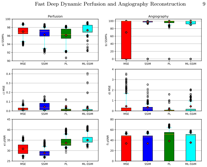
The central claim is that a 3D Dense-UNet trained with a multi-level SSIM loss can decode 4D perfusion and 4D angiography directly from interleaved 50-percent-sampled crushed and non-crushed Hadamard te-pCASL scans, without a separate full acquisition. The network uses an 8-row Hadamard encoding, takes four crushed and four non-crushed patches as input, and outputs 14 patches, seven perfusion and seven angiography time-points. With the ML-SSIM loss it achieves SSIM of 97.3 ± 1.1 on perfusion and 96.2 ± 11.1 on angiography for 313 test datasets, and reconstructs all time-points in about 205 ms. The paper also presents a data-generation framework based on the generalized Buxton kinetic model to create realistic training data from in vivo arterial arrival time, bolus arrival time, and simulated CBF maps.
Load-bearing premise
The synthetic te-pCASL data produced by the kinetic model from in vivo arrival-time maps and simulated anatomies is representative enough of real crushed and non-crushed Hadamard te-pCASL acquisitions; if real signals contain effects the model omits, the trained network's outputs may not match the reported SSIM.
Editorial extensions
If this is right
- If the central claim holds, simultaneous 4D MRA and perfusion can be acquired in one interleaved scan rather than two, halving the te-pCASL scan time.
- The network reconstructs all seven time-points of both output types in roughly 205 ms per patch set, suggesting a speed compatible with clinical workflow.
- The ML-SSIM loss gives a statistically significant improvement over MSE loss for perfusion ($p<0.05$), while matching the SSIM loss for angiography; this identifies the loss design as a key factor.
- The synthetic data-generation framework extends to higher-rank Hadamard matrices and other time-encoded ASL schemes, since the kinetic model equations generalize beyond Hadamard-8.
Reading between the lines
- The interleaved half-sampling idea could transfer to other dual-acquisition protocols where two scans are subtracted, with the network learning the unmixing directly from data.
- Because angiography is intrinsically sparse and dominated by elongated structures, explicit sparsity-aware or structure-aware loss terms could push early time-point angiography quality beyond the reported SNR.
- The synthetic-to-real gap is the largest open risk; fine-tuning on a small set of real in vivo crushed and non-crushed pairs would test whether the reported SSIM translates to clinical acquisitions.
- With inference under a quarter of a second, the network could be embedded into a real-time reconstruction loop, displaying both perfusion and angiography immediately after acquisition.
Signed reviews
Editorial analysis
A structured set of objections, weighed in public.
Referee Report
Summary. Hadamard time-encoded pCASL can provide both dynamic perfusion and 4D angiography when acquired with and without flow-crushing, but the two acquisitions double scan time. This paper proposes a 3D Dense-UNet that inputs interleaved 50%-sampled crushed and non-crushed Hadamard te-pCASL images (four of eight encodings each) and outputs seven-timepoint perfusion and angiographic volumes, achieving a factor-two acceleration relative to full sampling. Training and test data are generated entirely from a simulator built on a generalized Buxton kinetic model (Eqs. 3-4) using 6 BAT maps, 4 AAT maps, and 20 BrainWeb anatomies. The authors compare MSE, VGG perceptual, SSIM, and their proposed multi-level SSIM losses and report best perfusion SSIM of 97.3 +/- 1.1 and angiography SSIM of 96.2 +/- 11.1 on 313 synthetic test datasets. The paper claims this obviates the additional scan time in practice.
Significance. The contribution is potentially useful: an end-to-end 3D CNN with dense connectivity and a multi-level SSIM loss is a reasonable architecture for recovering both smooth perfusion and sparse vascular structures from undersampled Hadamard-encoded ASL, and the loss-function comparison with Wilcoxon tests is a strength. The data-generation pipeline is clearly specified and makes the method reproducible. The significance is conditional because the entire evaluation is in-distribution with respect to the authors' own simulator: no real crushed/non-crushed te-pCASL data are used for validation, and the metrics therefore estimate performance on synthetic signals that follow Eqs. (3)-(4), not on clinical acquisitions with dispersion, motion, partial volume, or B1/B0 effects. The abstract's clinical claim of 'negating the additional scan time' is stronger than the evidence supports.
major comments (3)
- [§2.3, Eqs. (3)-(4); Abstract; Conclusion] All training, validation, and test examples are generated by the authors' simulator from 6 BAT maps, 4 AAT maps, and 20 BrainWeb subjects; the only corruption added is Gaussian white noise with standard deviation between 0 and 5 (Section 3). The kinetic model omits transit-time dispersion, partial-volume effects, motion, B1/B0 inhomogeneities, T1/T2 variation, and labeling-efficiency errors. Because the network is trained and tested on this same distribution, the reported SSIM values (97.3 +/- 1.1 perfusion, 96.2 +/- 11.1 angiography) do not by themselves support the abstract's statement that the method 'negat[es] the additional scan time' in real acquisitions. The paper itself acknowledges this in the Conclusion: 'A further step of this study is enriching the training and validation datasets with in vivo data.' I ask the authors to either add an in vivo or at least a domain-shift experiment (e.g., realistic noise/artifact model or a held-out set from a different simulator/scanner) and to temper the abstract and conclusion so the claim is limited to the synthetic setting.
- [§2.3, final sentence] The sentence 'To evaluate the generated data, the signal evolution pattern was validated by the Buxton curve model [15]' is self-referential, because Eqs. (3)-(4) are themselves a generalization of the Buxton model. Matching the Buxton curve therefore only checks internal consistency of the generator, not agreement with real te-pCASL signals. This sentence should be removed or replaced by a comparison with measured crushed/non-crushed te-pCASL data.
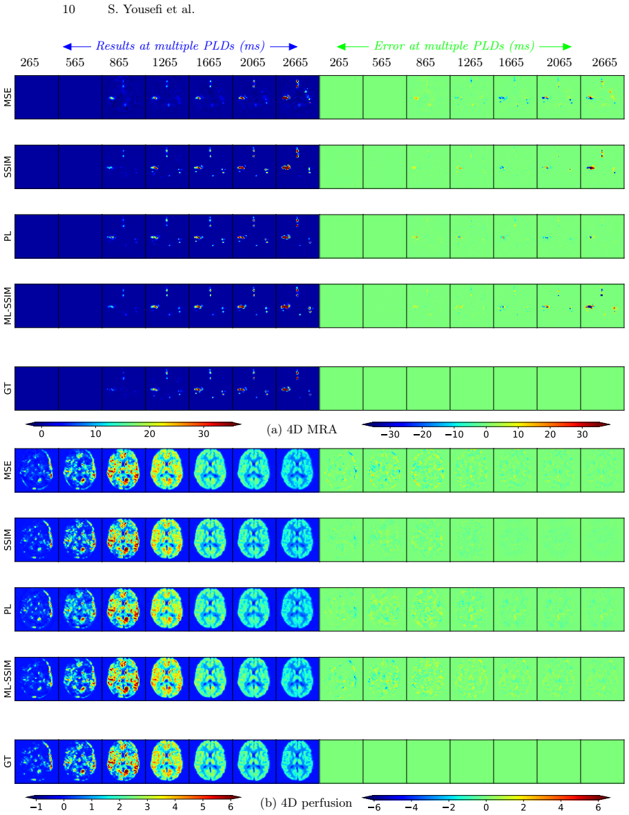
- [§1 and §4] The paper states the goal of 'accurate CBF quantification' but no quantitative CBF accuracy is reported. Table 1 and Fig. 3 contain only image-space metrics (SSIM, MSE, SNR, pSNR) on reconstructed perfusion volumes; there is no comparison of estimated CBF values (e.g., bias and limits of agreement in mL/100 g/min) against the known simulation parameters or against a reference method. Either add such an analysis or remove the CBF-quantification claim from the introduction and conclusion.
minor comments (4)
- [Table 1, Fig. 3] For the angiography rows with high variance (SSIM and ML-SSIM), the reported mean +/- SD is strongly skewed (ML-SSIM SSIM mean 96.2, median 99.7, SD 11.1); report median and IQR and state how many of the 313 test datasets drive the low mean.
- [Figure 1] The formula 'w1 x Loss level1 + w2 x Loss level1 + w3 x Loss level3' appears to have a typo; the second term should likely be 'w2 x Loss level2'.
- [Introduction, Reference [1]] Reference [1] (Ferlay et al., cancer epidemiology) does not correspond to the first sentence about ASL being non-invasive; please replace it with the intended ASL review or methods citation.
- [§2.3, Section 3] The Gaussian noise augmentation is specified as 'random standard deviation between 0 and 5' without stating the units or the signal scale to which it is applied; please clarify (e.g., as a percentage of the mean signal or in arbitrary intensity units).
Circularity Check
No significant circularity: the reconstruction target is full-sampled Hadamard decoding, which is independent of the network and the subsampled input.
full rationale
The paper's claimed derivation chain is the following: fully sampled crushed and non-crushed Hadamard te-pCASL data are decoded by the standard Hadamard decoding/subtraction operation M (Eq. 1) to produce ground-truth 4D perfusion and angiography; the network M' is trained to reproduce those same ground-truth outputs from interleaved 50%-subsampled inputs (Eq. 2). The target is therefore defined independently of the network, by the closed-form decoding of full data, and the subsampled inputs are strict subsets of those full data. This is a legitimate supervised regression task rather than a self-definitional or fitted-input-called-prediction loop: no parameter is fitted to the test outputs, and no uniqueness theorem or ansatz is imported from the authors' prior work to force the result. The only self-referential elements are minor: the synthetic generator is built from the Buxton kinetic model and then checked against the Buxton curve model, which is an internal consistency check rather than load-bearing evidence; and the decoding method is cited to the authors' own prior work, but that method is a standard published technique used as a fixed preprocessing operator, not a self-citation invoked to justify the paper's conclusion. The genuine limitation is external validity, not circularity: all training and test data come from the same in-silico generator, so reported SSIM estimates performance on simulator-like signals. The paper acknowledges this in the Conclusion: "A further step of this study is enriching the training and validation datasets with in vivo data." This is a correctness and generalization risk, not a circular-derivation risk. Therefore no circular step is exhibited.
Assumptions & free parameters
free parameters (4)
- GM/WM/CSF CBF values =
not stated (literature values assigned)
- aCBV (arterial cerebral blood volume) =
not stated
- ML-SSIM loss weights w1, w2, w3 =
not stated
- Arterial and tissue relaxation times T1a/T1b and M0a normalization =
not stated
assumptions (4)
- domain assumption Buxton general kinetic model for ASL signal
- domain assumption Voxel signal is the linear sum of tissue and arterial contributions
- domain assumption Synthetic data distribution matches real te-pCASL acquisitions
- standard math Hadamard decoding is the correct ground-truth estimator
Cite this review
Pith. "Pith review of Fast Dynamic Perfusion and Angiography Reconstruction using an end-to-end 3D Convolutional Neural Network." pith.science (2026). https://pith.science/paper/PPEOLTG6
@misc{pith2026190808947,
  author       = {Pith},
  title        = {Pith review of: Fast Dynamic Perfusion and Angiography Reconstruction using an end-to-end 3D Convolutional Neural Network},
  year         = {2026},
  howpublished = {\url{https://pith.science/paper/PPEOLTG6}},
  note         = {Machine review of arXiv:1908.08947}
}
abstract
Hadamard time-encoded pseudo-continuous arterial spin labeling (te-pCASL) is a signal-to-noise ratio (SNR)-efficient MRI technique for acquiring dynamic pCASL signals that encodes the temporal information into the labeling according to a Hadamard matrix. In the decoding step, the contribution of each sub-bolus can be isolated resulting in dynamic perfusion scans. When acquiring te-ASL both with and without flow-crushing, the ASL-signal in the arteries can be isolated resulting in 4D-angiographic information. However, obtaining multi-timepoint perfusion and angiographic data requires two acquisitions. In this study, we propose a 3D Dense-Unet convolutional neural network with a multi-level loss function for reconstructing multi-timepoint perfusion and angiographic information from an interleaved $50\%$-sampled crushed and $50\%$-sampled non-crushed data, thereby negating the additional scan time. We present a framework to generate dynamic pCASL training and validation data, based on models of the intravascular and extravascular te-pCASL signals. The proposed network achieved SSIM values of $97.3 \pm 1.1$ and $96.2 \pm 11.1$ respectively for 4D perfusion and angiographic data reconstruction for 313 test data-sets.
Figures


Figures from the paper (1 more)
Reference graph
Works this paper leans on
-
[15]
Richard B Buxton, Lawrence R Frank, Eric C Wong, Bettina Siewert, Steven Warach, and Robert R Edelman. A general kinetic model for quantitative perfusion imaging with arterial spin labeling.MRM, 40(3):383–396, 1998. 12 S. Yousefi et al
work page 1998
-
[1]
Cancer incidence and mortality worldwide: sources, methods and major patterns in globocan 2012
Jacques Ferlay, Isabelle Soerjomataram, Rajesh Dikshit, Sultan Eser, Colin Math- ers, Marise Rebelo, Donald Maxwell Parkin, David Forman, and Freddie Bray. Cancer incidence and mortality worldwide: sources, methods and major patterns in globocan 2012. International journal of cancer , 136(5):E359–E386, 2015
work page 2012
-
[2]
Matthias JP van Osch, Wouter M Teeuwisse, Zhensen Chen, Yuriko Suzuki, Michael Helle, and Sophie Schmid. Advances in arterial spin labelling mri meth- ods for measuring perfusion and collateral flow.Journal of Cerebral Blood Flow & Metabolism, 38(9):1461–1480, 2018
work page 2018
-
[3]
Esben Thade Petersen, Kim Mouridsen, and Xavier Golay. The quasar repro- ducibility study, part II: Results from a multi-center arterial spin labeling test– retest study. Neuroimage, 49(1):104–113, 2010
work page 2010
-
[4]
Esophageal gross tumor volume segmentation using a 3d convolutional neural network
SaharYousefi,HessamSokooti,MohamedSElmahdy,FemkePPeters,Mohammad T Manzuri Shalmani, Roel T Zinkstok, and Marius Staring. Esophageal gross tumor volume segmentation using a 3d convolutional neural network. InMICCAI, pages 343–351. Springer, 2018
work page 2018
-
[5]
Mohamed S Elmahdy, Thyrza Jagt, R Th Zinkstok, Yuchuan Qiao, Rahil Shahzad, Hessam Sokooti, Sahar Yousefi, Luca Incrocci, CAM Marijnen, Mischa Hoogeman, et al. Robust contour propagation using deep learning and image registration for online adaptive proton therapy of prostate cancer.Medical physics, 2019
work page 2019
-
[6]
Kuang Gong, Jiahui Guan, Kyungsang Kim, Xuezhu Zhang, Jaewon Yang, Youngho Seo, Georges El Fakhri, Jinyi Qi, and Quanzheng Li. Iterative pet image reconstruction using convolutional neural network representation.TMI, 38(3):675– 685, 2018
work page 2018
-
[7]
E Gong, J Pauly, and G Zaharchuk. Boosting snr and/or resolution of arterial spin label (ASL) imaging using multi-contrast approaches with multi-lateral guided filter and deep networks. InProceedings of the Annual Meeting of the International Society for Magnetic Resonance in Medicine, Honolulu, Hawaii , 2017
work page 2017
Show all 18 references
-
[8]
Fan, Mohammad M
Jia Guo, Enhao Gong, Maged Goubran, Audrey P. Fan, Mohammad M. Khalighi, and Greg Zaharchuk. Improving perfusion image quality and quantification accu- racy using multi-contrast mri and deep convolutional neural networks. InISMRM, Paris, France, 2018
2018
-
[9]
AtemporaldeeplearningapproachforMRperfusionparameterestimation in stroke
King Chung Ho, Fabien Scalzo, Karthik V Sarma, Suzie El-Saden, and Corey W Arnold. AtemporaldeeplearningapproachforMRperfusionparameterestimation in stroke. In23rd ICPR, pages 1315–1320. IEEE, 2016
2016
-
[10]
Densely connected convolutional networks
Gao Huang, Zhuang Liu, Laurens Van Der Maaten, and Kilian Q Weinberger. Densely connected convolutional networks. InCVPR, pages 4700–4708, 2017
2017
-
[11]
The one hundred layers tiramisu: Fully convolutional densenets for semantic segmentation
Simon Jégou, Michal Drozdzal, David Vazquez, Adriana Romero, and Yoshua Ben- gio. The one hundred layers tiramisu: Fully convolutional densenets for semantic segmentation. In CVPR, pages 11–19, 2017
2017
-
[12]
Rapid 3d dynamic arterial spin labeling with a sparse model-based image reconstruction
Li Zhao, Samuel W Fielden, Xue Feng, Max Wintermark, John P Mugler III, and Craig H Meyer. Rapid 3d dynamic arterial spin labeling with a sparse model-based image reconstruction. Neuroimage, 121:205–216, 2015
2015
-
[13]
Image super- resolution using deep convolutional networks.TPAMI, 38(2):295–307, 2015
Chao Dong, Chen Change Loy, Kaiming He, and Xiaoou Tang. Image super- resolution using deep convolutional networks.TPAMI, 38(2):295–307, 2015
2015
-
[14]
Perceptual losses for real-time style transfer and super-resolution
Justin Johnson, Alexandre Alahi, and Li Fei-Fei. Perceptual losses for real-time style transfer and super-resolution. InECCV, pages 694–711. Springer, 2016
2016
-
[16]
Brainweb: Online interface to a 3d mri simulated brain database
Chris A Cocosco, Vasken Kollokian, Remi K-S Kwan, G Bruce Pike, and Alan C Evans. Brainweb: Online interface to a 3d mri simulated brain database. In NeuroImage. Citeseer, 1997
1997
-
[17]
Elastix: a toolbox for intensity-based medical image registration
Stefan Klein, Marius Staring, Keelin Murphy, Max A Viergever, and Josien PW Pluim. Elastix: a toolbox for intensity-based medical image registration. TMI, 29(1):196–205, 2010
2010
-
[18]
Transit time mapping in the mouse brain using time- encoded pcasl
Lydiane Hirschler, Leon P Munting, Artem Khmelinskii, Wouter M Teeuwisse, Ernst Suidgeest, Jan M Warnking, Louise van der Weerd, Emmanuel L Barbier, and Matthias JP van Osch. Transit time mapping in the mouse brain using time- encoded pcasl. NMR in Biomedicine , 31(2):e3855, 2018
2018
Reviewed August 14, 2026 · model on record in the stance chip above.
Discussion (0). Continue with ORCID to comment.