REVIEW 4 major objections 4 minor 4 references
Interplay of Electrostatic Interaction and Steric Repulsion between Bacteria and Gold Surface Influences Raman Enhancement
T0 review · 4 major / 4 minor · reviewed 2026-08-10 · deepseek-v4-flash
Pith's one-line read Liquid bacterial SERS enhancement is governed by electrostatic cell–nanorod attraction, opposed by steric repulsion from surface polymers, with a zeta-potential sum as a practical predictor.
desk verdict Systematic SERS dataset with a real reproducibility story, but the DLVO model as written predicts attraction where the paper claims repulsion, so the central mechanism does not hold up. read the letter →
The pith
A machine-rendered reading of the paper's core claim, the machinery that carries it, and where it could break.
The reading
What carries the argument
The argument runs on a colloidal interaction model built from four free-energy terms: electrostatic double-layer interaction (as in DLVO theory), van der Waals attraction, acid–base repulsion, and a steric polymer term whose range is set by the extended length of surface biopolymers (200 nm for E. coli, 10 nm for S. epidermidis). The electrostatic term uses the measured zeta potentials as constant surface potentials under the Derjaguin approximation, and it is the term that sets the equilibrium separation distance. The paper's practical device is the simpler design rule $\Delta G_{ES} = \psi_{\mathrm{bacteria}} + \psi_{\mathrm{nanorod}}$, meant to let a researcher estimate SERS enhancement from two zeta-potential measurements alone.
What would settle it
A decisive check would be to measure the force–distance curve between a -9 mV gold surface and a single S. epidermidis cell with an atomic force microscope: the paper's constant-potential model predicts an attractive minimum near 2.8 nm separation, whereas a constant-charge model predicts strong repulsion below 10 nm, so the observed sign of the force at short separation would settle which electrostatics governs the SERS enhancement.
Extended reading notes
Core claim
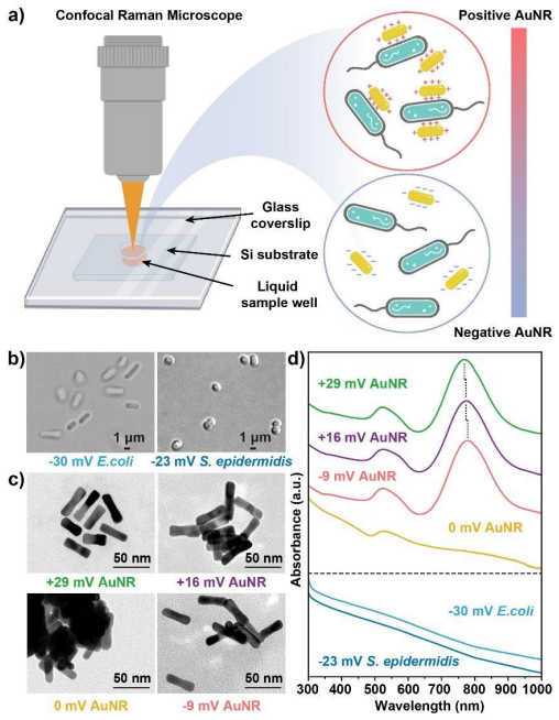
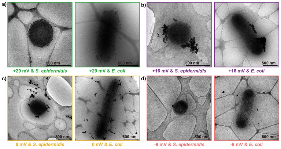
The paper's central discovery is that electrostatic interaction, not plasmonic hotspots alone, dominates the cell–nanorod proximity that sets SERS enhancement for bacteria in water. Using gold nanorods with zeta potentials of +29, +16, 0 and -9 mV, the authors measured enhancements of 7.2X, 3.6X, 4.2X and 1.3X for S. epidermidis and 3.9X, 2.8X, 2.9X and 1.1X for E. coli, respectively, and cryo-EM images show that this ordering tracks how closely nanorods adhere to the cell membranes. A DLVO-based calculation attributes the trends to an electrostatic double-layer attraction that weakens as nanorod charge drops, opposed at short range by steric repulsion from surface biopolymers; the longer polymer coat of E. coli is invoked to explain why S. epidermidis consistently gives stronger enhancement despite being less negatively charged. From this the authors distill the design rule $\Delta G_{ES} = \psi_{\mathrm{bacteria}} + \psi_{\mathrm{nanorod}}$ for quick estimation of SERS activity.
Load-bearing premise
The load-bearing premise, which the paper itself flags in its modeling section, is that the bacterial and nanorod surfaces stay at constant potential, which makes the electrostatic term purely attractive at close separations even when both zeta potentials have the same sign; if the surfaces were closer to constant charge, the short-range force would become strongly repulsive for like-signed pairs and the reading of the -9 mV nanorod data would change.
Editorial extensions
If this is right
- If electrostatics is the dominant control, liquid SERS sensitivity for a given bacterium can be rationally tuned by choosing nanorods with sufficiently positive zeta potential rather than by changing the laser or substrate.
- The zeta-potential sum rule gives a concrete threshold: enhancement is expected only when the sum exceeds the bacterium's own zeta potential, and negative sums predict weak or no SERS.
- The model predicts that removing or shortening surface biopolymers should raise enhancement, because steric repulsion is the main force preventing close nanorod–membrane contact.
- Because 0 mV nanorods aggregate and wrap cells, hotspot formation can partly compensate for missing surface charge, so aggregation state should be reported alongside zeta potential in SERS studies.
Reading between the lines
- Editorial inference: Because the model assumes constant-potential surfaces, its short-range electrostatic term is attractive for same-signed pairs; a constant-charge variant would likely predict stronger repulsion for the -9 mV nanorods, so the sign convention is a natural place to test the model's robustness.
- Editorial inference: The sum rule is dimensionally crude; a more quantitative extension would weight each zeta potential by Debye length or polymer thickness, and varying ionic strength in the same bacteria–nanorod system would reveal where the simple sum breaks down.
- Editorial inference: The polymer-length values of 10 nm and 200 nm are assigned to the two strains; replacing them with directly measured brush thicknesses from force spectroscopy would turn the steric-repulsion explanation into a predictive input rather than a fitted parameter.
- Editorial inference: If the sum rule generalizes, the same two-measurement recipe could be applied to other negatively charged bio-particles such as viruses or exosomes, as long as their surface polymer coats are accounted for, because the underlying physics is colloidal rather than bacterial-specific.
Signed reviews
Editorial analysis
A structured set of objections, weighed in public.
Referee Report
Summary. The manuscript reports a systematic study of the SERS enhancement of E. coli and S. epidermidis mixed with gold nanorods of controlled zeta potentials (+29, +16, 0, -9 mV). The experimental data are reproducible across 120 measurements and show a consistent ordering (+29 > 0 > +16 > -9 mV for both bacteria), with S. epidermidis generally enhanced more than E. coli. To explain this trend, the authors model cell-nanorod interactions with a DLVO-based calculation that includes electrostatic, van der Waals, acid-base, and steric polymer repulsion terms, and they propose a simple design rule based on the sum of the two zeta potentials. The central mechanistic claim is that electrostatics dominate cell-nanorod proximity and thus SERS enhancement, with steric repulsion from cell surface polymers as the key opposing force.
Significance. The experimental dataset is a useful contribution: the zeta-potential series of nanorods is well controlled, the Raman measurements are repeated and internally consistent, and the cryo-EM images are informative. The paper also provides its calculation code in the SI. However, the central theoretical claim is undermined by a direct contradiction inside the manuscript: the constant-potential DLVO model that the authors use predicts an attractive minimum for the supposedly repulsive -9 mV nanorod / S. epidermidis pair, as shown in Table S2. The proposed design rule is dimensionally inconsistent and not derived from the model. Because the mechanistic explanation is the paper's main claim, the current manuscript cannot be accepted as is.
major comments (4)
- [Section 2, theoretical paragraph; Table S2] The constant-potential DLVO model used for ΔG_ES is explicitly noted in the text to become purely attractive for κd << 1 irrespective of the signs of the two surface potentials. Consistent with that, Table S2 lists for the -9 mV nanorod with S. epidermidis an attractive minimum of -5.0 kBT at 2.8 nm separation, which is nearly as close as the +29 mV case (1.8 nm, -111.6 kBT). Yet the manuscript explains the measured 1.3X enhancement for this mixture as 'large electrostatic repulsion' (Cryo-EM paragraph, Fig. 3d). The model therefore does not produce the repulsion invoked to explain the data, so the central claim that electrostatics determine SERS enhancement via proximity is not supported by the model as written.
- [Section 7, Eq. for ΔG_ES] The proposed design rule ΔG_ES = ψ_bacteria + ψ_nanorod is asserted without derivation. The left-hand side has units of free energy while the right-hand side is a sum of electric potentials in millivolts, making the equality dimensionally inconsistent. It is not a simplification of the DLVO expression given earlier, and no quantitative relationship between this sum and the measured enhancement factors is provided. As presented, this rule cannot serve as a predictive design principle and should either be removed or replaced with a properly defined correlational metric.
- [Section 2 (steric repulsion) and Figure 6] The polymer extended lengths L (200 nm for E. coli, 10 nm for S. epidermidis) are chosen after the fact to make the predicted ordering match the SERS data, and the control calculation in Figure 6c/d assumes identical polymers and yields the opposite ordering. The values of L are inferred from the same cells whose SERS differences they are used to explain, so the steric-repulsion mechanism is not independently validated. The strong dependence of the conclusion on these two post hoc parameters should be explicitly acknowledged and, ideally, supported by independent measurements of the polymer layer thickness.
- [SI Table S1 (code) and Table S2] The calculation code in Table S1 uses zetab = -19 mV for S. epidermidis, whereas the main text and Figure S2 report the measured zeta potential as -23 mV. All quantitative results for S. epidermidis in Figure 4 and Table S2 are therefore based on an input that is inconsistent with the manuscript's stated experimental value. The code should be corrected to use -23 mV, or the text should be changed to explain the discrepancy; as written, this inconsistency makes the reported interaction energies for S. epidermidis unreliable.
minor comments (4)
- [Section 2, Eq. for ΔG_VDW] The van der Waals expression is written with the inverse Debye length κ in the denominator, i.e., ΔG_VDW = -A a1 a2 / (6 κ (a1+a2)); the denominator should be the separation distance d, not κ. As written, the equation incorrectly couples the VDW interaction to ionic strength.
- [Conclusion] The conclusion misidentifies the two bacteria as 'Gram-positive E. coli and Gram-negative S. epidermidis'; in fact E. coli is Gram-negative and S. epidermidis is Gram-positive. This error should be corrected.
- [Section 2, theoretical paragraph] The text refers to the 'permeability of water' where it means the permittivity, and the symbol π is used both for the numerical constant and for the correlation length in the acid-base term; the notation should be disambiguated for clarity.
- [Section 7] The statement that the prerequisite for obvious SERS enhancement is a sum value at least higher than the bacteria zeta potential itself is not supported by any quantitative analysis in the paper; it appears to be a qualitative observation and should be framed as such.
Circularity Check
DLVO prediction of bacterial SERS ordering is partly circular: the design rule sums its own inputs, and the polymer lengths are chosen to force the observed ordering.
-
self definitional
[Section 2, Figure 7 and Table S4 ('Finally, based on our findings...')]
"Finally, based on our findings we would like to propose a straightforward approach to qualitatively determine expected SERS intensities via determination of strength of electrostatic interactions by simple sum of the zeta potential of bacteria and nanorods ... 𝛥𝛥𝐺𝐺𝐸𝐸𝐸𝐸 = 𝜓𝜓𝐴𝐴𝐵𝐵𝐵𝐵𝐵𝐵𝐵𝐵𝐵𝐵𝐵𝐵𝐵𝐵 + 𝜓𝜓𝑁𝑁𝐵𝐵𝑁𝑁𝑁𝑁𝐵𝐵𝑁𝑁𝑁𝑁𝑁𝑁"
The proposed 'design principle' is literally the algebraic sum of the two measured inputs (bacteria and nanorod zeta potentials). No independent constant is fitted and no prediction is made that is not already contained in the experimental dataset from which it is extracted. Since the bacteria zeta potential is fixed for each species, the sum re-encodes exactly the nanorod zeta-potential rank that was used to sort the measured SERS enhancements. Calling this sum ΔG_ES does not derive it from the DLVO equations, and the expression is dimensionally a voltage, not an energy. The claim that positive sums give high SERS and negative sums give low SERS is a restatement of the observed trend, not a testable derivation from the model.
-
fitted input called prediction
[Section 2, theoretical paragraph, steric-repulsion expression (ΔG_SP) and Tables S2-S3]
"where 𝐿𝐿 is the extended length of the polymer layer on the bacterial surface (200 nm for E. coli but only 10 nm for S. epidermidis)"
The two polymer lengths are assigned after the SERS ordering (S. epidermidis > E. coli) was already known, and they are the only inputs that make the model reproduce that ordering. With L = 200 nm vs 10 nm, Table S2 gives S. epidermidis minimum separations of 1.8-2.8 nm and E. coli 89-194 nm. When the same polymer is assumed for both species (Table S3), the model predicts the opposite trend, with E. coli closer than S. epidermidis. Thus the cross-bacteria 'prediction' is not an output of electrostatic/steric theory but is installed through the chosen L values, which are inferred from images of the same cells whose SERS difference they are used to explain.
1 more flagged steps
-
other
[Section 2, constant-potential assumption and Cryo-EM paragraph]
"Note that we have assumed constant-potential surfaces, which leads to a purely attractive electrostatic potential for 𝜅𝜅𝜅𝜅 ≪ 1, irrespective of the signs of 𝜓𝜓1 and 𝜓𝜓2. ... In the case of -9 mV nanorods, there exists large repulsion between the cells and nanorods and very few nanorods are observed on the bacterial surface."
The repulsion invoked to explain the low SERS of -9 mV nanorods is not produced by the model's electrostatic term, which the paper itself states is purely attractive at small separations under constant-potential conditions. Table S2 confirms an attractive minimum (-5.0 kBT at 2.8 nm) for S. epidermidis with -9 mV nanorods. The 'large repulsion' explanation is therefore imported from the experimental observation of low enhancement rather than derived from the calculation; the mechanism is asserted by renaming the measured trend rather than by the model's predicted interaction energy.
full rationale
The experimental core of the paper is independent: zeta potentials and SERS enhancements were measured on the actual cell-nanorod mixtures, with multiple biological repeats, and the monotonic decrease of SERS with nanorod zeta potential is a real empirical pattern. The circularity concerns the mechanistic interpretation. First, the proposed design rule ΔG_ES = ψ_bacteria + ψ_nanorod is a rearrangement of the measured inputs; it cannot be falsified by the very data it summarizes, and it is presented as a derived equation although no derivation connects it to the DLVO free energy. Second, the theoretical ordering between the two bacterial species is forced by the post hoc assignment of polymer lengths (200 nm for E. coli, 10 nm for S. epidermidis); the control calculation with equal polymer lengths reverses the ordering, showing that the cross-species 'prediction' is an input, not an output, of the model. Third, the paper's own constant-potential assumption makes the electrostatic term attractive at short range regardless of sign, contradicting the 'large electrostatic repulsion' narrative used for the -9 mV nanorods. These issues do not invalidate the raw measurements, but they mean the central mechanistic claim that electrostatics determine proximity and enhancement is only partially derived from the model; part of the explanation is circular in the ways itemized above. No machine-checked verification or external benchmark is reported for the theoretical calculation.
Assumptions & free parameters
free parameters (6)
- Hamaker constant A =
3 x 10^-20 J
- Acid-base interaction strength ΔG0_AB =
1 x 10^-2 J/m^2
- Polymer grafting density Γ =
3 x 10^14 1/m^2
- Polymer extended length L =
200 nm (E. coli), 10 nm (S. epidermidis)
- Correlation length λ and closest approach d0 =
0.6 nm and 0.157 nm
- Effective bacterial radii =
650 nm (E. coli), 500 nm (S. epidermidis)
assumptions (5)
- domain assumption DLVO theory with the Derjaguin approximation is valid for cell-nanorod interaction.
- ad hoc to paper Constant-potential boundary condition for both surfaces.
- domain assumption Steric repulsion expression from Luo et al. for Pseudomonas putida biopolymers applies to E. coli and S. epidermidis.
- domain assumption Bacteria and nanorods behave as smooth spheres with surface charges at the zeta-potential plane.
- domain assumption SERS enhancement increases monotonically with closer cell-nanorod contact and hotspot density.
Cite this review
Pith. "Pith review of Interplay of Electrostatic Interaction and Steric Repulsion between Bacteria and Gold Surface Influences Raman Enhancement." pith.science (2026). https://pith.science/paper/RJKNXFWB
@misc{pith2026250106759,
  author       = {Pith},
  title        = {Pith review of: Interplay of Electrostatic Interaction and Steric Repulsion between Bacteria and Gold Surface Influences Raman Enhancement},
  year         = {2026},
  howpublished = {\url{https://pith.science/paper/RJKNXFWB}},
  note         = {Machine review of arXiv:2501.06759}
}
read the original abstract
Plasmonic nanostructures have wide applications in photonics including pathogen detection and diagnosis via Surface-Enhanced Raman Spectroscopy (SERS). Despite major role plasmonics play in signal enhancement, electrostatics in SERS is yet to be fully understood and harnessed. Here, we perform a systematic study of electrostatic interactions between 785 nm resonant gold nanorods designed to harbor zeta potentials of +29, +16, 0 and -9 mV spanning positive neutral and negative domains. SERS activity is tested on representative Gram-negative Escherichia coli and Gram-positive Staphylococcus epidermidis bacteria with zeta potentials of -30 and -23 mV respectively in water. Raman spectroscopy and Cryo-Electron microscopy reveal that +29, +16, 0 and -9 mV nanorods give SERS enhancement of 7.2X, 3.6X, 4.2X, 1.3X to Staphylococcus epidermidis and 3.9X, 2.8X, 2.9X, 1.1X to Escherichia coli. Theoretical results show that electrostatics play the major role among all interaction forces in determining cell-nanorod proximity and signal enhancement. We identify steric repulsion due to cell protrusions to be the critical opposing force. Finally, a design principle is proposed to estimate the electrostatic strength in SERS. Our work provides new insights into the principle of bacteria-nanorod interactions, enabling reproducible and precise biomolecular readouts, critical for next-generation point-of-care diagnostics and smart healthcare applications.
Figures

Reference graph
Works this paper leans on
-
[1]
J. R. Crockett, H. Win-Piazza, J. E. Doebler, T. Luan, Y. Bao, ACS Appl. Nano Mater. 2021, 4, 1654
work page 2021
-
[2]
L. F. Tadesse, C.-S. Ho, D.-H. Chen, H. Arami, N. Banaei, S. S. Gambhir, S. S. Jeffrey, A. A. E. Saleh, J. Dionne, Nano Lett. 2020, 20, 7655
work page 2020
-
[3]
H. J. Butler, L. Ashton, B. Bird, G. Cinque, K. Curtis, J. Dorney, K. Esmonde-White, N. J. Fullwood, B. Gardner, P. L. Martin-Hirsch, M. J. Walsh, M. R. McAinsh, N. Stone, F. L. Martin, Nat. Protoc. 2016, 11, 664
work page 2016
-
[4]
C. A. Lieber, A. Mahadevan-Jansen, Appl. Spectrosc. 2003, 57, 1363
work page 2003
Reviewed August 10, 2026 · model on record in the stance chip above.
Discussion (0). Continue with ORCID to comment.