REVIEW 4 major objections 5 minor 27 references
Surf2CT: Cascaded 3D Flow Matching Models for Torso 3D CT Synthesis from Skin Surface
T0 review · 4 major / 5 minor · reviewed 2026-08-07 · deepseek-v4-flash
Pith's one-line read Surf2CT generates full 3D torso CT volumes from external surface scans and demographics alone, with organ volumes mostly within about five percent of real CTs.
desk verdict New surface-to-CT synthesis task, but weak subject-specific organ correlation means the 'personalized' claim overreaches. read the letter →
The pith
A machine-rendered reading of the paper's core claim, the machinery that carries it, and where it could break.
The reading
What carries the argument
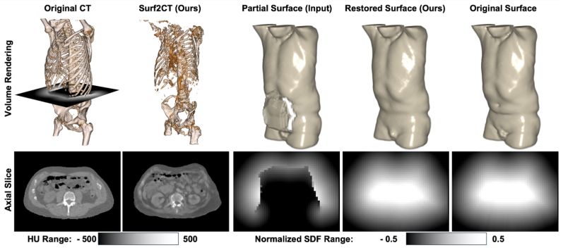
The load-bearing mechanism is a cascaded sequence of three flow-matching generative models, each a 3D-adapted convolutional network built on the EDM2 image-diffusion backbone and trained with a conditional optimal-transport flow-matching objective. Flow matching transports Gaussian noise to data by learning a velocity field for an ordinary differential equation, and the paper uses it three times: to estimate a complete signed distance function (SDF) from a partial scan plus demographics, to synthesize an 8 mm coarse CT from that SDF, and to generate high-resolution 2 mm CT patches conditioned on the upsampled coarse volume, positional encodings, and demographics. The SDF is the interface that turns mesh-like surface geometry into a volumetric condition usable by the CT generators, and the patch-wise third stage makes full-volume high-resolution synthesis computationally feasible.
What would settle it
Take subjects who have both a consumer depth-camera surface scan and a CT acquired within a few days, run Surf2CT on the scan, and compare surface-completion Chamfer distance and organ volumes to the CT: if errors move far outside the reported ranges (Chamfer distance near 2.7 mm, mean organ differences mostly within roughly 5 percent), the surface-to-CT mapping does not transfer to real acquisition conditions.
Extended reading notes
Core claim
On the paper's own terms, the discovery is that volumetric internal anatomy is sufficiently constrained by external body shape plus demographics that a generative model can produce realistic CT-like images without any internal imaging. Surf2CT implements this as a three-stage conditional flow-matching pipeline: a signed distance function (SDF) is completed from a partial frontal surface scan, a low-resolution CT volume is generated from that SDF and the demographic vector, and a patch-wise super-resolution stage upsamples it to 2 mm isotropic voxels. Evaluated on 700 paired surface-CT cases drawn from roughly 3,200 training CTs, the framework yields mean organ-volume differences from -11.1 percent to +4.4 percent, muscle and fat volume correlations with ground truth between R2 = 0.67 and 0.96, lung-localization bias of -2.5 mm, and surface-completion Chamfer distance improving from 521.8 mm to 2.7 mm.
Load-bearing premise
The whole pipeline is trained and tested on partial surfaces made by deleting the posterior half of skin meshes extracted from the ground-truth CTs, so the model assumes real-world depth scans will look like those clean frontal halves; real consumer scanners add noise, holes, and pose shifts that are absent from training, and the paper itself flags that deployment gap.
Editorial extensions
If this is right
- A partial frontal surface scan plus age, sex, height, and weight is enough input to synthesize a full 2 mm isotropic CT-like torso volume.
- Body-composition estimates (skeletal muscle, subcutaneous fat, visceral fat) track CT-derived values with R2 between 0.67 and 0.96, supporting radiation-free body-composition screening.
- Mean organ-volume errors stay mostly within about 5 percent (range -11.1 to +4.4 percent), so the volumes are useful for gross anatomical assessment but not voxel-level diagnosis.
- Surface completion reduces Chamfer distance from about 522 mm to 2.7 mm and raises surface IoU to 0.98, meaning the first stage turns unusable partial geometry into a reliable condition for CT synthesis.
- Lung localization bias of -2.5 mm indicates generated volumes preserve coarse anatomical landmarks, which matters for planning and navigation tasks.
Reading between the lines
- If applied to genuine consumer depth scans, performance will likely degrade; a natural extension is to train or fine-tune with synthetic noise, holes, and pose augmentation so that the distribution gap is closed.
- The low per-organ R2 values (heart 0.04-0.12, lung 0.01-0.06) suggest the model captures average anatomy better than subject-specific organ idiosyncrasies; the realistic use may be as a patient-specific prior or screening flag rather than a CT replacement.
- Because training data come largely from oncology CTs, the generated anatomy reflects that distribution; large discrepancies between predicted and actual volumes could be explored as an automated anomaly-screening signal, an idea the paper itself hints at.
- The same cascaded conditioning idea could extend beyond CT to MRI or PET synthesis, or accept additional inputs such as disease labels or text, to tighten the surface-to-volume mapping.
Editorial analysis
A structured set of objections, weighed in public.
Referee Report
Summary. Surf2CT proposes a three-stage cascaded flow-matching pipeline for synthesizing torso 3D CT volumes from a partial external body surface and demographic data (age, sex, height, weight). Stage 1 performs surface completion by estimating a signed distance function from a frontal-only partial surface; Stage 2 generates a coarse 8 mm CT volume conditioned on the completed SDF and demographics; Stage 3 refines the coarse volume patch-wise to 2 mm resolution. The models are trained on 3,198 torso CT scans from MGH and AutoPET and evaluated on 700 held-out paired surface-CT cases. The reported results include body-composition correlations (R2 0.74-0.96), organ-volume mean percentage differences within roughly ±11%, lung-localization bias of -2.5 mm, and surface-completion Chamfer distance improvement from 521.8 mm to 2.7 mm. The paper claims this is the first framework to synthesize clinically relevant, high-fidelity CT volumes from external surface data alone and discusses applications in preventive medicine and personalized assessment.
Significance. If the central claim held, this would be a notable contribution: non-invasive generation of internal anatomy from external scans could enable screening and monitoring without ionizing radiation. The manuscript has clear strengths: a large training dataset, a substantial held-out evaluation cohort (700 cases), a nontrivial three-stage generative pipeline, and quantitative evaluation of surface completion, body composition, and organ volumes. The surface-completion results in particular are impressive and well quantified. However, the evidence does not currently support the paper's stronger claims of 'strong anatomical fidelity' and 'personalized clinical assessments' of organ dimensions, because organ-level correlations with ground truth are near zero and the input surface is derived from the target CT rather than a real scanner. These issues are internal to the evaluation and would need to be addressed or substantially qualified before the paper can be recommended for publication.
major comments (4)
- [Table 3 and Appendix B] The organ-volume evaluation does not support the claim of subject-specific anatomical fidelity. Male lung R2 = 0.06, female lung R2 = 0.01, heart R2 = 0.12/0.11, and female kidney R2 = 0.04 indicate essentially no correlation with ground-truth organ volumes for the paired individual. The reported standard deviations reinforce this: generated male heart SD is 84.6 mL versus 133.2 mL in ground truth, and generated male lung SD is 439.8 mL versus 727.2 mL, showing marked variance compression. Small mean percentage differences are consistent with a model that outputs demographic-conditioned averages, so they cannot by themselves establish high-fidelity or personalized synthesis. This directly undercuts the abstract's claims of 'strong anatomical fidelity' and 'personalized clinical assessments' of organ dimensions.
- [Section 4, Dataset and Preprocessing; Section 6] The partial surface input is generated by removing the posterior half of the skin mesh derived from the ground-truth CT of the same subject. This makes the evaluation self-referential with respect to the surface-acquisition modality: real-world depth scans exhibit noise, holes, pose differences, and calibration artifacts that are not represented in training. The paper acknowledges this gap in Section 6, but the central claim of synthesizing CT 'from external surface scans' is not tested under realistic acquisition conditions. The authors should either evaluate on real surface scans or substantially qualify the claim to CT-derived partial surfaces.
- [Section 5 (Evaluation Setting and Results)] No quantitative comparison to any baseline method is provided. The paper claims to be the first approach of its kind, but it does not compare against even simple alternatives such as demographic-only conditional mean volumes, direct regression from SDF to CT, shape-only generation without demographics, or an existing digital-phantom method like XCAT or BOSS. Without such baselines, the contribution of the cascaded design and the conditioning variables cannot be isolated. Adding at least one or two baselines, even simplified ones, is necessary to support the claimed novelty and advantage.
- [Section 5, Organ-wise Volume Evaluation] The text states that lung-volume correlations were 'notably low' and attributes this to 'high inter-individual anatomical variability,' but this explanation is speculative and not tested. More importantly, the same paragraph earlier describes the organ results as demonstrating 'anatomically plausible organ volumes' and 'better performance in female subjects,' despite the female lung R2 of 0.01. The interpretation should be brought in line with the reported statistics: for the organs most affected by respiratory state and internal anatomy, the model currently provides population-level averages rather than subject-specific estimates.
minor comments (5)
- [Section 4, Dataset and Preprocessing] The description of how fpartial is constructed is imprecise: Section 3 says regions are 'selectively removed' or 'filled with heuristic guesses,' while Section 4 states the posterior half is removed. Please state the exact partial-surface generation procedure unambiguously in the method section.
- [Tables 2 and 3] The column header 'Slope (R2)' is unclear because it does not specify whether the slope is the ordinary least-squares slope of the regression of generated on original values, and whether R2 is the coefficient of determination of that regression. Please define these in the table captions.
- [Section 4, Implementation] The description of the network architecture is quite brief: it references a 3D-adapted EDM-img512-xxs backbone but does not specify the number of resolution levels, channel widths, or attention configurations used in the adaptation. More detail would improve reproducibility.
- [Throughout] There are several typographical and formatting issues, including 'V olume' in Table 2, 'Gflop sper step', and inconsistent use of 'Surf2CT' versus 'Surf2CT' in the text. These should be cleaned up.
- [Section 1, Introduction] The statement that this is 'the first approach capable of generating realistic volumetric internal anatomy images solely based on external body shape and demographics' is a strong claim that would benefit from a more thorough literature search and a precise definition of 'realistic' as used in the evaluation.
Circularity Check
No circular derivation: all three stages are trained on paired data and evaluated on held-out cases; the CT-derived partial surface is a generalization limitation, not a circular reduction.
full rationale
Surf2CT is a three-stage conditional generative pipeline. Each stage is trained with a flow-matching loss on paired data (partial SDF -> full SDF -> low-resolution CT -> high-resolution CT) and evaluated on reserved held-out cases. No stage's output is a fitted parameter that is then reported as a prediction: organ volumes, body composition, lung localization, and surface completion are all computed from generated volumes and compared to ground truth on held-out subjects. The only potentially self-referential element is that the partial surface input is manufactured by removing the posterior half of the mesh extracted from the ground-truth CT (Section 4, Dataset and Preprocessing). This means evaluation is conducted under an idealized input distribution and does not test real-world scanner noise; however, this is a dataset-construction and generalization limitation, not a derivational circularity. The model does not see the target at inference, and the output is not defined in terms of the target. The paper itself flags the real-world distribution shift in Section 6. There are no load-bearing self-citations: the cited methods (EDM2, TotalSegmentator, nnUnet2, AutoPET, flow matching literature) are external and are not invoked to justify the central claim. The low R2 values for organ volumes (Appendix B) are an empirical validity finding, not evidence of circularity; they indicate that the model may produce demographic-typical averages, but that is a performance limitation, not a reduction of the prediction to its inputs by construction. Therefore the derivation chain is self-contained and no circular step is present.
Assumptions & free parameters
free parameters (6)
- Learned weights, Stage 1 surface completion (v1_theta) =
~80.68M parameters
- Learned weights, Stage 2 coarse CT synthesis (v2_theta) =
~80.68M parameters
- Learned weights, Stage 3 super-resolution (v3_theta) =
~1.89M parameters
- Sampling hyperparameters (sigma_max, rho, ODE steps) =
80, 7, 200
- Patch size for Stage 3 =
56x56x88 voxels
- CT intensity normalization range =
-500 to 500 HU
assumptions (5)
- standard math The linear interpolation flow matching path with conditional optimal transport is a valid generative objective (Eqs. 4, 8, 13).
- domain assumption TotalSegmentator correctly identifies skin boundary, organs, and body composition on both real and synthetic CT.
- ad hoc to paper The frontal-only partial surface (posterior half removed from the CT-derived skin mesh) approximates real single-view capture.
- domain assumption A surface plus demographics carries enough information to predict the evaluated anatomical quantities.
- domain assumption Torso cropping from clavicle to sacrum yields a consistent coordinate system across subjects.
Cite this review
Pith. "Pith review of Surf2CT: Cascaded 3D Flow Matching Models for Torso 3D CT Synthesis from Skin Surface." pith.science (2026). https://pith.science/paper/SBD473PI
@misc{pith2026250522511,
  author       = {Pith},
  title        = {Pith review of: Surf2CT: Cascaded 3D Flow Matching Models for Torso 3D CT Synthesis from Skin Surface},
  year         = {2026},
  howpublished = {\url{https://pith.science/paper/SBD473PI}},
  note         = {Machine review of arXiv:2505.22511}
}
read the original abstract
We present Surf2CT, a novel cascaded flow matching framework that synthesizes full 3D computed tomography (CT) volumes of the human torso from external surface scans and simple demographic data (age, sex, height, weight). This is the first approach capable of generating realistic volumetric internal anatomy images solely based on external body shape and demographics, without any internal imaging. Surf2CT proceeds through three sequential stages: (1) Surface Completion, reconstructing a complete signed distance function (SDF) from partial torso scans using conditional 3D flow matching; (2) Coarse CT Synthesis, generating a low-resolution CT volume from the completed SDF and demographic information; and (3) CT Super-Resolution, refining the coarse volume into a high-resolution CT via a patch-wise conditional flow model. Each stage utilizes a 3D-adapted EDM2 backbone trained via flow matching. We trained our model on a combined dataset of 3,198 torso CT scans (approximately 1.13 million axial slices) sourced from Massachusetts General Hospital (MGH) and the AutoPET challenge. Evaluation on 700 paired torso surface-CT cases demonstrated strong anatomical fidelity: organ volumes exhibited small mean percentage differences (range from -11.1% to 4.4%), and muscle/fat body composition metrics matched ground truth with strong correlation (range from 0.67 to 0.96). Lung localization had minimal bias (mean difference -2.5 mm), and surface completion significantly improved metrics (Chamfer distance: from 521.8 mm to 2.7 mm; Intersection-over-Union: from 0.87 to 0.98). Surf2CT establishes a new paradigm for non-invasive internal anatomical imaging using only external data, opening opportunities for home-based healthcare, preventive medicine, and personalized clinical assessments without the risks associated with conventional imaging techniques.
Figures


Figures from the paper (6 more)


Reference graph
Works this paper leans on
-
[1]
Rebecca Smith-Bindman, Jafi Lipson, Ralph Marcus, Kwang-Pyo Kim, Mahadevappa Mahesh, Robert Gould, Amy Berrington De González, and Diana L Miglioretti. Radiation dose associated with common computed tomography examinations and the associated lifetime attributable risk of cancer.Archives of internal medicine, 169(22):2078–2086, 2009
work page 2009
-
[2]
3d body scanning and healthcare applications.Computer, 40(7):28–34, 2007
Philip Treleaven and Jonathan Wells. 3d body scanning and healthcare applications.Computer, 40(7):28–34, 2007
work page 2007
-
[3]
Yoon Seong Lee, Namki Hong, Joseph Nathanael Witanto, Ye Ra Choi, Junghoan Park, Pierre Decazes, Florian Eude, Chang Oh Kim, Hyeon Chang Kim, Jin Mo Goo, et al. Deep neural network for automatic volumetric segmentation of whole-body ct images for body composition assessment.Clinical Nutrition, 40(8):5038–5046, 2021
work page 2021
-
[4]
Jean-Pierre Després, Isabelle Lemieux, Jean Bergeron, Philippe Pibarot, Patrick Mathieu, Eric Larose, Josep Rodés-Cabau, Olivier F Bertrand, and Paul Poirier. Abdominal obesity and the metabolic syndrome: contribution to global cardiometabolic risk.Arteriosclerosis, thrombosis, and vascular biology, 28(6):1039–1049, 2008
work page 2008
-
[5]
Sarcopenia.The Lancet, 393(10191):2636–2646, 2019
Alfonso J Cruz-Jentoft and Avan A Sayer. Sarcopenia.The Lancet, 393(10191):2636–2646, 2019
work page 2019
-
[6]
Lisa Asciak, Justicia Kyeremeh, Xichun Luo, Asimina Kazakidi, Patricia Connolly, Frederic Picard, Kevin O’Neill, Sotirios A Tsaftaris, Grant D Stewart, and Wenmiao Shu. Digital twin assisted surgery, concept, opportunities, and challenges.npj Digital Medicine, 8(1):32, 2025
work page 2025
-
[7]
Generative modeling by estimating gradients of the data distribution.Advances in neural information processing systems, 32, 2019
Yang Song and Stefano Ermon. Generative modeling by estimating gradients of the data distribution.Advances in neural information processing systems, 32, 2019
2019
-
[8]
Flow matching for generative modeling.arXiv preprint arXiv:2210.02747, 2022
Yaron Lipman, Ricky TQ Chen, Heli Ben-Hamu, Maximilian Nickel, and Matt Le. Flow matching for generative modeling.arXiv preprint arXiv:2210.02747, 2022
arXiv 2022
Show all 27 references
-
[9]
Lion: Latent point diffusion models for 3d shape generation.Advances in Neural Information Processing Systems, 35:10021–10039, 2022
Arash Vahdat, Francis Williams, Zan Gojcic, Or Litany, Sanja Fidler, Karsten Kreis, et al. Lion: Latent point diffusion models for 3d shape generation.Advances in Neural Information Processing Systems, 35:10021–10039, 2022
2022
-
[10]
Sc-diff: 3d shape completion with latent diffusion models.arXiv preprint arXiv:2403.12470, 2024
Juan D Galvis, Xingxing Zuo, Simon Schaefer, and Stefan Leutengger. Sc-diff: 3d shape completion with latent diffusion models.arXiv preprint arXiv:2403.12470, 2024
2024 arXiv
-
[11]
Maisi: Medical ai for synthetic imaging
Pengfei Guo, Can Zhao, Dong Yang, Ziyue Xu, Vishwesh Nath, Yucheng Tang, Benjamin Simon, Mason Belue, Stephanie Harmon, Baris Turkbey, et al. Maisi: Medical ai for synthetic imaging. In2025 IEEE/CVF Winter Conference on Applications of Computer Vision (WACV), pages 4430–4441. ...
2025
-
[12]
Patient-specific reconstruction of volumetric computed tomography images from a single projection view via deep learning.Nature biomedical engineering, 3(11):880–888, 2019
Liyue Shen, Wei Zhao, and Lei Xing. Patient-specific reconstruction of volumetric computed tomography images from a single projection view via deep learning.Nature biomedical engineering, 3(11):880–888, 2019
2019
-
[13]
Synthetic ct generation from mri using 3d transformer-based denoising diffusion model.Medical Physics, 51(4):2538–2548, 2024
Shaoyan Pan, Elham Abouei, Jacob Wynne, Chih-Wei Chang, Tonghe Wang, Richard LJ Qiu, Yuheng Li, Junbo Peng, Justin Roper, Pretesh Patel, et al. Synthetic ct generation from mri using 3d transformer-based denoising diffusion model.Medical Physics, 51(4):2538–2548, 2024
2024
-
[14]
4d xcat phantom for multimodality imaging research.Medical physics, 37(9):4902–4915, 2010
W Paul Segars, G Sturgeon, S Mendonca, Jason Grimes, and Benjamin MW Tsui. 4d xcat phantom for multimodality imaging research.Medical physics, 37(9):4902–4915, 2010
2010
-
[15]
Boss: Bones, organs and skin shape model.Computers in Biology and Medicine, 165:107383, 2023
Karthik Shetty, Annette Birkhold, Srikrishna Jaganathan, Norbert Strobel, Bernhard Egger, Markus Kowarschik, and Andreas Maier. Boss: Bones, organs and skin shape model.Computers in Biology and Medicine, 165:107383, 2023
2023
-
[16]
X2ct-gan: reconstructing ct from biplanar x-rays with generative adversarial networks
Xingde Ying, Heng Guo, Kai Ma, Jian Wu, Zhengxin Weng, and Yefeng Zheng. X2ct-gan: reconstructing ct from biplanar x-rays with generative adversarial networks. InProceedings of the IEEE/CVF conference on computer vision and pattern recognition, pages 10619–10628, 2019. 10
2019
-
[17]
Difr3ct: Latent diffusion for probabilistic 3d ct reconstruc- tion from few planar x-rays.arXiv preprint arXiv:2408.15118, 2024
Yiran Sun, Hana Baroudi, Tucker Netherton, Laurence Court, Osama Mawlawi, Ashok Veer- araghavan, and Guha Balakrishnan. Difr3ct: Latent diffusion for probabilistic 3d ct reconstruc- tion from few planar x-rays.arXiv preprint arXiv:2408.15118, 2024
2024 arXiv
-
[18]
A geometry-informed deep learning framework for ultra-sparse 3d tomographic image reconstruction.Computers in Biology and Medicine, 148:105710, 2022
Liyue Shen, Wei Zhao, Dante Capaldi, John Pauly, and Lei Xing. A geometry-informed deep learning framework for ultra-sparse 3d tomographic image reconstruction.Computers in Biology and Medicine, 148:105710, 2022
2022
-
[19]
seg2med: a segmentation-based medical image generation framework using denoising diffusion probabilistic models.arXiv preprint arXiv:2504.09182, 2025
Zeyu Yang, Zhilin Chen, Yipeng Sun, Anika Strittmatter, Anish Raj, Ahmad Allababidi, Johann S Rink, and Frank G Zöllner. seg2med: a segmentation-based medical image generation framework using denoising diffusion probabilistic models.arXiv preprint arXiv:2504.09182, 2025
2025 arXiv
-
[20]
Diffcomplete: Diffusion-based generative 3d shape completion.Advances in neural information processing systems, 36:75951–75966, 2023
Ruihang Chu, Enze Xie, Shentong Mo, Zhenguo Li, Matthias Nießner, Chi-Wing Fu, and Jiaya Jia. Diffcomplete: Diffusion-based generative 3d shape completion.Advances in neural information processing systems, 36:75951–75966, 2023
2023
-
[21]
Patch diffusion: Faster and more data-efficient training of diffusion models.Advances in neural information processing systems, 36:72137–72154, 2023
Zhendong Wang, Yifan Jiang, Huangjie Zheng, Peihao Wang, Pengcheng He, Zhangyang Wang, Weizhu Chen, Mingyuan Zhou, et al. Patch diffusion: Faster and more data-efficient training of diffusion models.Advances in neural information processing systems, 36:72137–72154, 2023
2023
-
[22]
Analyzing and improving the training dynamics of diffusion models
Tero Karras, Miika Aittala, Jaakko Lehtinen, Janne Hellsten, Timo Aila, and Samuli Laine. Analyzing and improving the training dynamics of diffusion models. InProceedings of the IEEE/CVF Conference on Computer Vision and Pattern Recognition, pages 24174–24184, 2024
2024
-
[23]
Yaron Lipman, Marton Havasi, Peter Holderrieth, Neta Shaul, Matt Le, Brian Karrer, Ricky T. Q. Chen, David Lopez-Paz, Heli Ben-Hamu, and Itai Gat. Flow matching guide and code,
-
[24]
A whole-body fdg-pet/ct dataset with manually annotated tumor lesions.Scientific Data, 9(1):601, 2022
Sergios Gatidis, Tobias Hepp, Marcel Früh, Christian La Fougère, Konstantin Nikolaou, Christina Pfannenberg, Bernhard Schölkopf, Thomas Küstner, Clemens Cyran, and Daniel Rubin. A whole-body fdg-pet/ct dataset with manually annotated tumor lesions.Scientific Data, 9(1):601, 2022
2022
-
[25]
Totalseg- mentator: robust segmentation of 104 anatomic structures in ct images.Radiology: Artificial Intelligence, 5(5):e230024, 2023
Jakob Wasserthal, Hanns-Christian Breit, Manfred T Meyer, Maurice Pradella, Daniel Hinck, Alexander W Sauter, Tobias Heye, Daniel T Boll, Joshy Cyriac, Shan Yang, et al. Totalseg- mentator: robust segmentation of 104 anatomic structures in ct images.Radiology: Artificial Intel...
2023
-
[26]
Automated muscle and fat segmentation in computed tomography for comprehensive body composition analysis
Yaqian Chen, Hanxue Gu, Yuwen Chen, Jicheng Yang, Haoyu Dong, Joseph Y Cao, Adrian Camarena, Christopher Mantyh, Roy Colglazier, and Maciej A Mazurowski. Automated muscle and fat segmentation in computed tomography for comprehensive body composition analysis. arXiv preprint ar...
2025
-
[2024]
URLhttps://arxiv.org/abs/2412.06264
Reviewed August 7, 2026 · model on record in the stance chip above.
Discussion (0). Continue with ORCID to comment.