REVIEW 4 major objections 6 minor 51 references
Benchmarking and Explaining Deep Learning Cortical Lesion MRI Segmentation in Multiple Sclerosis
T0 review · 4 major / 6 minor · reviewed 2026-08-06 · deepseek-v4-flash
Pith's one-line read A standard nnU-Net with a blob-aware loss detects MS cortical lesions on routine 3T MRI, with F1 of 0.64 in-domain and 0.50 on a held-out scanner vendor.
desk verdict A genuinely useful multi-center benchmark and public model for cortical lesion segmentation, but the headline OOD number is confounded by annotation and cohort differences, so read the generalization claim with caution. read the letter →
The pith
A machine-rendered reading of the paper's core claim, the machinery that carries it, and where it could break.
The reading
What carries the argument
The carrying mechanism is the nnU-Net self-configuring segmentation framework, a 3D U-Net pipeline that automatically sets preprocessing, resolution, patch size, augmentation, ensembling, and post-processing from the data, combined with the Blob BCE + Dice loss, an instance-aware loss that reweights small connected components so that tiny lesions contribute as much as large ones. The benchmark design is the second mechanism: a stratified split of three in-domain sites into train and test sets, plus a deliberately out-of-domain fourth site with a different scanner vendor and a mix of MS and MS-mimic subjects, which gives the generalization numbers their meaning. The error typology, which classifies true-positive, false-positive, and false-negative lesions by lesion type and volume, and the bottleneck-feature analysis are the instruments that connect the model's behavior to clinically interpretable causes.
What would settle it
Have a single expert panel re-annotate a matched subset of scans from the in-domain sites and the out-of-domain site under one common protocol, then recompute per-site F1: if the out-of-domain F1 of about 0.50 rises toward the in-domain 0.64, the scanner-generalization claim is largely label heterogeneity, while if it stays near 0.50, true domain shift is confirmed. A complementary check is measuring inter-rater agreement between in-domain and out-of-domain annotators on identical scans, since model performance at or above that agreement means the network is near the annotation ceiling.
Extended reading notes
Core claim
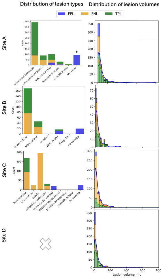
On its own terms, the paper's central discovery is that a vanilla 3D U-Net configured by nnU-Net, trained on MP2RAGE and MPRAGE data with the Blob BCE + Dice loss, segments cortical lesions across sites and protocols well enough to act as a practical baseline: lesion-detection F1-score of 0.641 in-domain and 0.502 on an out-of-domain site that differs in scanner manufacturer, acquisition protocol, patient cohort, and annotation workflow, including MS-mimic patients. The comparative experiments show that larger residual-encoder variants are not significantly better, that U-Mamba variants failed to converge, and that upsampling all data to 0.5 mm isotropic raises recall but collapses precision, cutting F1 by roughly 45 percent, so the authors reject it. Error analysis attributes most missed lesions to the smallest leukocortical lesions, most false positives to juxtacortical white matter lesions that raters themselves struggle to classify, and the worst performance to 7T subpial lesions that lower-field T1-weighted imaging cannot reliably show. Bottleneck-feature projections reveal that the network encodes site- and modality-specific information alongside task-relevant features, and the authors conclude that a standard nnU-Net with task-specific loss adjustments effectively segments cortical lesions across centers and protocols, with the models released for public use.
Load-bearing premise
The benchmark treats the manual lesion masks from the four sites as interchangeable ground truth even though each site used different raters, different source sequences, and different lesion-inclusion criteria, so if those label differences are large, the reported site-to-site gaps and the out-of-domain drop may reflect annotation mismatch rather than scanner or protocol shift.
Editorial extensions
If this is right
- The released vanilla nnU-Net with Blob loss can be applied directly to new 3T MPRAGE/MP2RAGE data as an off-the-shelf cortical lesion detection baseline, with expected performance near the reported in-domain F1 of 0.64.
- Clinical sites gain nothing from the largest architectures: since the vanilla model matches or beats the residual-encoder M, L, and XL variants, lighter models with lower memory and energy costs are the justified choice.
- The Blob BCE + Dice loss gives a modest but consistent precision gain, most notable on out-of-domain data, making it the recommended default for small-lesion segmentation tasks.
- Upsampling heterogeneous data to the finest available resolution is counterproductive for cortical lesion segmentation, since the false-positive explosion outweighs the recall gain.
- Because missed lesions are mostly small leukocortical ones and false positives overlap juxtacortical white matter lesions, further progress depends on clearer annotation guidelines and possibly soft labels, not on bigger networks.
Reading between the lines
- If the out-of-domain site's scans were re-annotated by the in-domain sites' raters, the 0.50 out-of-domain F1 could shift measurably in either direction; part of what the paper attributes to scanner-shift generalization may be rater and lesion-definition shift, so the true cross-vendor gap is not yet pinned down.
- Because many false positives fall inside white-matter lesion masks, the measured F1 likely understates clinical usefulness: some detections counted as wrong are real lesions that the ground truth did not label, which is exactly what a second-opinion reading tool would want to surface.
- The sharp site and modality clustering in the bottleneck features suggests that lightweight domain adaptation, such as feature-statistics alignment or test-time adaptation, could recover a large share of the out-of-domain gap without any new annotated data.
- The paper's own table reports no recall for the MS-mimic subjects in the out-of-domain set, so the headline 0.50 F1 blends detection on MS patients with false-positive control on mimics; a site planning deployment should read the MS-only and mimic-only numbers separately.
Editorial analysis
A structured set of objections, weighed in public.
Referee Report
Summary. The paper presents a multi-center benchmark of deep learning cortical lesion (CL) segmentation in multiple sclerosis, built on 656 scans from four institutions with expert-consensus annotations. The authors systematically compare nnU-Net architecture variants, loss functions (standard BCE+Dice vs. Blob BCE+Dice), and input resampling strategies, report that the vanilla nnU-Net with Blob loss performs best, and characterize its per-site, per-modality, and out-of-domain behavior. They also analyze lesion-level errors and bottleneck features for interpretability and release the implementation and model weights. The central claim is that a standard nnU-Net with task-specific adjustments such as Blob BCE+Dice loss effectively segments cortical lesions across different imaging centers and MRI protocols, supported by reported in-domain F1 of 0.64 and out-of-domain F1 of 0.50.
Significance. The study is valuable as a large, multi-center benchmark for CL segmentation, a clinically relevant but technically challenging task. Its strengths include the size and heterogeneity of the dataset (656 scans, 3T/7T, multiple vendors and protocols), the use of standard errors and FDR-corrected statistical tests, the inclusion of negative results (upsampling degrades precision, U-Mamba fails to converge), and the public release of models and code. If the out-of-domain claim were clean, the paper would provide a practical baseline for clinical deployment. However, the central generalization claim rests on a confounded out-of-domain comparison and on model selection performed on the test set, so the significance is conditional on addressing these issues.
major comments (4)
- [Section 3.1.2, Table 5, Section 6] The out-of-domain generalization claim is confounded. Site D differs from sites A-C not only in scanner vendor but also in annotation protocol (three raters, detection on 0.7 mm DIR plus MP2RAGE, manual delineation by a trained neuroscientist, as described in Section 3.1.1), cohort composition (40 MS-mimic patients), preprocessing (registration to 3D EPI, skull-stripping with HD-BET to MPRAGE plus dilation), and the set of available modalities. Therefore, the reported out-of-domain F1 of 0.50 in Table 5 cannot be attributed to scanner shift alone. The abstract and conclusions should either reframe this as evaluation on a held-out site with multiple simultaneous differences, or the authors should isolate the scanner effect, for example by analyzing the MS-only subset of site D or by having an independent annotation protocol applied to a subset of site D data. As written, the central claim in Section 6 that the model 'effectively segments cortical lesions across different imaging centers and MRI protocols' is not fully supported.
- [Sections 4.1 and 4.2, Tables 3 and 4] The selection of the 'best model' (Vanilla nnU-Net with Blob loss) is based on the same Test-in set that is later used to report final performance. The architecture and loss comparisons in Tables 3 and 4 are performed on Test-in, and the model with the highest metrics on that set is then analyzed in detail. No separate validation split or nested cross-validation is used. This procedure introduces an optimistic bias in the reported in-domain F1, DSC, and related metrics. The authors should disclose this selection-on-test issue explicitly and, if possible, re-evaluate the model selection with a proper validation partition or cross-validation. At minimum, the reported Test-in numbers should be labeled as model-selected estimates rather than unbiased performance estimates.
- [Section 4.2.1 and Section 5.2, Table 5] The cross-site performance comparison is interpreted primarily as a property of the model, but the reference masks from different sites were generated under different annotation protocols, rater expertise, and lesion criteria. For example, the much larger number of false-negative lesions per scan in site D (6.5, versus roughly 3 in sites A and B) and the very high FNL count in site C 7T (55.5) may reflect differences in annotation recall or lesion definition rather than model failure. The discussion in Section 5.2 should temper claims about site-specific model limitations and should explicitly acknowledge that without a common reference standard, the observed gaps are not directly attributable to the segmentation model. A quantitative analysis of inter-rater or inter-protocol variability would strengthen this point.
- [Section 4.1.2 and Table 4] The benefit of Blob BCE+Dice loss is presented as a positive finding, but the only statistically significant improvement is out-of-domain precision (p<0.05), while in-domain improvements in nDSC, DSC, F1, and precision are not reported as significant. Given the multiple comparisons performed, the evidence for the loss-function advantage is weaker than the text and abstract imply. The authors should state clearly which comparisons reached significance after FDR correction and avoid implying a general improvement from Blob loss based on non-significant trends.
minor comments (6)
- [Section 3.2, references] The text 'We employed the nnU-Net framework [24, 27]' cites reference [24], which is the HD-BET brain extraction paper, not the nnU-Net paper. The nnU-Net method paper is reference [22] (Isensee et al., 2021). Please correct the citation.
- [Table 2] The column header 'A B D C' appears inconsistent with the row totals and with the text stating that site D is used for out-of-domain testing. The table is difficult to parse, especially the rows marked with a checkmark for 7T and the MS-mimic rows. Please reformat the table so that the site columns, the 7T indicator, and the MS-mimic indicator are unambiguous.
- [Section 3.3.1, Eq. (1)] The definition of h in the nDSC formula is ambiguous: 'h is the ratio between the positive and the negative classes in the ground truth scan segmentation' does not specify which class is in the numerator. Please clarify whether h = N_negative / N_positive or the inverse, and define all symbols explicitly.
- [Table 5] In the MS-mimic rows, F1-score is listed but Recall is shown as '-'. Please explain why recall is undefined for these subjects and how F1 is computed without it, or add a footnote clarifying that the MS-mimic patients have no cortical lesion ground truth and that the reported value is precision under a different definition.
- [Section 4.2.2] The sentence 'the median FNL volume is 15mL' is implausible for cortical lesions, which are typically on the order of cubic millimeters. If the value is in mm^3, please correct the unit; if it is truly 15 mL, please explain how such large lesions are classified as cortical.
- [Throughout] There are several minor language issues, including 'widespread in adoption' in the Contributions section, 'U-Mamba architectures could not finally be included' in Section 4.1.1, and 'leuko-/intra-cortical' hyphenation inconsistencies. A careful proofreading pass is recommended.
Circularity Check
No significant circularity: the central claim is supported by held-out training/test splits and an untouched OOD site.
full rationale
This is an empirical benchmark paper, not a derivation, so the circularity patterns that involve equations reducing to their inputs do not apply. The nnU-Net variants and Blob BCE + Dice loss are trained on a Train split and evaluated on a held-out Test-in split plus an untouched site D (Test-out); the reported F1 values in Tables 3-5 are predictions on data not used to fit the model. The main conclusion that a vanilla nnU-Net with Blob loss is a practical CL segmentation baseline follows directly from these held-out numbers. Self-citations such as [32] for nDSC and [48] for PSU provide explicit metric and uncertainty definitions rather than load-bearing evidence for the segmentation result. The choice of the vanilla architecture based on Test-in metrics is a model-selection issue that can inflate in-domain performance, and the site D OOD comparison is confounded by annotation protocol, cohort composition, and preprocessing differences, but these are statistical-validity and generalizability concerns, not circular reductions where an output is equivalent to an input by construction. No step in the paper's chain equates a claimed prediction with a fitted parameter or with a self-citation-derived postulate.
Assumptions & free parameters
free parameters (3)
- Adam learning rate =
3e-4
- Upsampling target resolution =
0.5 mm isotropic
- Stratification bins for data split =
5 quantile bins
assumptions (3)
- domain assumption Manual annotations across the four sites are comparable enough to serve as benchmark ground truth
- domain assumption Site D is a valid out-of-distribution test for scanner generalization
- domain assumption Multiple scans per patient (timepoints and modalities) can be treated as independent samples for statistical tests
Cite this review
Pith. "Pith review of Benchmarking and Explaining Deep Learning Cortical Lesion MRI Segmentation in Multiple Sclerosis." pith.science (2026). https://pith.science/paper/SSHTLKQY
@misc{pith2026250712092,
  author       = {Pith},
  title        = {Pith review of: Benchmarking and Explaining Deep Learning Cortical Lesion MRI Segmentation in Multiple Sclerosis},
  year         = {2026},
  howpublished = {\url{https://pith.science/paper/SSHTLKQY}},
  note         = {Machine review of arXiv:2507.12092}
}
read the original abstract
Cortical lesions (CLs) have emerged as valuable biomarkers in multiple sclerosis (MS), offering high diagnostic specificity and prognostic relevance. However, their routine clinical integration remains limited due to subtle magnetic resonance imaging (MRI) appearance, challenges in expert annotation, and a lack of standardized automated methods. We propose a comprehensive multi-centric benchmark of CL detection and segmentation in MRI. A total of 656 MRI scans, including clinical trial and research data from four institutions, were acquired at 3T and 7T using MP2RAGE and MPRAGE sequences with expert-consensus annotations. We rely on the self-configuring nnU-Net framework, designed for medical imaging segmentation, and propose adaptations tailored to the improved CL detection. We evaluated model generalization through out-of-distribution testing, demonstrating strong lesion detection capabilities with an F1-score of 0.64 and 0.5 in and out of the domain, respectively. We also analyze internal model features and model errors for a better understanding of AI decision-making. Our study examines how data variability, lesion ambiguity, and protocol differences impact model performance, offering future recommendations to address these barriers to clinical adoption. To reinforce the reproducibility, the implementation and models will be publicly accessible and ready to use at https://github.com/Medical-Image-Analysis-Laboratory/ and https://doi.org/10.5281/zenodo.15911797.
Figures


Figures from the paper (5 more)



Reference graph
Works this paper leans on
-
[1]
D. S. Reich, C. F. Lucchinetti, P. A. Calabresi, Multiple Scle- rosis, New England Journal of Medicine 378 (2018) 169–180. URL: http://www.nejm.org/doi/10.1056/NEJMra1401483. doi: 10. 1056/NEJMra1401483
-
[2]
E. Portaccio, M. Magyari, E. K. Havrdova, A. Ruet, B. Brochet, A. Scalfari, M. Di Filippo, C. Tur, X. Montalban, M. P. Amato, Multiple sclerosis: emerging epidemiological trends and redefining the clinical course, The Lancet Regional Health - Europe 44 (2024) 100977. URL: https://www.sciencedirect.com/science/article/ pii/S2666776224001443. doi:10.1016/j....
-
[3]
C. C. Hemond, R. Bakshi, Magnetic resonance imaging in multi- ple sclerosis, Cold Spring Harbor Perspectives in Medicine 8 (2018). doi:10.1101/cshperspect.a028969, publisher: Cold Spring Harbor Laboratory Press
-
[4]
M.Filippi, W.Brück, D.Chard, F.Fazekas, J.J.G.Geurts, C.Enzinger, S. Hametner, T. Kuhlmann, P. Preziosa, A. Rovira, K. Schmierer, C. Stadelmann, M. A. Rocca, Attendees of the Correlation between Pathological and MRI findings in MS workshop, Association between pathological and MRI findings in multiple sclerosis, The Lancet. Neu- rology 18 (2019) 198–210. ...
-
[5]
M. P. Wattjes, O. Ciccarelli, D. S. Reich, B. Banwell, N. de Stefano, C. Enzinger, F. Fazekas, M. Filippi, J. Frederiksen, C. Gasperini, Y. Ha- cohen, L. Kappos, D. K. B. Li, K. Mankad, X. Montalban, S. D. New- some, J. Oh, J. Palace, M. A. Rocca, J. Sastre-Garriga, M. Tintoré, A. Traboulsee, H. Vrenken, T. Yousry, F. Barkhof, A. Rovira, M. P. Wattjes, O....
work page 2021
-
[6]
H. Kolb, O. Al-Louzi, E. S. Beck, P. Sati, M. Absinta, D. S. Reich, From pathology to MRI and back: clinically relevant biomarkers of multiple sclerosis lesions, NeuroImage: Clinical (2022) 103194. URL: https://linkinghub.elsevier.com/retrieve/ pii/S2213158222002595. doi:10.1016/j.nicl.2022.103194
-
[7]
E. S. Beck, J. Maranzano, N. J. Luciano, P. Parvathaneni, S. Filip- pini, M. Morrison, D. J. Suto, T. Wu, P. van Gelderen, J. A. de Zwart, S. Antel, D. Fetco, J. Ohayon, F. Andrada, Y. Mina, C. Thomas, S. Ja- cobson, J. Duyn, I. Cortese, S. Narayanan, G. Nair, P. Sati, D. S. Reich, Cortical lesion hotspots and association of subpial lesions with disabil- ...
work page 2022
-
[8]
A. Cagol, R. Cortese, M. Barakovic, S. Schaedelin, E. Ruberte, M. Ab- sinta, F. Barkhof, M. Calabrese, M. Castellaro, O. Ciccarelli, S. Co- cozza, N. De Stefano, C. Enzinger, M. Filippi, M. Jurynczyk, P. Maggi, N. Mahmoudi, S. Messina, X. Montalban, J. Palace, G. Pontillo, A.- K. Probstel, M. A. Rocca, S. Ropele, A. Rovira, M. M. Schoonheim, P. Sowa, E. S...
Show all 51 references
-
[9]
Absinta, P
M. Absinta, P. Sati, F. Masuzzo, G. Nair, V. Sethi, H. Kolb, J. Ohayon, T. Wu, I. C. M. Cortese, D. S. Reich, Association of Chronic Active Multiple Sclerosis Lesions With Disability In Vivo, JAMA neurology 76 (2019) 1474–1483. doi:10.1001/jamaneurol.2019.2399
2019
-
[10]
Maggi, P
P. Maggi, P. Sati, G. Nair, I. C. M. Cortese, S. Jacobson, B. R. Smith, A. Nath, J. Ohayon, V. v. Pesch, G. Perrotta, C. Pot, M. Théaudin, V. Martinelli, R. Scotti, T. Wu, R. D. Pasquier, P. A. Calabresi, M. Fil- ippi, D. S. Reich, M. Absinta, Paramagnetic Rim Lesions are Spec...
2020 doi
-
[11]
P. Sati, J. Oh, R. T. Constable, N. Evangelou, C. R. G. Guttmann, R. G. Henry, E. C. Klawiter, C. Mainero, L. Massacesi, H. McFarland, F. Nel- son, D. Ontaneda, A. Rauscher, W. D. Rooney, A. P. R. Samaraweera, R. T. Shinohara, R. A. Sobel, A. J. Solomon, C. A. Treaba, J. Wuer-...
2016
-
[12]
E. S. Beck, W. A. Mullins, J. dos Santos Silva, S. Filippini, P. Parvatha- neni, J. Maranzano, M. Morrison, D. J. Suto, C. Donnay, H. Dieckhaus, N. J. Luciano, K. Sharma, M. I. Gaitán, J. Liu, J. A. de Zwart, P. van Gelderen, I. Cortese, S. Narayanan, J. H. Duyn, G. Nair, P. S...
2024 doi
-
[13]
A. J. Thompson, B. Banwell, F. Barkhof, W. M. Carroll, T. Coet- zee, G. Comi, J. Correale, F. Fazekas, M. Filippi, M. S. Freedman, K. Fujihara, S. Galetta, H. P. Hartung, L. Kappos, F. Lublin, R. A. Marrie, A. Miller, D. H. Miller, X. Montalbán, E. M. Mowry, P. S. Sørensen, M....
2018
-
[14]
F. L. Rosa, A. Abdulkadir, M. J. Fartaria, R. Rahmanzadeh, P. J. Lu, R. Galbusera, M. Barakovic, J. P. Thiran, C. Granziera, M. B. Cuadra, Multiple sclerosis cortical and WM lesion segmentation at 3T MRI: a deep learning method based on FLAIR and MP2RAGE, NeuroImage: Clinical ...
2020
-
[15]
Ronneberger, P
O. Ronneberger, P. Fischer, T. Brox, U-NET: Convolutional Networks 34 for Biomedical Image Segmentation, 2015. URL: https://doi.org/ 10.1007/978-3-319-24574-4_28. doi:10.1007/978-3-319-24574-4_ 28, publication Title: Lecture notes in computer science
2015 doi
-
[16]
Çiçek, A
O. Çiçek, A. Abdulkadir, S. S. Lienkamp, T. Brox, O. Ronneberger, 3D U-Net: Learning Dense Volumetric Segmentation from Sparse Annota- tion, in: S. Ourselin, L. Joskowicz, M. R. Sabuncu, G. Unal, W. Wells (Eds.), Medical Image Computing and Computer-Assisted Intervention – MIC...
2016 doi
-
[17]
Kober, C
T. Kober, C. Granziera, D. Ribes, P. Browaeys, M. Schluep, R. Meuli, R. Frackowiak, R. Gruetter, G. Krueger, MP2RAGE multiple sclerosis magnetic resonance imaging at 3 T, Investigative Radiology 47 (2012) 346–352. doi:10.1097/RLI.0b013e31824600e9
2012 doi
-
[18]
M. J. Fartaria, T. Kober, C. Granziera, M. B. Cuadra, Longitudinal analysis of white matter and cortical lesions in multiple sclero- sis, NeuroImage: Clinical 23 (2019) 101938. URL: https://www. sciencedirect.com/science/article/pii/S2213158219302888. doi:https://doi.org/10.10...
2019
-
[19]
E. Beck, P. Sati, V. Sethi, T. Kober, B. Dewey, P. Bhargava, G. Nair, I. Cortese, D. Reich, Improved Visualization of Cortical Lesions in Mul- tiple Sclerosis Using 7T MP2RAGE, AJNR: American Journal of Neuro- radiology 39 (2018) 459–466. URL:https://www.ncbi.nlm.nih.gov/ pmc/...
2018 doi
-
[20]
La Rosa, E
F. La Rosa, E. S. Beck, J. Maranzano, R.-A. Todea, P. van Gelderen, J. A. de Zwart, N. J. Luciano, J. H. Duyn, J.-P. Thiran, C. Granziera, D. S. Reich, P. Sati, M. Bach Cuadra, Multiple sclerosis cortical le- sion detection with deep learning at ultra-high-field MRI, NMR in Bi...
2022 doi
-
[21]
P. M. Gordaliza, J. Muller, A. Cagol, N. Molchanova, F. L. Rosa, C. Tsagkas, C. Granziera, M. B. Cuadra, Fluid and White Matter Suppression Contrasts MRI Improves Deep Learning Detection of Mul- tiple Sclerosis Cortical Lesions, 2025. URL: https://www.medrxiv. 35 org/content/1...
2025 doi
-
[22]
Isensee, P
F. Isensee, P. F. Jaeger, S. A. A. Kohl, J. Petersen, K. H. Maier-Hein, nnU-Net: a self-configuring method for deep learning-based biomedical image segmentation, Nature Meth- ods 18 (2021) 203–211. URL: https://www.nature.com/articles/ s41592-020-01008-z. doi: 10.1038/s41592-0...
2021 doi
-
[23]
M. G. Dwyer, N. Bergsland, A. Bartnik, D. Jakimovski, S. Noteboom, M. M. Schoonheim, M. D. Steenwijk, J. Pei, D. Clayton, R. Zivadi- nov, Quantifying cortical lesions in large legacy multiple sclerosis clin- ical trial MRI datasets using multi-contrast post-processing and deep...
2025 doi
-
[24]
Isensee, M
F. Isensee, M. Schell, I. Pflueger, G. Brugnara, D. Bonekamp, U. Neu- berger, A. Wick, H.-P. Schlemmer, S. Heiland, W. Wick, M. Bendszus, K. Maier-Hein, P. Kickingereder, Automated brain extraction of multi- sequence MRI using artificial neural networks, Human Brain Mapping 40...
2019 doi
-
[25]
Billot, D
B. Billot, D. N. Greve, O. Puonti, A. Thielscher, K. Van Leem- put, B. Fischl, A. V. Dalca, J. E. Iglesias, SynthSeg: Seg- mentation of brain MRI scans of any contrast and resolu- tion without retraining, Medical Image Analysis 86 (2023) 102789. URL: https://www.sciencedirect....
2023
-
[26]
M. A. J. Madsen, V. Wiggermann, S. Bramow, J. R. Christensen, F. Sellebjerg, H. R. Siebner, Imaging cortical multiple sclerosis le- sions with ultra-high field MRI, NeuroImage: Clinical 32 (2021) 102847. URL: https://www.sciencedirect.com/science/article/ pii/S2213158221002916...
2021
-
[27]
Isensee, T
F. Isensee, T. Wald, C. Ulrich, M. Baumgartner, S. Roy, K. Maier-Hein, P. F. Jäger, nnU-Net Revisited: A Call for Rigorous Validation in 3D Medical Image Segmentation, in: M. G. Linguraru, Q. Dou, A. Feragen, 36 S. Giannarou, B. Glocker, K. Lekadir, J. A. Schnabel (Eds.), Medi...
2024
-
[28]
D. P. Kingma, J. Ba, Adam: A Method for Stochastic Optimization,
- [29]
-
[30]
Kofler, S
F. Kofler, S. Shit, I. Ezhov, L. Fidon, I. Horvath, R. Al-Maskari, H. Li, H. Bhatia, T. Loehr, M. Piraud, A. Erturk, J. Kirschke, J. C. Peeken, T. Vercauteren, C. Zimmer, B. Wiestler, B. Menze, blob loss: instance imbalance aware loss functions for semantic segmenta- tion, 202...
-
[31]
La Rosa, M
F. La Rosa, M. Wynen, O. Al-Louzi, E. S. Beck, T. Huelnhagen, P. Maggi, J.-P. Thiran, T. Kober, R. T. Shinohara, P. Sati, D. S. Reich, C. Granziera, M. Absinta, M. Bach Cuadra, Cortical lesions, central vein sign, and paramagnetic rim le- sions in multiple sclerosis: Emerging ...
2022
-
[33]
C. M. Bishop, Pattern recognition and machine learning, Information science and statistics, Springer, New York, 2006
2006
- [34]
-
[35]
Jiang, M
J. Jiang, M. Wang, H. Tian, L. Cheng, Y. Liu, LV-UNet: A Lightweight and Vanilla Model for Medical Image Segmenta- tion, 2024. URL: http://arxiv.org/abs/2408.16886. doi:10.48550/ arXiv.2408.16886, arXiv:2408.16886 [eess]
2024 doi
-
[36]
Javed, T
S. Javed, T. M. Khan, A. Qayyum, H. Alinejad-Rokny, A. Sowmya, I. Razzak, Advancing Medical Image Segmentation with Mini-Net: A Lightweight Solution Tailored for Efficient Segmentation of Medical Im- ages, 2024. URL:http://arxiv.org/abs/2405.17520. doi:10.48550/ arXiv.2405.175...
-
[37]
E. J. Topol, High-performance medicine: the convergence of hu- man and artificial intelligence, Nature Medicine 25 (2019) 44–
2019
-
[38]
E. J. Topol, As artificial intelligence goes multimodal, medical appli- cations multiply, Science 381 (2023) eadk6139. URL: https://www. science.org/doi/10.1126/science.adk6139. doi:10.1126/science. adk6139, publisher: American Association for the Advancement of Sci- ence
2023 doi
-
[39]
Maranzano, M
J. Maranzano, M. Dadar, D. A. Rudko, D. D. Nigris, C. Elliott, J. S. Gati, S. A. Morrow, R. S. Menon, D. L. Collins, D. L. Arnold, S. Narayanan, Comparison of Multiple Sclerosis Cortical Lesion Types Detected by Multicontrast 3T and 7T MRI, American Journal of Neuroradiology 4...
2019 doi
- [40]
- [41]
-
[42]
Raina, N
V. Raina, N. Molchanova, M. Graziani, A. Malinin, H. Muller, M. B. Cuadra, M. Gales, Tackling Bias in the Dice Similarity Coefficient: In- troducing NDSC for White Matter Lesion Segmentation, in: 2023 IEEE 20th International Symposium on Biomedical Imaging (ISBI), 2023, pp. 1–...
2023
-
[43]
Spagnolo, N
F. Spagnolo, N. Molchanova, M. O. Pineda, L. Melie-Garcia, M. B. Cuadra, C. Granziera, V. Andrearczyk, A. Depeursinge, Exploit- ing XAI maps to improve MS lesion segmentation and detec- tion in MRI, 2024. URL: https://www.biorxiv.org/content/10. 1101/2024.08.29.610090v1. doi:1...
2024 doi
-
[44]
La Rosa, A
F. La Rosa, A. Abdulkadir, J.-P. Thiran, C. Granziera, M. Bach Cuadra, Software: Multiple sclerosis cortical and WM lesion segmentation at 3T MRI: A deep learning method based on FLAIR and MP2RAGE, 2020. URL: https://zenodo.org/records/3942042. doi:10.5281/zenodo. 3942042
2020
- [45]
-
[46]
Zhang, C
X. Zhang, C. Liu, N. Ou, X. Zeng, Z. Zhuo, Y. Duan, X. Xiong, Y. Yu, Z. Liu, Y. Liu, C. Ye, CarveMix: A simple data augmen- tation method for brain lesion segmentation, NeuroImage 271 (2023) 120041. URL: https://www.sciencedirect.com/science/article/ pii/S1053811923001878. doi...
2023
-
[47]
J. M. J. Valanarasu, P. Guo, V. Vs, V. M. Patel, On-the-Fly Test-time Adaptation for Medical Image Segmentation, in: Medi- cal Imaging with Deep Learning, PMLR, 2024, pp. 586–598. URL: 39 https://proceedings.mlr.press/v227/valanarasu24a.html, iSSN: 2640-3498
2024
-
[48]
Molchanova, V
N. Molchanova, V. Raina, A. Malinin, F. L. Rosa, A. De- peursinge, M. Gales, C. Granziera, H. Muller, M. Graziani, M. B. Cuadra, Structural-based uncertainty in deep learning across anatomical scales: Analysis in white matter lesion seg- mentation, Computers in Biology and Med...
2025
-
[56]
doi:10.1038/s41591-018-0300-7, publisher: Nature Publishing Group
URL: https://www.nature.com/articles/s41591-018-0300-7. doi:10.1038/s41591-018-0300-7, publisher: Nature Publishing Group
-
[722]
doi:10.1038/nrneurol.2016.166, publisher: NaturePublishingGroup
URL: https://www.nature.com/articles/nrneurol.2016.166. doi:10.1038/nrneurol.2016.166, publisher: NaturePublishingGroup
2016 doi
-
[1363]
URL: https://doi.org/10.1177/13524585211069167. doi:10. 1177/13524585211069167, publisher: SAGE Publications Ltd STM
-
[2014]
URL: http://arxiv.org/abs/1412.6980, arXiv: 1412.6980
Reviewed August 6, 2026 · model on record in the stance chip above.
Discussion (0). Sign in to comment.