REVIEW 6 minor 89 references
Objective Task-based Evaluation of Quantitative Medical Imaging Methods: Emerging Frameworks and Future Directions
T0 review · 0 major / 6 minor · reviewed 2026-08-06 · deepseek-v4-flash
Pith's one-line read This review argues that four emerging frameworks—virtual imaging trials, no-gold-standard evaluation, joint detection-quantification assessment, and multidimensional-parameter evaluation—together provide a practical route to objectively…
desk verdict A clear and honest review of four evaluation frameworks for quantitative imaging; the NGSE linearity caveat is real but already self-flagged. read the letter →
The pith
A machine-rendered reading of the paper's core claim, the machinery that carries it, and where it could break.
The reading
What carries the argument
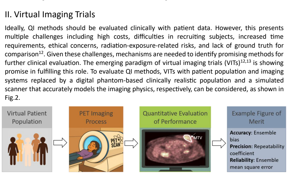
The load-bearing objects are the four frameworks themselves. Virtual imaging trials replace patients and scanners with digital anthropomorphic phantoms and Monte-Carlo or analytical PET simulators, providing known ground truth and figures of merit such as bias, repeatability, and ensemble mean squared error. No-gold-standard evaluation builds on regression-without-truth, which assumes measured value equals slope times true value plus bias plus zero-mean Gaussian noise, with true values drawn from a bounded parametric distribution, and uses maximum likelihood to estimate the noise-to-slope ratio (NSR) as the precision ranking metric. Joint detection and quantification evaluation uses estimation receiver operating characteristic (EROC) curves and the area under them (AEROC), computed from utility scores and false-positive fraction, with anthropomorphic and ideal observers performing the task. Multidimensional-parameter evaluation anchors on the clinical decision task, with study-type selection, representative test data, reference standards, and task-appropriate figures of merit such as AUC or Kaplan-Meier estimates.
What would settle it
A direct test would be to run the no-gold-standard framework on a clinical dataset for which ground truth is also available—for example, PET images with known lesion volumes from surgical pathology or from physical phantoms scanned on a real PET scanner—and compare the NSR-based ranking with rankings from bias, precision, and ensemble mean squared error computed against the known truth; disagreement would show that the linearity or bounded-distribution assumptions fail for that task. A second observation would come from comparing a virtual imaging trial's predicted ranking of two reconstruction or segmentation methods with the ranking from a prospective clinical reader study; inversion would indicate insufficient realism in the digital phantom or simulator.
Extended reading notes
Core claim
On the paper's own terms, the discovery is that four existing but fragmented evaluation strategies can be assembled into a comprehensive structure for task-based evaluation of QI methods. The virtual imaging trial substitutes a digital patient population and a simulated scanner for real patients and hardware, giving access to ground truth at low cost. The no-gold-standard framework estimates, without any truth values, the linear relationship between measured and true values for each candidate method and ranks methods by noise-to-slope ratio, a precision-based figure of merit. The joint detection and quantification framework evaluates methods on the realistic two-step task of detecting a signal and estimating a parameter, summarized by the area under the estimation receiver operating characteristic curve. The multidimensional-parameter framework evaluates radiomics-style methods through their impact on diagnostic, prognostic, or predictive clinical decisions, following RELAINCE-style study design. The paper holds that together these cover evaluation in virtual and clinical settings, for unidimensional and multidimensional outputs, and with or without ground truth.
Load-bearing premise
The no-gold-standard ranking is only as sound as the assumptions that each method's measured values are linearly related to the true values, the true-value distribution has known bounds, and noise across methods is uncorrelated or correctly modeled; the virtual-imaging framework likewise assumes digital phantoms and simulated scanners faithfully reproduce clinical reality.
Editorial extensions
If this is right
- Promising QI methods can be screened in virtual imaging trials before committing to expensive clinical studies, and the same trials can supply the ground truth needed to check estimability and noise behavior.
- Clinical datasets without any gold standard can still be used to rank candidate QI methods on precision, provided the linearity and distributional assumptions are checked and bootstrap confidence intervals are computed for the NSR differences.
- Evaluation of PET methods that require lesion detection first can be summarized by AEROC, capturing both the detection and the quantification error in one number.
- Radiomic and other multidimensional QI methods should be judged by their effect on the clinical decision, such as classification AUC or survival separation, rather than by per-feature accuracy alone.
- When these frameworks are applied to AI-based methods, the resulting performance reports should follow the RELAINCE guidelines so claims are stated consistently.
Reading between the lines
- The four frameworks could be assembled into a staged translational pipeline—VIT screening, NGSE ranking on clinical data, then JDQ and multidimensional validation—so that only methods that pass earlier gates proceed to more expensive evaluation; the paper outlines the parts but not this explicit workflow.
- The NGSE linearity assumption could likely be relaxed to known monotonic nonlinear links if the true-value bounds remain available, but the paper does not develop this extension; testing it on simulated PET data would be straightforward.
- If VITs are validated through VVUQ, in silico evidence could eventually support regulatory or reimbursement claims for QI methods, a consequence the paper points toward but leaves implicit.
- A concrete testable extension is to apply the NGSE bootstrap-ranking procedure to deep-learning-based segmentation and quantification methods, where the linearity assumption is less obviously satisfied; where it fails, precision-only ranking would need replacement by a bias-aware figure of merit.
Signed reviews
Editorial analysis
A structured set of objections, weighed in public.
Referee Report
Summary. This manuscript is a review article that outlines four emerging frameworks for objective task-based evaluation of quantitative imaging (QI) methods, with a focus on PET applications: (1) virtual imaging trials (VITs), (2) no-gold-standard evaluation (NGSE) of QI methods without ground truth, (3) evaluation of joint detection and quantification (JDQ) tasks, and (4) evaluation of QI methods that output multidimensional parameters such as radiomic features. For each framework, the paper describes the key components, provides illustrative figures, discusses figures of merit, and identifies areas of future research. The Discussion extends the scope to physical phantoms, human-in-the-loop AI algorithms, per-patient evaluation, and digital twins. The paper is explicitly based on previous literature and presents itself as a roadmap for clinical translation rather than a new methodological contribution.
Significance. The manuscript provides a useful, well-organized synthesis of emerging evaluation methodologies for quantitative medical imaging, particularly in the context of PET. It accurately represents the cited literature, including the underlying assumptions and limitations of each framework, such as the need for validation of virtual imaging trials and the linearity assumption in no-gold-standard evaluation. The paper also gives appropriate credit to prior work, including the authors' own contributions, and clearly distinguishes between what is established and what remains open. If adopted, these frameworks could help standardize evaluation practices and improve the reporting of QI method performance, which is timely given advances in long axial field-of-view PET and AI-based methods. The review is not a new methodological development, but it fulfills an important educational and catalytic role for the field.
minor comments (6)
- [III. Evaluating quantitative imaging methods without ground truth] The discussion under 'Check Linearity Between True and Measured Values' states that the linearity assumption can be verified through inter-method comparisons, realistic simulations, and phantom studies, but it does not specify what should be done if the linearity check fails; adding a brief statement about the consequences of violation would make the framework more complete.
- [III. Evaluating quantitative imaging methods without ground truth] The text notes that the noise-to-slope ratio (NSR) is a figure of merit based on precision, but it does not explicitly acknowledge that a method with favorable NSR may still have poor accuracy; a sentence recommending complementary evaluation of bias would be helpful for readers planning clinical translation.
- [I. Introduction] In the paragraph on recent advances in PET, there is a typo with a double comma after 'PET'; the sentence should read 'PET, including.'
- [III. Evaluating quantitative imaging methods without ground truth] The word 'summerized' in the sentence 'with key components summerized below' should be 'summarized.'
- [V. Evaluation of QI Methods for Quantifying Multi-dimensional Parameters] In the sentence 'Typical research studies using muti-dimensional parameters', 'muti-dimensional' should be 'multi-dimensional.'
- [References] In reference 31, 'Mont Carlo' should be 'Monte Carlo.'
Circularity Check
No significant circularity: this is a review article that explicitly synthesizes prior literature; the frameworks are presented, not derived, and no prediction reduces to a fitted input.
full rationale
The paper is a narrative review, not a derivation chain. Its abstract states 'based on previous literature, we outline four emerging frameworks' and the body repeatedly frames the content as a review of existing techniques rather than as new predictions. The NGSE discussion, the most heavily self-cited portion, restates assumptions from RWT/NGSE (linear relationship between true and measured values, known bounds, uncorrelated noise) and presents NSR as a figure of merit from prior publications; it does not fit a parameter to data and then rename that fit as a prediction. The paper is also explicit about a key limitation: NGSE validations 'rely on simulations due to the need for ground truth.' That stated dependence is the opposite of circularity, since it concedes the framework's clinical validation remains open. The self-citations in the NGSE and RELAINCE sections are normal for a review authored by the group that developed those methods, and the load-bearing references include independent prior work (Hoppin/Kupinski RWT, Clarkson EROC, XCAT phantoms, and multi-institutional guidelines). No equation in this paper is equal to its own input by construction, and no fitted value is presented as an independent prediction. The skeptical concern that the linearity assumption is clinically untested is a correctness/validity risk, not a circularity per the review rules.
Assumptions & free parameters
assumptions (3)
- domain assumption Measured values are linearly related to true values for each QI method.
- domain assumption The true values are sampled from a parametric distribution with known bounds.
- domain assumption Virtual imaging trials provide clinically realistic simulations of patient populations and imaging systems.
Cite this review
Pith. "Pith review of Objective Task-based Evaluation of Quantitative Medical Imaging Methods: Emerging Frameworks and Future Directions." pith.science (2026). https://pith.science/paper/SXH4G2SB
@misc{pith2026250704591,
  author       = {Pith},
  title        = {Pith review of: Objective Task-based Evaluation of Quantitative Medical Imaging Methods: Emerging Frameworks and Future Directions},
  year         = {2026},
  howpublished = {\url{https://pith.science/paper/SXH4G2SB}},
  note         = {Machine review of arXiv:2507.04591}
}
read the original abstract
Quantitative imaging (QI) is demonstrating strong promise across multiple clinical applications. For clinical translation of QI methods, objective evaluation on clinically relevant tasks is essential. To address this need, multiple evaluation strategies are being developed. In this paper, based on previous literature, we outline four emerging frameworks to perform evaluation studies of QI methods. We first discuss the use of virtual imaging trials (VITs) to evaluate QI methods. Next, we outline a no-gold-standard evaluation framework to clinically evaluate QI methods without ground truth. Third, a framework to evaluate QI methods for joint detection and quantification tasks is outlined. Finally, we outline a framework to evaluate QI methods that output multi-dimensional parameters, such as radiomic features. We review these frameworks, discussing their utilities and limitations. Further, we examine future research areas in evaluation of QI methods. Given the recent advancements in PET, including long axial field-of-view scanners and the development of artificial-intelligence algorithms, we present these frameworks in the context of PET.
Figures


Reference graph
Works this paper leans on
-
[1]
Clinical utility of quantitative imaging
Rosenkrantz AB, Mendiratta-Lala M, Bartholmai BJ, et al. Clinical utility of quantitative imaging. Acad Radiol. 2015;22(1):33-49
2015
-
[2]
Quantitative imaging in cancer clinical trials
Yankeelov TE, Mankoff DA, Schwartz LH, et al. Quantitative imaging in cancer clinical trials. Clin Cancer Res. 2016;22(2):284-290
2016
-
[3]
Prognostic value of volumetric parameters of 18F -FDG PET in non-small-cell lung cancer: a meta-analysis
Im HJ, Pak K, Cheon GJ, et al. Prognostic value of volumetric parameters of 18F -FDG PET in non-small-cell lung cancer: a meta-analysis. Eur J Nucl Med Mol Imaging. 2015;42(2):241-251
2015
-
[4]
Predicting and monitoring cancer treatment response with diffusion-weighted MRI
Thoeny HC, Ross BD. Predicting and monitoring cancer treatment response with diffusion-weighted MRI. J Magn Reson Imaging. 2010;32(1):2-16
2010
-
[5]
Strategies to Perform Joint Detection and Quantification Task : JDQ task s can be performed sequentially or simultaneously
Quantification, as defined in the previous section, refers to the estimation of parameters such as MTV and TLG. Strategies to Perform Joint Detection and Quantification Task : JDQ task s can be performed sequentially or simultaneously. The sequential process typically involves first detection and then quantification, while the simultaneous process execute...
-
[6]
The clinical value of myocardial blood flow measurement
Camici PG, Rimoldi OE. The clinical value of myocardial blood flow measurement. J Nucl Med . 2009;50(7):1076-1087
2009
-
[7]
Dynamic whole-body PET parametric imaging: I
Karakatsanis NA, Lodge MA, Tahari AK, Zhou Y , Wahl RL, Rahmim A. Dynamic whole-body PET parametric imaging: I. concept, acquisition protocol optimization and clinical application. Phys Med Biol . 2013;58(20):7391-7418
2013
-
[8]
The impact of PET and SPECT on dosimetry for targeted radionuclide therapy
Flux G, Bardies M, Monsieurs M, Savolainen S, Strand SE, Lassmann M. The impact of PET and SPECT on dosimetry for targeted radionuclide therapy. Z Med Phys. 2006;16(1):47-59
2006
Show all 89 references
-
[9]
Radiomics: the bridge between medical imaging and personalized medicine
Lambin P , Leijenaar RTH, Deist TM, et al. Radiomics: the bridge between medical imaging and personalized medicine. Nat Rev Clin Oncol. 2017;14(12):749-762
2017
-
[10]
Objective task -based evaluation of artificial intelligence -based medical imaging methods: framework, strategies, and role of the physician
Jha AK, Myers KJ, Obuchowski NA, et al. Objective task -based evaluation of artificial intelligence -based medical imaging methods: framework, strategies, and role of the physician. PET Clin. 2021;16(4):493-511
2021
-
[11]
Total-body imaging: transforming the role of positron emission tomography
Cherry SR, Badawi RD, Karp JS, Moses WW, Price P , Jones T. Total-body imaging: transforming the role of positron emission tomography. Sci Transl Med. 2017;9(381):eaaf6169
2017
-
[12]
Applications of artificial intelligence and deep learning in molecular imaging and radiotherapy
Arabi H, Zaidi H. Applications of artificial intelligence and deep learning in molecular imaging and radiotherapy. European J Hybrid Imaging. 2020;4(1):17
2020
-
[13]
Virtual clinical trials in medical imaging: a review
Abadi E, Segars WP , Tsui BMW, et al. Virtual clinical trials in medical imaging: a review. J Med Imag . 2020;7(04):1
2020
-
[14]
Evaluation of Digital Breast Tomosynthesis as Replacement of Full-Field Digital Mammography Using an In Silico Imaging Trial
Badano A, Graff CG, Badal A, et al. Evaluation of Digital Breast Tomosynthesis as Replacement of Full-Field Digital Mammography Using an In Silico Imaging Trial. JAMA Netw Open. 2018;1(7):e185474
2018
-
[15]
4D XCAT phantom for multimodality imaging research
Segars WP , Sturgeon G, Mendonca S, Grimes J, Tsui BMW. 4D XCAT phantom for multimodality imaging research. Med Phys. 2010;37(9):4902-4915
2010
-
[16]
Population of anatomically variable 4D XCAT adult phantoms for imaging research and optimization
Segars WP , Bond J, Frush J, et al. Population of anatomically variable 4D XCAT adult phantoms for imaging research and optimization. Medical Physics. 2013;40(4):043701
2013
-
[17]
The development of a population of 4D pediatric XCAT phantoms for imaging research and optimization
Segars WP , Norris H, Sturgeon GM, et al. The development of a population of 4D pediatric XCAT phantoms for imaging research and optimization. Medical Physics. 2015;42(8):4719-4726
2015
-
[18]
A Bayesian approach to tissue -fraction estimation for oncological PET segmentation
Liu Z, Mhlanga JC, Laforest R, Derenoncourt PR, Siegel BA, Jha AK. A Bayesian approach to tissue -fraction estimation for oncological PET segmentation. Phys Med Biol. 2021;66(12):124002
2021
-
[19]
A physics -guided modular deep -learning based automated framework for tumor segmentation in PET
Leung KH, Marashdeh W, Wray R, et al. A physics -guided modular deep -learning based automated framework for tumor segmentation in PET. Phys Med Biol. 2020;65(24):245032
2020
-
[20]
GATE: a simulation toolkit for PET and SPECT
Jan S, Santin G, Strul D, et al. GATE: a simulation toolkit for PET and SPECT. Phys Med Biol. 2004;49(19):4543- 4561
2004
-
[21]
Monte Carlo simulation and scatter correction of the GE Advance PET scanner with SimSET and Geant4
Barret O, Carpenter TA, Clark JC, Ansorge RE, Fryer TD. Monte Carlo simulation and scatter correction of the GE Advance PET scanner with SimSET and Geant4. Phys Med Biol. 2005;50(20):4823-4840
2005
-
[22]
SMART (SiMulAtion and ReconsTruction) PET: an efficient PET simulation-reconstruction tool
Pfaehler E, De Jong JR, Dierckx RAJO, Van Velden FHP , Boellaard R. SMART (SiMulAtion and ReconsTruction) PET: an efficient PET simulation-reconstruction tool. EJNMMI Phys. 2018;5(1):16
2018
-
[23]
ASIM: An Analytic PET Simulator
Elston BF, Comtat C, Harrison RL, Kinahan P . ASIM: An Analytic PET Simulator . In: Monte Carlo Calculations in Nuclear Medicine. 2nd ed. CRC Press; 2012
2012
-
[24]
FAST (fast analytical simulator of tracer) -PET: an accurate and efficient PET analytical simulation tool
Li S, Hamdi M, Dutta K, et al. FAST (fast analytical simulator of tracer) -PET: an accurate and efficient PET analytical simulation tool. Phys Med Biol. 2024;69(16):165020
2024
-
[25]
STIR: software for tomographic image reconstruction release 2
Thielemans K, Tsoumpas C, Mustafovic S, et al. STIR: software for tomographic image reconstruction release 2. Phys Med Biol. 2012;57(4):867-883
2012
-
[26]
CASToR: a generic data organization and processing code framework for multi-modal and multi-dimensional tomographic reconstruction
Merlin T, Stute S, Benoit D, et al. CASToR: a generic data organization and processing code framework for multi-modal and multi-dimensional tomographic reconstruction. Phys Med Biol. 2018;63(18):185005
2018
-
[27]
PyTomography: A python library for medical image reconstruction
Polson LA, Fedrigo R, Li C, et al. PyTomography: A python library for medical image reconstruction. SoftwareX. 2025;29:102020
2025
-
[28]
A Maximum Likelihood Method for Region -of-Interest Evaluation in Emission Tomography
Carson RE. A Maximum Likelihood Method for Region -of-Interest Evaluation in Emission Tomography. J Comp Assist Tomography. 1986;10(4):654-663
1986
-
[29]
WIN -PDQ: a Wiener -estimator-based projection -domain quantitative SPECT method that accounts for intra -regional uptake heterogeneity
Li Z, Benabdallah N, Thorek DLJ, Jha AK. WIN -PDQ: a Wiener -estimator-based projection -domain quantitative SPECT method that accounts for intra -regional uptake heterogeneity. Proc SPIE Med Imag . 2024;12925:1292512
2024
-
[30]
Estimating random signal parameters from noisy images with nuisance parameters: linear and scanning-linear methods
Whitaker MK, Clarkson E, Barrett HH. Estimating random signal parameters from noisy images with nuisance parameters: linear and scanning-linear methods. Opt Express. 2008;16(11):8150
2008
-
[31]
Fast and Accurate Amyloid Brain PET Quantification Without MRI Using Deep Neural Networks
Kang SK, Kim D, Shin SA, Kim YK, Choi H, Lee JS. Fast and Accurate Amyloid Brain PET Quantification Without MRI Using Deep Neural Networks. J Nucl Med. 2023;64(4):659-666
2023
-
[32]
Development of a GPU-Based Mont Carlo Simulation Tool for PET
Lai Y , Zhong Y, Chalise A, et al. Development of a GPU-Based Mont Carlo Simulation Tool for PET. In: 2019 IEEE Nuclear Science Symposium and Medical Imaging Conference (NSS/MIC). IEEE; 2019:1-3
2019
-
[33]
MCGPU -PET: An open-source real-time Monte Carlo PET simulator
Herraiz JL, Lopez-Montes A, Badal A. MCGPU -PET: An open-source real-time Monte Carlo PET simulator . Comput Phys Commun. 2024;296:109008
2024
-
[34]
Observer -study-based approaches to quantitatively evaluate the realism of synthetic medical images
Liu Z, Wolfe S, Yu Z, et al. Observer -study-based approaches to quantitatively evaluate the realism of synthetic medical images. Phys Med Biol. 2023;68(7):074001
2023
-
[35]
Observer study-based evaluation of TGAN architecture used to generate oncological PET images
Fedrigo R, Yousefirizi F, Liu Z, et al. Observer study-based evaluation of TGAN architecture used to generate oncological PET images. Proc SPIE Med Imag. 2023;12467:124670S
2023
-
[36]
How much realism is needed?
Badano A. “How much realism is needed?” — the wrong question in silico imagers have been asking. Med Phys. 2017;44(5):1607-1609
2017
-
[37]
A Framework for In Silico Clinical Trials for Medical Devices Using Concepts From Model Verification, Validation, and Uncertainty Quantification (VVUQ)
Bodner J, Kaul V. A Framework for In Silico Clinical Trials for Medical Devices Using Concepts From Model Verification, Validation, and Uncertainty Quantification (VVUQ). In: ASME 2021 Verification and Validation Symposium. American Society of Mechanical Engineers; 2021:V001T02A001
2021
-
[38]
40-Assessing Credibility of Computational Modeling through Verification and Validation: Application to Medical Devices
V&V. 40-Assessing Credibility of Computational Modeling through Verification and Validation: Application to Medical Devices. American Society of Mechanical Engineers; 2018
2018
-
[39]
Toward widespread use of virtual trials in medical imaging innovation and regulatory science
Abadi E, Barufaldi B, Lago M, et al. Toward widespread use of virtual trials in medical imaging innovation and regulatory science. Medical Physics. 2024;51(12):9394-9404
2024
-
[40]
Quantitative imaging biomarkers: a review of statistical methods for computer algorithm comparisons
Obuchowski NA, Reeves AP , Huang EP , et al. Quantitative imaging biomarkers: a review of statistical methods for computer algorithm comparisons. Stat Methods Med Res. 2015;24(1):68-106
2015
-
[41]
Objective comparison of quantitative imaging modalities without the use of a gold standard
Hoppin JW, Kupinski MA, Kastis GA, Clarkson E, Barrett HH. Objective comparison of quantitative imaging modalities without the use of a gold standard. IEEE Trans Med Imaging. 2002;21(5)
2002
-
[42]
Estimation in medical imaging without a gold standard
Kupinski MA, Hoppin JW, Clarkson E, Barrett HH, Kastis GA. Estimation in medical imaging without a gold standard. Acad Radiol. 2002;9(3):290-297
2002
-
[43]
Comparing cardiac ejection fraction estimation algorithms without a gold standard
Kupinski MA, Hoppin JW, Krasnow J, et al. Comparing cardiac ejection fraction estimation algorithms without a gold standard. Acad Radiol. 2006;13(3):329-337
2006
-
[44]
Nonsupervised ranking of different segmentation approaches: application to the estimation of the left ventricular ejection fraction from cardiac cine MRI sequences
Lebenberg J, Buvat I, Lalande A, et al. Nonsupervised ranking of different segmentation approaches: application to the estimation of the left ventricular ejection fraction from cardiac cine MRI sequences. IEEE Trans Med Imaging. 2012;31(8):1651-1660
2012
-
[45]
A no -gold-standard technique for objective assessment of quantitative nuclear - medicine imaging methods
Jha AK, Caffo B, Frey EC. A no -gold-standard technique for objective assessment of quantitative nuclear - medicine imaging methods. Phys Med Biol. 2016;61(7):2780-2800
2016
-
[46]
Task -based evaluation of segmentation algorithms for diffusion-weighted MRI without using a gold standard
Jha AK, Kupinski MA, Rodríguez JJ, Stephen RM, Stopeck AT. Task -based evaluation of segmentation algorithms for diffusion-weighted MRI without using a gold standard. Phys Med Biol. 2012;57(13):4425
2012
-
[47]
No-gold-standard evaluation of quantitative imaging methods in the presence of correlated noise
Liu Z, Li Z, Mhlanga JC, Siegel BA, Jha AK. No-gold-standard evaluation of quantitative imaging methods in the presence of correlated noise. Proc SPIE Med Imag. 2022;12035:12035M
2022
-
[48]
A no -gold-standard technique for objective evaluation of quantitative nuclear -medicine imaging methods in the presence of correlated noise
Liu J, Liu Z, Moon HS, Mhlanga J, Jha A. A no -gold-standard technique for objective evaluation of quantitative nuclear -medicine imaging methods in the presence of correlated noise. J Nucl Med . 2020;61(supplement 1):523-523
2020
-
[49]
No-gold-standard evaluation of quantitative SPECT methods for alpha-particle radiopharmaceutical therapy
Liu Y , Liu Z, Li Z, Thorek D, Siegel B, Jha A. No-gold-standard evaluation of quantitative SPECT methods for alpha-particle radiopharmaceutical therapy. J Nucl Med. 2023;64(supplement 1):P1331-P1331
2023
-
[50]
Practical no -gold-standard evaluation framework for quantitative imaging methods: application to lesion segmentation in positron emission tomography
Jha AK, Mena E, Caffo B, et al. Practical no -gold-standard evaluation framework for quantitative imaging methods: application to lesion segmentation in positron emission tomography. J Med Imag . 2017;4(1):011011
2017
-
[51]
Incorporating prior information in a no -gold-standard technique to assess quantitative SPECT reconstruction methods
Jha AK, Frey EC. Incorporating prior information in a no -gold-standard technique to assess quantitative SPECT reconstruction methods. International Meeting on Fully 3D reconstruction in Radiology and Nuclear Medicine. Published online 2015:47-51
2015
-
[52]
Estimation receiver operating characteristic curve and ideal observers for combined detection/estimation tasks
Clarkson E. Estimation receiver operating characteristic curve and ideal observers for combined detection/estimation tasks. J Opt Soc Am A. 2007;24(12):B91
2007
-
[53]
Shannon information for joint estimation/detection tasks and complex imaging systems
Clarkson E, Cushing JB. Shannon information for joint estimation/detection tasks and complex imaging systems. J Opt Soc Am A. 2016;33(3):286
2016
-
[54]
Human - and model -observer performance in ramp -spectrum noise: effects of regularization and object variability
Abbey CK, Barrett HH. Human - and model -observer performance in ramp -spectrum noise: effects of regularization and object variability. J Opt Soc Am A. 2001;18(3):473
2001
-
[55]
Addition of a channel mechanism to the ideal -observer model
Myers KJ, Barrett HH. Addition of a channel mechanism to the ideal -observer model. J Opt Soc Am A . 1987;4(12):2447
1987
-
[56]
Foundations of Image Science
Barrett HH, Myers KJ. Foundations of Image Science. John Wiley & Sons; 2004
2004
-
[57]
Clinical impact of time -of-flight and point response modeling in PET reconstructions: a lesion detection study
Schaefferkoetter J, Casey M, Townsend D, El Fakhri G. Clinical impact of time -of-flight and point response modeling in PET reconstructions: a lesion detection study. Phys Med Biol. 2013;58(5):1465-1478
2013
-
[58]
Model Observers in Medical Imaging Research
He X, Park S. Model Observers in Medical Imaging Research. Theranostics. 2013;3(10):774-786
2013
-
[59]
A Perceptually Relevant Channelized Joint Observer (PCJO) for the Detection -Localization of Parametric Signals
Lu Zhang, Cavaro -Menard C, Le Callet P , Tanguy JY. A Perceptually Relevant Channelized Joint Observer (PCJO) for the Detection -Localization of Parametric Signals. IEEE Trans Med Imaging . 2012;31(10):1875- 1888
2012
-
[60]
Channelized model observer for the detection and estimation of signals with unknown amplitude, orientation, and size
Zhang L, Goossens B, Cavaro-Ménard C, Callet PL, Ge D. Channelized model observer for the detection and estimation of signals with unknown amplitude, orientation, and size. J Opt Soc Am A. 2013;30(11):2422
2013
-
[61]
Combination of detection and estimation tasks using channelized scanning linear observer for CT imaging systems
Tseng HW, Fan J, Kupinski MA. Combination of detection and estimation tasks using channelized scanning linear observer for CT imaging systems. Proc SPIE Med Imag. 2015;9416:94160H
2015
-
[62]
A Hybrid Approach for Approximating the Ideal Observer for Joint Signal Detection and Estimation Tasks by Use of Supervised Learning and Markov -Chain Monte Carlo Methods
Li K, Zhou W, Li H, Anastasio MA. A Hybrid Approach for Approximating the Ideal Observer for Joint Signal Detection and Estimation Tasks by Use of Supervised Learning and Markov -Chain Monte Carlo Methods. IEEE Trans Med Imaging. 2022;41(5):1114-1124
2022
-
[63]
Optimal Joint Detection and Estimation That Maximizes ROC -Type Curves
Wunderlich A, Goossens B, Abbey CK. Optimal Joint Detection and Estimation That Maximizes ROC -Type Curves. IEEE Trans Med Imaging. 2016;35(9):2164-2173
2016
-
[64]
Noise properties of the EM algorithm
Barrett HH, Wilson DW, Tsui BMW. Noise properties of the EM algorithm. I. Theory. Phys Med Biol . 1994;39(5):833
1994
-
[65]
Exploration of PET and MRI radiomic features for decoding breast cancer phenotypes and prognosis
Huang S ying, Franc BL, Harnish RJ, et al. Exploration of PET and MRI radiomic features for decoding breast cancer phenotypes and prognosis. NPJ Breast Cancer. 2018;4(1):1-13
2018
-
[66]
A framework for evaluating the technical performance of multiparameter quantitative imaging biomarkers (mp-QIBs)
Obuchowski NA, Huang E, deSouza NM, et al. A framework for evaluating the technical performance of multiparameter quantitative imaging biomarkers (mp-QIBs). Acad Radiol. 2023;30(2):147-158
2023
-
[67]
The effect of SUV discretization in quantitative FDG -PET Radiomics: the need for standardized methodology in tumor texture analysis
Leijenaar RTH, Nalbantov G, Carvalho S, et al. The effect of SUV discretization in quantitative FDG -PET Radiomics: the need for standardized methodology in tumor texture analysis. Sci Rep. 2015;5(1):11075
2015
-
[68]
Robustness of radiomic features in [11C]Choline and [18F]FDG PET/CT imaging of nasopharyngeal carcinoma: impact of segmentation and discretization
Lu L, Lv W, Jiang J, et al. Robustness of radiomic features in [11C]Choline and [18F]FDG PET/CT imaging of nasopharyngeal carcinoma: impact of segmentation and discretization. Mol Imaging Biol. 2016;18(6):935- 945
2016
-
[69]
Quantitative radiomics: impact of stochastic effects on textural feature analysis implies the need for standards
Nyflot MJ, Yang F, Byrd D, Bowen SR, Sandison GA, Kinahan PE. Quantitative radiomics: impact of stochastic effects on textural feature analysis implies the need for standards. J Med Imag. 2015;2(4):041002
2015
-
[70]
Joint EANM/SNMMI guideline on radiomics in nuclear medicine: jointly supported by the EANM Physics Committee and the SNMMI Physics, Instrumentation and Data Sciences Council
Hatt M, Krizsan AK, Rahmim A, et al. Joint EANM/SNMMI guideline on radiomics in nuclear medicine: jointly supported by the EANM Physics Committee and the SNMMI Physics, Instrumentation and Data Sciences Council. Eur J Nucl Med Mol Imaging. 2023;50(2):352-375
2023
-
[71]
Nuclear medicine and artificial intelligence: best practices for evaluation (the RELAINCE guidelines)
Jha AK, Bradshaw TJ, Buvat I, et al. Nuclear medicine and artificial intelligence: best practices for evaluation (the RELAINCE guidelines). J Nucl Med. 2022;63(9):1288-1299
2022
-
[72]
AI in medical physics: guidelines for publication
El Naqa I, Boone JM, Benedict SH, et al. AI in medical physics: guidelines for publication. Med Phys . 2021;48(9):4711-4714
2021
-
[73]
MRI-based radiomics models to assess prostate cancer, extracapsular extension and positive surgical margins
He D, Wang X, Fu C, et al. MRI-based radiomics models to assess prostate cancer, extracapsular extension and positive surgical margins. Cancer Imaging. 2021;21(1):46
2021
-
[74]
Use of expert panels to define the reference standard in diagnostic research: a systematic review of published methods and reporting
Bertens LCM, Broekhuizen BDL, Naaktgeboren CA, et al. Use of expert panels to define the reference standard in diagnostic research: a systematic review of published methods and reporting. PLoS Med . 2013;10(10):e1001531
2013
-
[75]
PET parametric imaging: past, present, and future
Wang G, Rahmim A, Gunn RN. PET parametric imaging: past, present, and future. IEEE Trans Radiat Plasma Med Sci. 2020;4(6):663-675
2020
-
[76]
Recent advances on the development of phantoms using 3D printing for imaging with CT, MRI, PET, SPECT, and ultrasound
Filippou V, Tsoumpas C. Recent advances on the development of phantoms using 3D printing for imaging with CT, MRI, PET, SPECT, and ultrasound. Med Phys. 2018;45(9):e740-e760
2018
-
[77]
Abdo -Man: a 3D -printed anthropomorphic phantom for validating quantitative SIRT
Gear JI, Cummings C, Craig AJ, et al. Abdo -Man: a 3D -printed anthropomorphic phantom for validating quantitative SIRT. EJNMMI Phys. 2016;3(1):17
2016
-
[78]
Design of an anthropomorphic PET phantom with elastic lungs and respiration modeling
Black DG, Yazdi YO, Wong J, et al. Design of an anthropomorphic PET phantom with elastic lungs and respiration modeling. Med Phys. 2021;48(8):4205-4217
2021
-
[79]
Anthropomorphic thorax phantom for cardio - respiratory motion simulation in tomographic imaging
Bolwin K, Czekalla B, Frohwein LJ, Büther F, Schäfers KP . Anthropomorphic thorax phantom for cardio - respiratory motion simulation in tomographic imaging. Phys Med Biol. 2018;63(3):035009
2018
-
[80]
PET textural features stability and pattern discrimination power for radiomics analysis: An “ad-hoc” phantoms study
Presotto L, Bettinardi V, De Bernardi E, et al. PET textural features stability and pattern discrimination power for radiomics analysis: An “ad-hoc” phantoms study. Physica Medica. 2018;50:66-74
2018
-
[81]
Phantom and methodology for comparison of small lesion detectability in PET
Wollenweber SD, Alessio AM, Kinahan PE. Phantom and methodology for comparison of small lesion detectability in PET. Med Phys. 2023;50(5):2998-3007
2023
-
[82]
A Study on the Basic Criteria for Selecting Heterogeneity Parameters of F18-FDG PET Images
Forgacs A, Pall Jonsson H, Dahlbom M, et al. A Study on the Basic Criteria for Selecting Heterogeneity Parameters of F18-FDG PET Images. Chen K, ed. PLoS ONE. 2016;11(10):e0164113
2016
-
[83]
Human-in-the- loop machine learning: a state of the art
Mosqueira-Rey E, Hernández-Pereira E, Alonso-Ríos D, Bobes-Bascarán J, Fernández-Leal Á. Human-in-the- loop machine learning: a state of the art. Artif Intell Rev. 2023;56(4):3005-3054
2023
-
[84]
Meeting the Moment: Addressing Barriers and Facilitating Clinical Adoption of Artificial Intelligence in Medical Diagnosis
University of California, San Francisco, Adler-Milstein J, Aggarwal N, et al. Meeting the Moment: Addressing Barriers and Facilitating Clinical Adoption of Artificial Intelligence in Medical Diagnosis. NAM Perspectives. 2022;22(9)
2022
-
[85]
Adaptive SPECT
Barrett HH, Furenlid LR, Freed M, et al. Adaptive SPECT. IEEE Trans Med Imaging. 2008;27(6):775-788
2008
-
[86]
Statistics for radiology research
Obuchowski N, Subhas N, Polster J. Statistics for radiology research. Semin Musculoskelet Radiol . 2017;21(01):023-031
2017
-
[87]
Testing for interchangeability of imaging tests
Obuchowski NA, Subhas N, Schoenhagen P . Testing for interchangeability of imaging tests. Acad Radiol. 2014;21(11):1483-1489
2014
-
[88]
Digital twins for predictive oncology will be a paradigm shift for precision cancer care
Hernandez-Boussard T, Macklin P , Greenspan EJ, et al. Digital twins for predictive oncology will be a paradigm shift for precision cancer care. Nat Med. 2021;27(12):2065-2066
2021
-
[89]
Integrating mechanism -based modeling with biomedical imaging to build practical digital twins for clinical oncology
Wu C, Lorenzo G, Hormuth DA, et al. Integrating mechanism -based modeling with biomedical imaging to build practical digital twins for clinical oncology. Biophys Rev. 2022;3(2):021304
2022
Reviewed August 6, 2026 · model on record in the stance chip above.
Discussion (0). Continue with ORCID to comment.