Neural electron backscatter diffraction
Pith reviewed 2026-06-27 12:58 UTC · model grok-4.3
The pith
A coordinate-based neural network represents EBSD scans as continuous four-dimensional fields of Kikuchi diffraction intensity.
A machine-rendered reading of the paper's core claim, the machinery that carries it, and where it could break.
Core claim
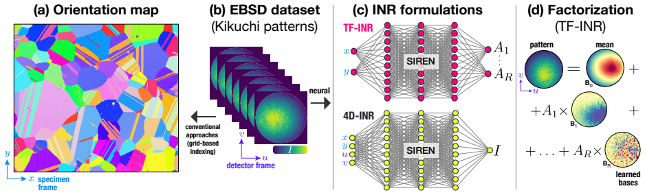
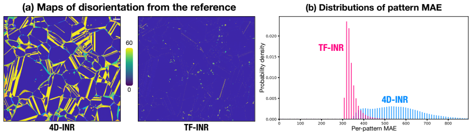
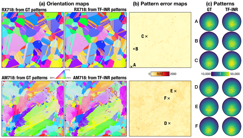
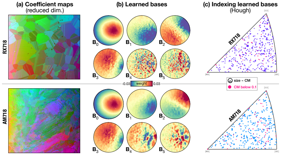
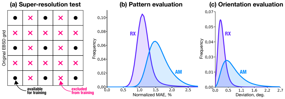
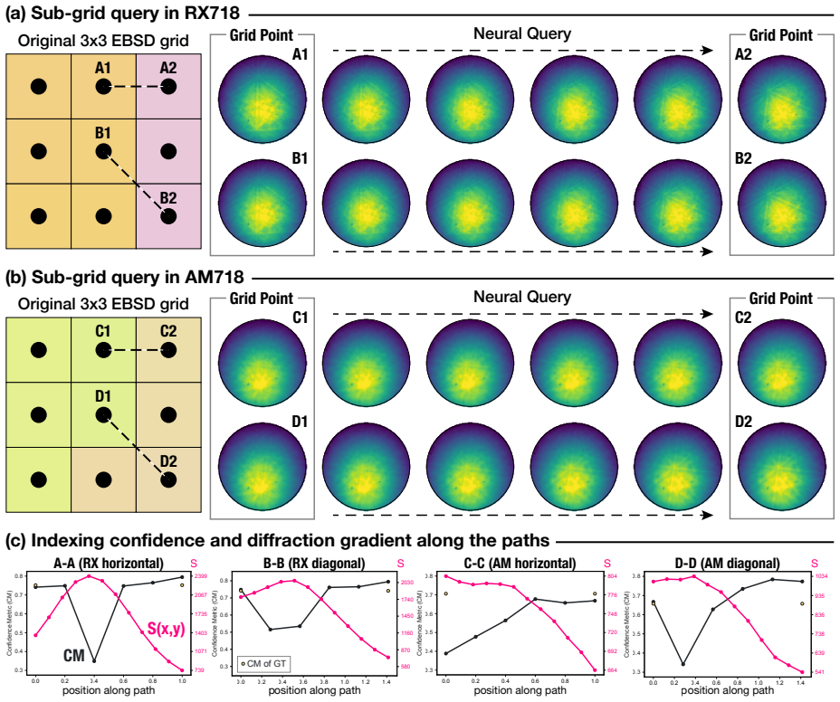
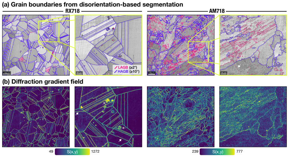
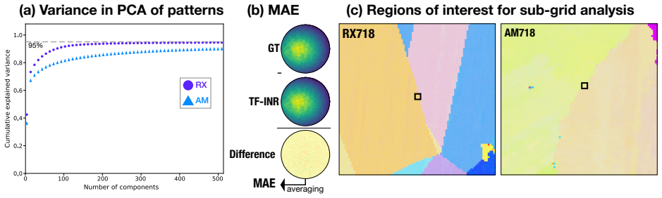
We introduce neural EBSD, which treats an EBSD scan as a continuous, differentiable four-dimensional field of Kikuchi diffraction intensity (in specimen--detector domain) and then represents it with a coordinate-based neural network. We develop and compare two formulations: a joint formulation that maps all four coordinates to intensity, and a factorized formulation that combines continuous specimen-domain coefficient fields with learned detector-domain basis patterns. Tested on recrystallized and additively manufactured Inconel 718, the factorized formulation shows better accuracy in reconstructing Kikuchi patterns that have map-averaged errors below 1% of the maximum intensity.
What carries the argument
Factorized coordinate-based neural network separating continuous specimen-domain coefficient fields from learned detector-domain basis patterns to represent the four-dimensional Kikuchi intensity field.
If this is right
- Full-pattern super-resolution in the specimen frame
- Continuous querying along arbitrary off-grid paths
- Spatially continuous boundary and heterogeneity localization from analytical spatial derivatives
- 700-fold compression by storing network weights and learned bases instead of raw patterns
Where Pith is reading between the lines
- The continuous differentiable representation could couple directly with deformation simulations that require smooth fields rather than gridded data.
- Off-grid querying opens the possibility of adaptive, non-uniform EBSD scanning strategies that concentrate measurements where heterogeneity is highest.
- The compression and on-demand access approach could apply to other large diffraction or imaging datasets in materials science where grid storage becomes prohibitive.
Load-bearing premise
Kikuchi diffraction intensity varies sufficiently smoothly in the specimen-detector domain and factorizes into continuous specimen-domain coefficient fields plus learned detector-domain basis patterns to allow accurate neural representation without loss of orientation or dislocation information.
What would settle it
Reconstruction errors exceeding 1% of maximum intensity on the Inconel 718 samples, or loss of fidelity when the network is queried along continuous off-grid paths, would show the representation fails to capture the underlying field.
Figures
read the original abstract
In a polycrystalline microstructure, orientation and dislocation content vary smoothly within grains, and the grain boundaries between them are continuous curves. Electron backscatter diffraction (EBSD) records this continuum on a discrete grid with every subsequent analysis (from indexing to advanced pattern processing) confined to that grid. We introduce neural EBSD, which treats an EBSD scan as a continuous, differentiable four-dimensional field of Kikuchi diffraction intensity (in specimen--detector domain) and then represents it with a coordinate-based neural network. We develop and compare two formulations: a joint formulation that maps all four coordinates to intensity, and a factorized formulation that combines continuous specimen-domain coefficient fields with learned detector-domain basis patterns. Tested on recrystallized and additively manufactured Inconel 718, the factorized formulation shows better accuracy in reconstructing Kikuchi patterns that have map-averaged errors below 1% of the maximum intensity. Beyond reconstruction, it provides full-pattern super-resolution in the specimen frame, continuous querying along arbitrary off-grid paths, as well as spatially continuous boundary and heterogeneity localization from analytical spatial derivatives. Storing the network weights and learned bases in place of the raw patterns in a large dataset offers a 700-fold compression while preserving on-demand access to the full patterns for downstream analyses.
Editorial analysis
A structured set of objections, weighed in public.
Referee Report
Summary. The paper introduces neural EBSD, representing EBSD scans as a continuous, differentiable 4D Kikuchi intensity field via coordinate-based neural networks. It compares a joint formulation (mapping all four coordinates to intensity) against a factorized formulation (specimen-domain coefficient fields multiplied by learned detector-domain bases). On recrystallized and additively manufactured Inconel 718, the factorized version achieves map-averaged reconstruction errors below 1% of peak intensity, while enabling full-pattern super-resolution in the specimen frame, continuous off-grid querying, analytical spatial derivatives for boundary/heterogeneity localization, and 700-fold compression via stored weights and bases.
Significance. If the reconstruction accuracy and preservation of downstream information hold, the work provides a practical continuous representation for EBSD data that could reduce storage demands in large datasets while enabling new analyses such as derivative-based boundary detection. The empirical testing on two distinct Inconel 718 microstructures and the explicit comparison of the two formulations are strengths; the compression factor and on-demand pattern access are concrete advantages for practical adoption.
major comments (2)
- [Abstract / Results] Abstract and results section: The central claim that the representation 'preserves on-demand access to the full patterns for downstream analyses' rests on map-averaged reconstruction error <1% of maximum intensity, but this metric does not establish that orientation indexing or GND density maps remain accurate. Grain boundaries and dislocations violate the smoothness premise of the factorized formulation (continuous coefficient fields imes fixed bases), and averaging can mask localized errors at those sites; a direct comparison of indexed orientations or GND densities computed from the neural patterns versus the original grid is required to support the claim.
- [Methods] Methods / factorized formulation: The assumption that 4D Kikuchi intensity admits a low-rank separable decomposition into specimen-domain coefficients and detector-domain bases is load-bearing for the reported accuracy advantage of the factorized model, yet no quantitative assessment (e.g., singular-value spectrum of the data tensor or sensitivity to rank choice) is supplied to show that the decomposition does not discard orientation or dislocation information.
minor comments (2)
- [Abstract] The abstract states '700-fold compression' without specifying the baseline (raw pattern storage size, bit depth, or whether it includes the network weights plus bases); a precise definition and comparison table would clarify the claim.
- [Results] No error bars, dataset sizes, or cross-validation procedure are mentioned for the <1% error figure; adding these in the results section would strengthen reproducibility.
Simulated Author's Rebuttal
We thank the referee for the constructive feedback. We address each major comment below, agreeing that the current evidence for downstream preservation is indirect and that additional quantitative support for the separability assumption would strengthen the manuscript.
read point-by-point responses
-
Referee: [Abstract / Results] Abstract and results section: The central claim that the representation 'preserves on-demand access to the full patterns for downstream analyses' rests on map-averaged reconstruction error <1% of maximum intensity, but this metric does not establish that orientation indexing or GND density maps remain accurate. Grain boundaries and dislocations violate the smoothness premise of the factorized formulation (continuous coefficient fields imes fixed bases), and averaging can mask localized errors at those sites; a direct comparison of indexed orientations or GND densities computed from the neural patterns versus the original grid is required to support the claim.
Authors: We agree that map-averaged reconstruction error alone does not fully establish preservation of derived quantities such as indexed orientations or GND densities, particularly near grain boundaries where the smoothness assumption is most stressed. The manuscript demonstrates sub-1% error on both recrystallized and AM Inconel 718 (which contain boundaries and defects) and shows that the factorized model enables analytical derivatives for boundary localization, but we acknowledge the need for explicit validation. We will add direct comparisons of orientation indexing accuracy and GND density maps computed from neural-reconstructed patterns versus the original data in the revised results section. revision: yes
-
Referee: [Methods] Methods / factorized formulation: The assumption that 4D Kikuchi intensity admits a low-rank separable decomposition into specimen-domain coefficients and detector-domain bases is load-bearing for the reported accuracy advantage of the factorized model, yet no quantitative assessment (e.g., singular-value spectrum of the data tensor or sensitivity to rank choice) is supplied to show that the decomposition does not discard orientation or dislocation information.
Authors: The factorized formulation is motivated by the physical separation between specimen coordinates (encoding orientation and defect variation) and detector coordinates (fixed diffraction geometry), and its superior reconstruction accuracy relative to the joint model provides empirical support that essential information is retained. However, we did not include a singular-value spectrum of the 4D data tensor or a rank-sensitivity study. We will add both analyses to the methods section in revision to quantify the effective rank and confirm that orientation/dislocation content is not discarded. revision: yes
Circularity Check
No circularity; empirical performance claims are independent
full rationale
The paper introduces coordinate-based neural representations (joint and factorized) for 4D Kikuchi intensity fields in EBSD data. Central claims rest on measured reconstruction errors (<1% map-averaged) and derived capabilities (super-resolution, continuous querying, compression) evaluated directly against input patterns from Inconel 718 samples. No equations or steps reduce by construction to inputs, no self-citations are load-bearing, and no fitted parameters are relabeled as predictions. The smoothness premise is an explicit modeling assumption whose validity is checked empirically rather than assumed tautologically. The derivation chain is self-contained against external data benchmarks.
Axiom & Free-Parameter Ledger
axioms (1)
- domain assumption EBSD intensity varies smoothly within grains and can be represented as a continuous differentiable 4D field in specimen-detector coordinates.
Reference graph
Works this paper leans on
-
[1]
A. J. Schwartz, M. Kumar, B. L. Adams, D. P. Field, Electron backscatter diffraction in materials science, Vol. 2, Springer, 2009
2009
-
[2]
A. J. Wilkinson, T. B. Britton, Strains, planes, and EBSD in materials science, Materials today 15 (9) (2012) 366–376
2012
-
[3]
Kunze, S
K. Kunze, S. Wright, B. L. Adams, D. J. Dingley, Ad- vances in automatic ebsp single orientation measure- ments, Texture, Stress, and Microstructure 20 (1-4) (1993) 41–54
1993
-
[4]
Krieger Lassen, Automatic localisation of electron backscattering pattern bands from hough transform, Materials Science and Technology 12 (10) (1996) 837– 843
N. Krieger Lassen, Automatic localisation of electron backscattering pattern bands from hough transform, Materials Science and Technology 12 (10) (1996) 837– 843
1996
-
[5]
Y. H. Chen, S. U. Park, D. Wei, G. Newstadt, M. A. Jackson, J. P. Simmons, M. De Graef, A. O. Hero, A dictionary approach to electron backscatter diffraction indexing, Microscopy and Microanalysis 21 (3) (2015) 739–752
2015
-
[6]
Bachmann, R
F. Bachmann, R. Hielscher, H. Schaeben, Grain de- tection from 2D and 3D EBSD data—specification of the MTEX algorithm, Ultramicroscopy 111 (12) (2011) 1720–1733
2011
-
[7]
Pantleon, Resolving the geometrically necessary dis- location content by conventional electron backscatter- ing diffraction, Scripta Materialia 58 (11) (2008) 994– 997
W. Pantleon, Resolving the geometrically necessary dis- location content by conventional electron backscatter- ing diffraction, Scripta Materialia 58 (11) (2008) 994– 997
2008
-
[8]
S. I. Wright, M. M. Nowell, D. P. Field, A review of strain analysis using electron backscatter diffraction, Microscopy and microanalysis 17 (3) (2011) 316–329
2011
-
[9]
Kamaya, Assessment of local deformation using EBSD: Quantification of local damage at grain bound- aries, Materials characterization 66 (2012) 56–67
M. Kamaya, Assessment of local deformation using EBSD: Quantification of local damage at grain bound- aries, Materials characterization 66 (2012) 56–67
2012
-
[10]
D. M. Saylor, B. S. El-Dasher, B. L. Adams, G. S. Rohrer, Measuring the five-parameter grain-boundary distribution from observations of planar sections, Met- allurgical and Materials Transactions A 35 (7) (2004) 1981–1989. 15
2004
-
[11]
Wang, J.-C
F. Wang, J.-C. Stinville, M. Charpagne, M. P. Ech- lin, S. R. Agnew, T. M. Pollock, M. De Graef, D. S. Gianola, Dislocation cells in additively manufactured metallic alloys characterized by electron backscatter diffraction pattern sharpness, Materials Characteriza- tion 197 (2023) 112673
2023
-
[12]
Thome, S
P. Thome, S. Medghalchi, J. Frenzel, J. Schreuer, G. Eggeler, Ni-base superalloy single crystal (sx) mo- saicity characterized by the rotation vector base line electron back scatter diffraction (rvb-ebsd) method, Ul- tramicroscopy 206 (2019) 112817
2019
-
[13]
L. N. Brewer, P. G. Kotula, J. R. Michael, Multivariate statistical approach to electron backscattered diffrac- tion, Ultramicroscopy 108 (6) (2008) 567–578
2008
-
[14]
A. J. Wilkinson, D. M. Collins, Y. Zayachuk, R. Ko- rla, A. Vilalta-Clemente, Applications of multivariate statistical methods and simulation libraries to anal- ysis of electron backscatter diffraction and transmis- sion kikuchi diffraction datasets, Ultramicroscopy 196 (2019) 88–98
2019
-
[15]
T. P. McAuliffe, D. Dye, T. B. Britton, Spherical- angular dark field imaging and sensitive microstructural phase clustering with unsupervised machine learning, Ultramicroscopy 219 (2020) 113132
2020
-
[16]
Chauniyal, P
A. Chauniyal, P. Thome, M. Stricker, Employing constrained nonnegative matrix factorization for mi- crostructure segmentation, Microscopy and Microanal- ysis 30 (4) (2024) 712–723
2024
-
[17]
Z. T. Varley, G. S. Rohrer, M. De Graef, Accelerating dictionary indexing of electron backscatter diffraction patterns with pca and quantization, Scientific Reports 16 (2026) 4382
2026
-
[18]
Calvat, C
M. Calvat, C. Bean, D. Anjaria, H. Park, H. Wang, K. Vecchio, J. Stinville, Learning metal microstructural heterogeneity through spatial mapping of diffraction la- tent space features, npj Computational Materials 11 (1) (2025) 284
2025
-
[19]
Liu, C.-K
Y.-C. Liu, C.-K. Yeh, S.-P. Tsai, P.-Y. Tung, Learning crystallographic orientations from electron backscatter diffraction patterns using variational autoencoder, Cell Reports Physical Science 6 (10)
-
[20]
J. Wang, M. Calvat, J. C. Stinville, M. I. Latypov, Lattice genome: representation and analysis of hetero- geneous crystalline microstructures, arXiv:2606.09611 (2026)
Pith/arXiv arXiv 2026
-
[21]
M. Calvat, D. Anjaria, H. Wang, K. Vecchio, J.-C. Stinville, Kikuchi pattern dataset from wrought and as- built additively manufactured superalloys (Sep. 2025). doi:10.5061/DRYAD.ZCRJDFNR9
-
[22]
M. I. Latypov, M. K¨ uhbach, I. J. Beyerlein, J.-C. Stinville, L. S. Toth, T. M. Pollock, S. R. Kalidindi, Application of chord length distributions and principal component analysis for quantification and representa- tion of diverse polycrystalline microstructures, Materi- als Characterization 145 (2018) 671–685
2018
-
[23]
Mildenhall, P
B. Mildenhall, P. P. Srinivasan, M. Tancik, J. T. Bar- ron, R. Ramamoorthi, R. Ng, Nerf: Representing scenes as neural radiance fields for view synthesis, Communi- cations of the ACM 65 (1) (2021) 99–106
2021
-
[24]
J. J. Park, P. Florence, J. Straub, R. Newcombe, S. Lovegrove, Deepsdf: Learning continuous signed dis- tance functions for shape representation, in: Proceed- ings of the IEEE/CVF conference on computer vision and pattern recognition, 2019, pp. 165–174
2019
-
[25]
Sitzmann, J
V. Sitzmann, J. Martel, A. Bergman, D. Lindell, G. Wetzstein, Implicit neural representations with peri- odic activation functions, Advances in neural informa- tion processing systems 33 (2020) 7462–7473
2020
-
[26]
Str¨ umpler, J
Y. Str¨ umpler, J. Postels, R. Yang, L. Van Gool, F. Tombari, Implicit neural representations for image compression, in: Computer Vision – ECCV 2022: 17th European Conference, Tel Aviv, Israel, October 23–27, 2022, Proceedings, Part XXVI, Springer-Verlag, 2022, pp. 74–91
2022
-
[27]
Pezzoli, F
M. Pezzoli, F. Antonacci, A. Sarti, Implicit neural rep- resentation with physics-informed neural networks for the reconstruction of the early part of room impulse responses, in: Proceedings of the 10th Convention of the European Acoustics Association Forum Acusticum 2023, European Acoustics Association, 2023, pp. 2177– 2184
2023
-
[28]
P. Xie, Y. Liu, C. Liu, C. Song, X. Zhang, Simulta- neous suppression of seismic random and erratic noise using PINN with high-frequency preservation, Journal of Geophysics and Engineering 22 (6) (2025) 1796–1808
2025
-
[29]
Mildenhall, P
B. Mildenhall, P. Hedman, R. Martin-Brualla, P. P. Srinivasan, J. T. Barron, Nerf in the dark: High dy- namic range view synthesis from noisy raw images, in: Proceedings of the IEEE/CVF conference on computer vision and pattern recognition, 2022, pp. 16190–16199
2022
-
[30]
J. Xu, D. Moyer, B. Gagoski, J. E. Iglesias, P. E. Grant, P. Golland, E. Adalsteinsson, Nesvor: implicit neural representation for slice-to-volume reconstruction in mri, IEEE transactions on medical imaging 42 (6) (2023) 1707–1719
2023
-
[31]
S. Ye, L. Shen, M. T. Islam, L. Xing, Super-resolution biomedical imaging via reference-free statistical implicit neural representation, Physics in Medicine & Biology 68 (20) (2023) 205020
2023
- [32]
-
[33]
Y. Moussaoui, D. Mateus, N. Taheri, S. Moussaoui, S. Carlier, Thomas an Stute, Implicit neural repre- sentations for end-to-end pet reconstruction, in: 2025 IEEE 22nd International Symposium on Biomedical Imaging (ISBI), 2025, pp. 1–5, hAL Id: hal-04999198. doi:10.1109/ISBI60581.2025.10980961
-
[34]
C.-M. Hsu, S. Xu, M. J. Lynch, Y. Wang, X. Liu, C. Williams, X. Wang, L. Qian, K. G. Field, X. Qian, INR-TEM: Robust cavity detection in multifocus tem images via implicit neural representations, APL Ma- chine Learning 4 (1)
-
[35]
A. Chen, Z. Xu, A. Geiger, J. Yu, H. Su, TensoRF: Tensorial radiance fields, in: Computer Vision – ECCV 2022, Springer, 2022, pp. 333–350
2022
-
[36]
Y. Xie, T. Takikawa, S. Saito, O. Litany, S. Yan, N. Khan, F. Tombari, J. Tompkin, V. Sitzmann, S. Sridhar, Neural fields in visual computing and be- yond, Computer Graphics Forum 41 (2) (2022) 641– 676
2022
-
[37]
Y. Chen, S. Liu, X. Wang, Learning continuous image representation with local implicit image function, in: 2021 IEEE/CVF Conference on Computer Vision and Pattern Recognition (CVPR), 2021, pp. 8624–8634
2021
-
[38]
Mescheder, M
L. Mescheder, M. Oechsle, M. Niemeyer, S. Nowozin, A. Geiger, Occupancy networks: Learning 3D recon- struction in function space, in: 2019 IEEE/CVF Con- ference on Computer Vision and Pattern Recognition 16 (CVPR), 2019, pp. 4455–4465
2019
-
[39]
M¨ uller, A
T. M¨ uller, A. Evans, C. Schied, A. Keller, Instant neu- ral graphics primitives with a multiresolution hash en- coding, ACM transactions on graphics (TOG) 41 (4) (2022) 1–15
2022
-
[40]
H. W. ˚Anes, L. A. H. Lervik, O. Natlandsmyr, T. Bergh, E. Prestat, A. Gerlt, A. V. Bugten, E. M. Østvold, Z. Xu, C. Francis, M. Nord, pyxem/kikuchipy: kikuchipy 0.11.5 (2026)
2026
-
[41]
S. I. Wright, B. L. Adams, Automatic analysis of electron backscatter diffraction patterns, Metallurgical Transactions A 23 (1992) 759–767
1992
-
[42]
Bachmann, R
F. Bachmann, R. Hielscher, H. Schaeben, Texture anal- ysis with MTEX–free and open source software toolbox, Solid state phenomena 160 (2010) 63–68
2010
-
[43]
Krakow, R
R. Krakow, R. J. Bennett, D. N. Johnstone, Z. Vuk- manovic, W. Solano-Alvarez, S. J. Lain´ e, J. F. Einsle, P. A. Midgley, C. M. F. Rae, R. Hielscher, On three- dimensional misorientation spaces, Proceedings of the Royal Society A: Mathematical, Physical and Engineer- ing Sciences 473 (2206) (2017) 20170274
2017
-
[44]
Z. Ding, E. Pascal, M. De Graef, Indexing of electron back-scatter diffraction patterns using a convolutional neural network, Acta Materialia 199 (2020) 370–382
2020
-
[45]
Q. Shi, Y. Zhou, H. Zhong, D. Loisnard, C. Dan, F. Zhang, Z. Chen, H. Wang, S. Roux, Indexation of electron diffraction patterns at grain boundaries, Mate- rials Characterization 182 (2021) 111553
2021
-
[46]
V. Tong, J. Jiang, A. J. Wilkinson, T. B. Britton, The effect of pattern overlap on the accuracy of high res- olution electron backscatter diffraction measurements, Ultramicroscopy 155 (2015) 62–73
2015
-
[47]
Marquardt, M
K. Marquardt, M. De Graef, S. Singh, H. Mar- quardt, A. Rosenthal, S. Koizuimi, Quantitative elec- tron backscatter diffraction (ebsd) data analyses us- ing the dictionary indexing (di) approach: Overcoming indexing difficulties on geological materials, American Mineralogist 102 (9) (2017) 1843–1855
2017
-
[48]
Patala, C
S. Patala, C. A. Schuh, A continuous and one-to-one coloring scheme for misorientations, Acta materialia 59 (2) (2011) 554–562
2011
-
[49]
M. I. Latypov, J.-C. Stinville, J. R. Mayeur, J. M. He- stroffer, T. M. Pollock, I. J. Beyerlein, Insight into microstructure-sensitive elastic strain concentrations from integrated computational modeling and digital im- age correlation, Scripta Materialia 192 (2021) 78–82
2021
-
[50]
Stinville, W
J.-C. Stinville, W. C. Lenthe, M. P. Echlin, P. G. Calla- han, D. Texier, T. M. Pollock, Microstructural statistics for fatigue crack initiation in polycrystalline nickel-base superalloys, International Journal of Fracture 208 (1) (2017) 221–240
2017
-
[51]
Fotos, A
G. Fotos, A. Campbell, P. Murray, E. Yakushina, Deep learning enhanced watershed for microstructural analy- sis using a boundary class semantic segmentation, Jour- nal of Materials Science 58 (2023) 14390–14410
2023
-
[52]
Beucher, The watershed transformation applied to image segmentation, Scanning Microscopy 1992 (6)
S. Beucher, The watershed transformation applied to image segmentation, Scanning Microscopy 1992 (6)
1992
-
[53]
Zaefferer, On the formation mechanisms, spatial res- olution and intensity of backscatter Kikuchi patterns, Ultramicroscopy 107 (2–3) (2007) 254–266
S. Zaefferer, On the formation mechanisms, spatial res- olution and intensity of backscatter Kikuchi patterns, Ultramicroscopy 107 (2–3) (2007) 254–266
2007
-
[54]
F. J. Humphreys, Review grain and subgrain charac- terisation by electron backscatter diffraction, Journal of Materials Science 36 (2001) 3833–3854
2001
-
[55]
P. W. Trimby, Orientation mapping of nanostruc- tured materials using transmission kikuchi diffraction in the scanning electron microscope, Ultramicroscopy 120 (2012) 16–24
2012
-
[56]
R. R. Keller, R. H. Geiss, Transmission ebsd from 10 nm domains in a scanning electron microscope, Journal of Microscopy 245 (3) (2012) 245–251. 17
2012
discussion (0)
Sign in with ORCID, Apple, or X to comment. Anyone can read and Pith papers without signing in.