REVIEW 4 major objections 4 minor 33 references
Relative Afferent Pupillary Defect Screening through Transfer Learning
T0 review · 4 major / 4 minor · reviewed 2026-08-14 · deepseek-v4-flash
Pith's one-line read A headset-based automated swinging flashlight test using a transfer-learned pupil detector screens for relative afferent pupillary defect at 90.6% sensitivity and specificity.
desk verdict A useful new RAPD dataset and a plausible comparative benchmark, but the headline accuracy numbers are in-sample and should not be taken as prospective performance. read the letter →
The pith
A machine-rendered reading of the paper's core claim, the machinery that carries it, and where it could break.
The reading
What carries the argument
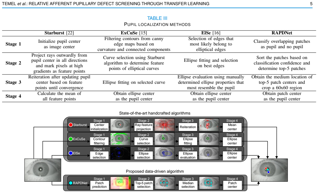
The central object is RAPDNet, a transfer-learned pupil detector. A convolutional feature extractor pretrained for generic object recognition is kept fixed; its final classifier is replaced with one fully connected layer trained from scratch on 50x50 pupil/no-pupil patches from existing eye-tracking datasets, converging in a few epochs. During screening, overlapping patches are classified, the median of the top-5 confident pupil patches gives the pupil center, and a circular Hough transform measures pupil radius in a cropped patch. The RAPD index, $1 - \min(|\Delta R|, |\Delta L|) / \max(|\Delta R|, |\Delta L|)$, converts the left and right absolute pupil-size changes into a 0-to-1 dissimilarity score, and a per-algorithm threshold turns that score into a binary RAPD call. The transfer-learning machinery does the work of avoiding handcrafted feature engineering: the pretrained features already encode curved and intensity edges that distinguish the dark pupil from the surrounding iris.
What would settle it
Run the full pipeline on a new cohort in which each subject's RAPD status is established by masked agreement of at least two experienced examiners using standardized neutral-density-filter quantification, or by an independent automated pupillograph, and compare RAPDNet's thresholded scores against those labels; a large drop from 90.6% sensitivity/specificity, or substantial disagreement with the original clinical labels, would weaken the central claim.
Extended reading notes
Core claim
RAPDNet—a pupil detector made by taking a pretrained AlexNet convolutional network, discarding its original classifier, and training a single fully connected layer to label overlapping image patches as pupil or no pupil—can drive an automated swinging-flashlight RAPD test. Combined with circular Hough transform pupil-size measurement and a proposed RAPD index, the system classifies RAPD-positive versus no-RAPD cases with 90.6% sensitivity and 90.6% specificity and an AUC of 0.929 over 64 balanced test cases from a headset-collected HD video dataset. The paper reports that RAPDNet beats the Starburst, ExCuSe, and ElSe pupil-localization baselines on every one of the nine metrics considered, with the best handcrafted AUC at 0.848. It also reports that the handcrafted algorithms need per-dataset parameter tuning, whereas RAPDNet inherits generic visual features and learns only the final mapping.
Load-bearing premise
The clinical RAPD labels that define ground truth come from subjective swinging-flashlight and neutral-density-filter examinations by practitioners, with no reported inter-rater reliability or repeatability; if those labels are partly wrong, the measured sensitivity and specificity are not meaningful.
Editorial extensions
If this is right
- If replicated, a headset-based automated swinging flashlight test with RAPDNet can produce objective RAPD screening without relying on a practitioner's subjective judgment, which matters where ophthalmology specialists are scarce.
- Because only a single fully connected layer is trained, adapting the pupil detector to a new headset or camera geometry may require modest new labeled data rather than retraining a full network.
- The RAPD index is a continuous 0-to-1 score, not just a binary label, so the same framework could support repeated monitoring of a patient's pupillary asymmetry over time.
- The benchmark suggests that choosing the detection threshold separately per algorithm is necessary; a deployment-ready system would need to fix that threshold before use on new patients.
Reading between the lines
- Editorial inference: the reported 90.6% figures are likely optimistic because the detection threshold was selected on the same 64 test cases that produced the ROC curve; an independent test set would probably show lower accuracy.
- Editorial inference: if the transfer-learning recipe generalizes, the same frozen-features-plus-one-layer approach could be applied to other pupillary measurements, such as constriction latency, amplitude, or velocity, and to anisocoria screening, since all share the same pupil-localization front end.
- Editorial inference: the headset isolates each eye and controls the light stimulus, so the validation covers this specific geometry; performance on video from off-axis or handheld cameras remains untested.
Signed reviews
Editorial analysis
A structured set of objections, weighed in public.
Referee Report
Summary. The paper presents an automated framework for screening relative afferent pupillary defect (RAPD) using video sequences recorded by a custom headset. Pupils are localized either by handcrafted algorithms (Starburst, ExCuSe, ElSe) or by a proposed transfer-learned pupil detector based on AlexNet with a single new fully connected layer, trained on ElSe dataset patches. Pupil diameters are estimated via Circular Hough Transform, and a dissimilarity index (RAPD index) is computed from left/right pupil size changes. On 64 balanced clinical test cases, the proposed RAPDNet achieves 90.6% sensitivity and specificity and AUC 0.929, outperforming the handcrafted algorithms. The authors conclude that transfer learning from generic object recognition provides a practical objective RAPD screening method.
Significance. If the reported performance transfers to independent clinical data, this would be a practically valuable contribution: a low-cost, portable, automated RAPD screening tool. The paper's strengths include the development of a new clinical dataset (RAPD-HD), a careful comparison of existing pupil datasets, a benchmark of three established pupil localization algorithms, and a transfer-learning scheme that requires training only one fully connected layer. The pupil detector itself is evaluated on a separate (ElSe) test set, which is a sound use of external data. However, the headline RAPD detection metrics are compromised by test-set selection of the threshold and configuration, and the claimed dominance across all metrics relies on different configurations per metric. These issues need to be addressed before the quantitative claims can be accepted.
major comments (4)
- [V-B, IV-D] The classification threshold is selected on the same 64 test cases by measuring sensitivity and specificity for all possible threshold values and choosing the one that maximizes both (Section IV-D), and the patch size and smoothing configuration are selected per algorithm by the highest AUC on the same test set (Section V-B). Consequently, the reported sensitivity of 90.6%, specificity of 90.6%, and AUC of 0.929 are in-sample optimized values rather than unbiased estimates of performance on new patients. The abstract's claim that RAPDNet 'can achieve' these numbers is not supported as a prospective accuracy. The authors should provide a held-out validation cohort, nested cross-validation, or at least bootstrap confidence intervals, and should explicitly state that the current numbers are the result of test-set selection.
- [Table VI] The claim that 'RAPDNet outperforms all other algorithms in every performance category' is based on highlighting the best value per metric across rows, and different RAPDNet configurations attain the best value for different metrics: for example, the highest AUC ROC (0.929) uses image size/2 with moving average, while the highest accuracy (92.2%) and specificity (93.8%) use 60x60 without smoothing. Thus no single RAPDNet configuration simultaneously achieves all of the best values, and the comparison does not establish that a fixed RAPDNet pipeline dominates all handcrafted algorithms on all metrics. The authors should either compare configurations on a fixed basis (e.g., a single chosen configuration per algorithm, selected a priori) or clearly state that different configurations are used for different metrics.
- [III] The ground-truth RAPD labels were obtained from clinical diagnoses based on the swinging flashlight test and neutral density filter test, which are subjective. The paper reports no inter-rater agreement, no repeatability analysis, and no description of how many practitioners performed the diagnoses or whether they were masked to the algorithm results. If the reference standard is unreliable, the reported sensitivity and specificity are not anchored to a trustworthy ground truth. The authors should add a reliability analysis (e.g., repeated examinations or agreement between clinicians) or at minimum discuss this limitation explicitly.
- [IV-D] The definition of the RAPD index is not fully specified. The equation uses ΔR and ΔL as the 'pupil size change in the right eye' and 'pupil size change in the left eye,' but the manuscript does not define over which temporal interval or stimulus condition these changes are measured, nor how the multiple direct and consensual light reflex instances are aggregated. Without this information, the framework is not reproducible and the index could be computed in multiple inequivalent ways. Please specify the exact signal-processing steps that produce the scalar ΔR and ΔL for each test case.
minor comments (4)
- [I] The word 'propse' should be 'propose'.
- [V] The word 'hancrafted' should be 'handcrafted'.
- [IV-B] The training images are reported as '3,620 images from datasets XII, X, XIII, XIV, XVI, XVII, and XVIII in the ElSe study'; please clarify whether these are individual frames and how the dataset labels (e.g., 'XII') map to the ElSe publication.
- [IV-D] The phrase 'select the values that correspond to highest sensitivity and specificity' is ambiguous; it should state how ties between sensitivity and specificity are resolved.
Circularity Check
Threshold and configuration are fit on the same 64 test cases used to report sensitivity, specificity, and AUC.
-
fitted input called prediction
[Section IV-D, RAPD Assessment; results in Table VI, Section V-B]
"To determine the classification threshold for all the benchmarked algorithms, we measure the sensitivity and specificity for all possible threshold values and select the values that correspond to highest sensitivity and specificity for each algorithm."
The threshold that separates RAPD-positive from no-RAPD is chosen by maximizing sensitivity and specificity on the same 64 test cases that are later used to report those exact metrics. Therefore the headline 90.6% sensitivity and 90.6% specificity are the optimized in-sample operating point, not an independent prediction on new patients. The procedure is a resubstitution estimate: the parameter (threshold) is fitted to the evaluation set and then evaluated on the same set, forcing the reported point to be the best achievable on this cohort.
-
fitted input called prediction
[Section V-B, RAPD Detection]
"In the remaining of this study, we utilize the patch size and smoothing configuration that leads to the highest AUC in ROC analysis for each algorithm."
The algorithm configuration (patch size and smoothing) is selected by comparing AUC values computed on the same 64 RAPD-HD test cases. Thus the reported AUC of 0.929 for RAPDNet is the maximum over the tried configurations on the test set, not an unbiased estimate for a held-out set. This configuration selection is part of the same test-set-fitting procedure as the threshold choice.
full rationale
The central circularity is in the evaluation protocol, not in the derivation of the algorithm. The paper fits two kinds of parameters directly to the 64 test cases: the classification threshold is chosen to maximize sensitivity and specificity, and the patch size plus smoothing configuration is chosen to maximize AUC. The headline numbers in the abstract are then reported as what the algorithm 'can achieve' on those same 64 cases, which is an in-sample, optimized value rather than a prospective prediction. The AUC is threshold-independent, but it is still the maximum over configurations selected on the test set, so the point estimate 0.929 is also optimistically biased. No load-bearing self-citation or definitional circularity was found: the transfer-learning pupil detector is trained on the external ElSe dataset, and the RAPD index and handcrafted baselines are evaluated through the same protocol. The comparison among algorithms may be internally consistent because all methods receive the same test-set fitting, but the absolute claimed performance is not an independent validation. The paper would need a separate hold-out set, nested cross-validation, or explicit reporting of the selection procedure as in-sample fitting to support the abstract's prospective wording.
Assumptions & free parameters
free parameters (4)
- RAPD classification threshold =
not reported
- Patch size configuration =
60x60 for RAPDNet, image size/2 or 60x60 for others
- Smoothing configuration (moving average) =
applied or not per algorithm
- CHT Canny and accumulator thresholds =
not reported
assumptions (4)
- domain assumption Pupil size measured by Circular Hough Transform with radius 5-30 pixels corresponds to physical pupil diameters of 2-8 mm.
- domain assumption AlexNet ImageNet features transfer to pupil detection on a different imaging setup.
- domain assumption The clinical RAPD diagnosis by swinging flashlight and neutral density filter tests is an accurate gold standard.
- domain assumption There is at most one pupil in the field of view.
Cite this review
Pith. "Pith review of Relative Afferent Pupillary Defect Screening through Transfer Learning." pith.science (2026). https://pith.science/paper/U4WII6AF
@misc{pith2026190802300,
  author       = {Pith},
  title        = {Pith review of: Relative Afferent Pupillary Defect Screening through Transfer Learning},
  year         = {2026},
  howpublished = {\url{https://pith.science/paper/U4WII6AF}},
  note         = {Machine review of arXiv:1908.02300}
}
read the original abstract
Abnormalities in pupillary light reflex can indicate optic nerve disorders that may lead to permanent visual loss if not diagnosed in an early stage. In this study, we focus on relative afferent pupillary defect (RAPD), which is based on the difference between the reactions of the eyes when they are exposed to light stimuli. Incumbent RAPD assessment methods are based on subjective practices that can lead to unreliable measurements. To eliminate subjectivity and obtain reliable measurements, we introduced an automated framework to detect RAPD. For validation, we conducted a clinical study with lab-on-a-headset, which can perform automated light reflex test. In addition to benchmarking handcrafted algorithms, we proposed a transfer learning-based approach that transformed a deep learning-based generic object recognition algorithm into a pupil detector. Based on the conducted experiments, proposed algorithm RAPDNet can achieve a sensitivity and a specificity of 90.6% over 64 test cases in a balanced set, which corresponds to an AUC of 0.929 in ROC analysis. According to our benchmark with three handcrafted algorithms and nine performance metrics, RAPDNet outperforms all other algorithms in every performance category.
Figures


Figures from the paper (3 more)


Reference graph
Works this paper leans on
-
[1]
National Eye Institute, “Eye Disease Statistics ,” [Online] Available: https://nei.nih.gov/sites/default/files/ nei-pdfs/NEI_Eye_Disease_Statistics_Factsheet_ 2014_V10.pdf. [Accessed: 30-May-2018]
work page 2018
-
[2]
American Academy of Ophthalmology, “Eye Health Statis- tics ,” [Online] Available: https://www.aao.org/newsroom/ eye-health-statistics, [Accessed: 30-May-2018]
work page 2018
-
[3]
Lab-on-a- heaset: Multi-purpose ocular monitoring,
G. AlRegib, Y . M. Khalifa, M. J. Mathew, and D. Temel, “Lab-on-a- heaset: Multi-purpose ocular monitoring,” in U.S. Provisional Patent, No: 62/642,279 , March 2018
work page 2018
-
[4]
G. AlRegib, Y . M. Khalifa, M. J. Mathew, and D. Temel, “Ocular monitoring headset,” in U.S. Non-Provisional Patent, No: 16/382,363 , March 2019
work page 2019
-
[5]
Transfer learning for medical applications using limited data,
G. AlRegib, M. J. Mathew, and D. Temel, “Transfer learning for medical applications using limited data,” in U.S. Provisional Patent, No: 62/853,753 , May 2019
work page 2019
-
[6]
Eyeing up the Future of the Pupillary Light Reflex in Neurodiagnostics,
C. A. Hall and R. P. Chilcott, “Eyeing up the Future of the Pupillary Light Reflex in Neurodiagnostics,” Diagnostics, vol. 8, no. 1, Mar 2018
work page 2018
-
[7]
How to test for a relative afferent pupillary defect (RAPD),
D. C. Broadway, “How to test for a relative afferent pupillary defect (RAPD),” Community Eye Health , vol. 25, no. 79-80, pp. 58–59, 2012
work page 2012
-
[8]
Chapter 16 - disorders of the pupil,
H. Wilhelm, “Chapter 16 - disorders of the pupil,” in Neuro- ophthalmology, C. Kennard and R. J. Leigh, Eds., vol. 102 of Handbook of Clinical Neurology , pp. 427 – 466. Elsevier, 2011
work page 2011
Show all 33 references
-
[9]
High variance in pupillary examination findings among postanesthesia care unit nurses,
L. Omburo et al. , “High variance in pupillary examination findings among postanesthesia care unit nurses,” Journal of PeriAnesthesia Nursing, vol. 32, no. 3, pp. 219 – 224, 2017
2017
-
[10]
Iris Mechanics I. Influences of TEMEL et al.: RELATIVE AFFERENT PUPILLARY DEFECT SCREENING THROUGH TRANSFER LEARNING 9 Pupil Size on Dynamics of Pupillary Movements,
I. E. Loewenfeld and D. A. Newsome, “Iris Mechanics I. Influences of TEMEL et al.: RELATIVE AFFERENT PUPILLARY DEFECT SCREENING THROUGH TRANSFER LEARNING 9 Pupil Size on Dynamics of Pupillary Movements,” American Journal of Ophthalmology , vol. 71, no. 1, pp. 347–362, 1971
1971
-
[11]
Variability of the relative afferent pupillary defect,
A. Kawasaki, P. Moore, and R. H. Kardon, “Variability of the relative afferent pupillary defect,” American Journal of Ophthalmology , vol. 120, no. 5, pp. 622 – 633, 1995
1995
-
[12]
Robust real-time pupil tracking in highly off-axis images,
L. ´Swirski, A. Bulling, and N. Dodgson, “Robust real-time pupil tracking in highly off-axis images,” in Proceedings of the Symposium on Eye Tracking Research and Applications , 2012, pp. 173–176
2012
-
[13]
Driving with binocular visual field loss? a study on a supervised on-road parcours with simultaneous eye and head tracking,
E. Kasneci et al., “Driving with binocular visual field loss? a study on a supervised on-road parcours with simultaneous eye and head tracking,” PLOS ONE , vol. 9, no. 2, pp. 1–13, 02 2014
2014
-
[14]
Binocular glaucomatous visual field loss and its impact on visual exploration - a supermarket study,
K. Sippel et al., “Binocular glaucomatous visual field loss and its impact on visual exploration - a supermarket study,” PLOS ONE, vol. 9, no. 8, pp. 1–7, 08 2014
2014
-
[15]
Excuse: Robust pupil detection in real-world scenarios,
W. Fuhl et al., “Excuse: Robust pupil detection in real-world scenarios,” in Computer Analysis of Images and Patterns , George Azzopardi and Nicolai Petkov, Eds., Cham, 2015, pp. 39–51, Springer International Publishing
2015
-
[16]
Else: Ellipse selection for robust pupil detection in real-world environments,
W. Fuhl, T. C. Santini, T. K ¨ubler, and E. Kasneci, “Else: Ellipse selection for robust pupil detection in real-world environments,” in Proceedings of the Ninth Biennial ACM Symposium on Eye Tracking Research & Applications, New York, NY , USA, 2016, pp. 123–130
2016
-
[17]
Non-intrusive practitioner pupil detection for unmodified microscope oculars,
W. Fuhl et al., “Non-intrusive practitioner pupil detection for unmodified microscope oculars,” Computers in Biology and Medicine , vol. 79, pp. 36 – 44, 2016
2016
-
[18]
Labelled pupils in the wild: A dataset for studying pupil detection in unconstrained environments,
M. Tonsen, X. Zhang, Y . Sugano, and A. Bulling, “Labelled pupils in the wild: A dataset for studying pupil detection in unconstrained environments,” in Proceedings of the Ninth Biennial ACM Symposium on Eye Tracking Research & Applications , New York, NY , USA, 2016, ETRA ’16...
2016
-
[19]
Portable pupillography of the swinging flashlight test to detect afferent pupillary defects,
N. J. V olpe et al. , “Portable pupillography of the swinging flashlight test to detect afferent pupillary defects,” Ophthalmology, vol. 107, no. 10, pp. 1913 – 1921, 2000
1913
-
[20]
Pupillography of automated swinging flashlight test in amblyopia,
A. Miki et al. , “Pupillography of automated swinging flashlight test in amblyopia,” Clinical Ophthalmology , vol. 2, no. 4, pp. 781–786, 2008
2008
-
[21]
Detection of asymmetric glaucomatous damage using automated pupillography, the swinging flashlight method and the magnified-assisted swinging flashlight method,
M Waisbourd et al. , “Detection of asymmetric glaucomatous damage using automated pupillography, the swinging flashlight method and the magnified-assisted swinging flashlight method,” Nature Eye, vol. 29, pp. 1321–1328, 2015
2015
-
[22]
Starburst: A hybrid algorithm for video-based eye tracking combining feature-based and model-based approaches,
D. Li, D. Winfield, and D. J. Parkhurst, “Starburst: A hybrid algorithm for video-based eye tracking combining feature-based and model-based approaches,” in IEEE Computer Society Conference on Computer Vision and Pattern Recognition Workshops , June 2005, pp. 79–79
2005
-
[23]
Automated pupillary light reflex assessment on a portable platform,
D. Temel, M. J. Mathew, G. AlRegib, and Y . M. Khalifa, “Automated pupillary light reflex assessment on a portable platform,” IEEE Inter- national Symposium on Medical Robotics, , 2019
2019
-
[24]
Pupilnet: Convolu- tional neural networks for robust pupil detection,
W. Fuhl, T. Santini, G. Kasneci, and E. Kasneci, “Pupilnet: Convolu- tional neural networks for robust pupil detection,” in arXiv:1601.04902, 2016
2016 arXiv
-
[25]
Pupilnet v2.0: Convolutional neural networks for cpu based real time robust pupil detection,
W. Fuhl, T. Santini, W. Rosenstiel, and E. Kasneci, “Pupilnet v2.0: Convolutional neural networks for cpu based real time robust pupil detection,” in arXiv:1711.00112, 2017
2017 arXiv
-
[26]
Least-squares fitting of circles and ellipses,
W. Gander, G. H. Golub, and R. Strebel, “Least-squares fitting of circles and ellipses,” 1994
1994
-
[27]
Robust circle detection,
B. Lamiroy, L. Fritz, and O. Gaucher, “Robust circle detection,” in Ninth International Conference on Document Analysis and Recognition , Sept 2007, vol. 1, pp. 526–530
2007
-
[28]
Least squares fitting of circles and lines,
N. Chernov and C. Lesort, “Least squares fitting of circles and lines,” in arXiv:0301001, 2008
2008
-
[29]
Imagenet: A large-scale hierarchical image database,
J. Deng et al. , “Imagenet: A large-scale hierarchical image database,” in 2009 IEEE Conference on Computer Vision and Pattern Recognition , June 2009, pp. 248–255
2009
-
[30]
Imagenet classification with deep convolutional neural networks,
A. Krizhevsky, I. Sutskever, and G. E. Hinton, “Imagenet classification with deep convolutional neural networks,” in Advances in Neural Information Processing Systems , F. Pereira, C. J. C. Burges, L. Bottou, and K. Q. Weinberger, Eds., pp. 1097–1105. 2012
2012
-
[31]
Torchvision Models Package,
PyTorch, “Torchvision Models Package,” [Online] Available: https://pytorch.org/docs/stable/torchvision/ models.html. [Accessed: 7-March-2019]
2019
-
[32]
The OpenCV Library,
G. Bradski, “The OpenCV Library,” Dr . Dobb’s Journal of Software Tools, 2000
2000
-
[33]
R. H. Spector, Clinical Methods: The History, Physical, and Laboratory Examinations. 3rd edition. , chapter 58: The Pupils, Walker HK, Hall WD, Hurst JW, editors. Boston: Butterworths, 1990. Dogancan Temel (M’13) received the M.S. and the Ph.D. degrees in electrical and comput...
2017
Reviewed August 14, 2026 · model on record in the stance chip above.
Discussion (0). Continue with ORCID to comment.