REVIEW 4 major objections 6 minor 64 references
Insights into experimental evaluation of the non-fourier heat transfer model in biological tissues
T0 review · 4 major / 6 minor · reviewed 2026-08-06 · deepseek-v4-flash
Pith's one-line read Bovine skin experiments show the TPL heat model's phase lags are under 0.5 seconds, much smaller than many values used in bioheat simulations.
desk verdict Real experimental effort, but the phase-lag bounds are not yet quantitatively supported; as written, the paper overclaims its headline result. read the letter →
The pith
A machine-rendered reading of the paper's core claim, the machinery that carries it, and where it could break.
The reading
What carries the argument
The central object is the Two-Dimensional Three-Phase Lag (TPL) bioheat equation, a constitutive law relating heat flux and temperature gradient through four parameters: the heat-flux phase lag $\tau_q$, the temperature-gradient phase lag $\tau_\theta$, the thermal displacement coefficient $k^*$, and the thermal displacement phase lag $\tau_v$ (thermal displacement $v$ has time derivative equal to temperature). The paper solves this equation with an implicit finite-difference scheme using central spatial and forward temporal differences, couples it to a Beer-Lambert Gaussian laser heat source, and evaluates each parameter by comparing simulated temperature histories to experimental infrared-sensor readings. The load-bearing mechanism is the decoupled experiment: two laser pulses of equal energy but different delivery rates isolate $\tau_q$; pulsed irradiation with varying periods isolates $\tau_\theta$ via heating/cooling slope asymmetry; a 3 s pulse with two sensors monitors for the sensor-sign reversal that would signal $k^*$ wave propagation; and Equation (13) shows $k^* \tau_v$ acts as an additive conductivity, motivating $\tau_v = 0$.
What would settle it
Measure the thermal conductivity and heat capacity of the actual bovine sample (e.g., with a guarded hot-plate or differential scanning calorimeter) and rerun the two-rate laser experiment; if the $\tau_q$ value that matches the measured temperature difference shifts outside the reported <0.5 s range, the phase-lag estimates are artifacts of the assumed properties. Alternatively, repeat the irradiation with a temperature sensor sampling faster than 0.1 s; if the maximum temperature difference between the two laser cases keeps shrinking, $\tau_q$ is below 0.1 s and the current setup cannot resolve it.
Extended reading notes
Core claim
On the paper's own terms, the central discovery is that the phase-lag parameters governing non-Fourier heat conduction in bovine skin are sub-second and partially redundant. By matching a two-dimensional axisymmetric finite-difference TPL simulation to temperature histories measured with MLX90614 infrared sensors, the authors estimate $\tau_q < 0.5$ s (simulation at $\tau_q = 0.1$ matches the measured maximum temperature difference between two laser delivery rates), $\tau_\theta < 0.25$ s, $k^* \approx 0$ (Sensor 1 never falls below Sensor 2, so no thermal-wave reversal appears), and $\tau_v = 0$ (because $k^* \tau_v$ merely adds to effective thermal conductivity). The paper concludes that several large phase-lag values reported in the literature are unrealistic and can produce thermal instabilities and undershoots of tens of kelvin, and it positions these experiments as a correction toward more precise, parameter-isolated validation.
Load-bearing premise
The extracted phase lags rest on treating the bovine skin as a thermally homogeneous material with literature values for thermal conductivity (0.45 W/m·K), specific heat (3500 J/kg·K), density (1000 kg/m³), and convection coefficient (15 W/m²·K) rather than values measured on the actual sample, and the paper states that no thorough experimental studies have exactly ascertained these characteristics.
Editorial extensions
If this is right
- Published phase-lag values of tens of seconds are likely unphysical for skin; bioheat simulations should use sub-second lags to avoid non-physical temperature undershoots of tens of kelvin.
- The thermal displacement coefficient $k^*$ is negligible at the mm-to-cm spatial scale and second timescales probed here, so the TPL model effectively reduces to a dual-phase-lag model for this setting.
- The thermal displacement phase lag $\tau_v$ is redundant: the product $k^* \tau_v$ adds to the measured thermal conductivity, so it can be set to zero whenever $k^*$ is negligible.
- A two-rate laser protocol (same total energy, different delivery rates) gives an operational estimate of $\tau_q$, but only down to the sensor's 0.1 s temporal resolution.
Reading between the lines
- If the same protocol were applied to perfused human skin in vivo, the extracted lags could shift because blood perfusion and metabolic heating are absent from the excised sample.
- The 0.1 s resolution floor means $\tau_q$ could be near zero; higher-bandwidth thermal imaging would reveal whether the non-Fourier lag terms are physically needed at all or whether a Fourier model with corrected surface boundary conditions matches the data.
- Because $\tau_\theta$ and $\tau_v$ both mimic increased thermal conductivity, temperature-only inversions are systematically confounded; jointly estimating conductivity and phase lags from the same data (for instance with a Bayesian approach) would tighten the reported ranges.
Signed reviews
Editorial analysis
A structured set of objections, weighed in public.
Referee Report
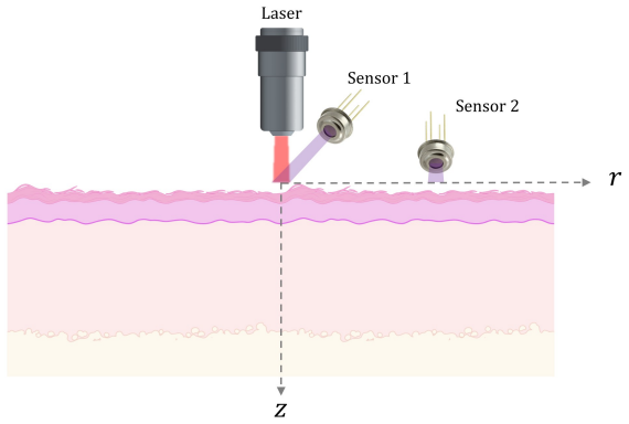
Summary. This paper implements a two-dimensional axisymmetric finite-difference solution of the Three-Phase Lag (TPL) heat conduction model and combines it with 450 nm laser experiments on excised bovine skin monitored by three infrared sensors. The stated goal is to extract four model parameters: the heat flux phase lag τ_q, the temperature gradient phase lag τ_θ, the thermal displacement coefficient k*, and the thermal displacement phase lag τ_v. The extraction strategy is a decoupled protocol: τ_q is inferred by matching the maximum temperature difference between two heating-rate cases (Cases 1 and 2), τ_θ is proposed to be inferred from heating/cooling slope asymmetry under periodic pulses, k* is inferred from the presence or absence of a wave-like reversal between Sensor 1 and Sensor 2, and τ_v is argued to be redundant because k*τ_v acts as an additional effective conductivity. The headline conclusion (Section 7, Table 3) is that the parameters are substantially lower than many literature values: τ_q < 0.5 s, τ_θ < 0.25 s, k* ≈ 0, and τ_v = 0.
Significance. If the quantitative claims were supported, the paper would provide useful experimental constraints on non-Fourier bioheat parameters, which are scattered and often weakly justified in the literature. The strengths are genuine: the idea of decoupling the four parameters is sound and clearly motivated by the model structure; repeated trials (10-20 per condition) are reported; the sensor field-of-view is modeled rather than treated as point measurements; and a thermal-conductivity sensitivity study is initiated. However, only τ_q is subjected to a quantitative comparison, and even that comparison lacks uncertainty quantification. The entries for τ_θ, k*, and τ_v in Table 3 are proposed protocols, qualitative null observations, or explicit assumptions rather than measured values. The paper is therefore best viewed as a promising protocol with pilot data, not as a validated parameter set; the requested revisions are primarily analysis and reporting tasks rather than new experiments.
major comments (4)
- [§6.1, Table 3] The upper bound τ_q < 0.5 s is not quantitatively established. The extraction is a parameter search: simulations with different τ_q are compared to the experimental maximum temperature difference between Cases 1 and 2, but the manuscript never reports the measured value of that maximum difference, the standard deviation across the 10-20 repeats, or a fitting metric. Figure 22 shows two measured temperature curves without a difference trace, and the text only states that τ_q = 0.1 'yields results most consistent' with the data. Because the sensor sampling interval is 0.1 s, the claimed best-fit value sits at the resolution limit, so the statement that τ_q values below 0.1 cannot be resolved does not by itself justify an upper bound of 0.5 s. Please report the experimental ΔT_max with its uncertainty and show the mismatch (e.g., residual or RMS error) for τ_q = 0, 0.1, 0.25, 0.5, and 1 s.
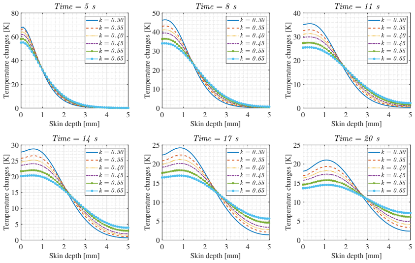
- [§3 and §5.5] The promised sensitivity propagation is missing. Section 3 states that simulations with 'a range of various thermal conductivity values' will ensure that uncertainty in the thermal properties 'does not compromise the accuracy of the derived phase lag estimates,' but Section 5.5 only displays temperature profiles for different k values; the τ_q extraction is never re-run under perturbed k. This is load-bearing because k = 0.45 W/m·K, c = 3500 J/kg·K, and ρ = 1000 kg/m³ (Table 1) are literature values admitted to be unmeasured (Section 3). The inferred τ_q is derived from differences between simulated temperature curves, and k uncertainty shifts those curves directly. Please recompute the Case 1/Case 2 comparison for k in, say, the range 0.3-0.6 W/m·K and report how the inferred τ_q range changes.
- [§6.2, Table 3] The value τ_θ < 0.25 s is not derived anywhere in the manuscript. Section 6.2 describes a proposed periodic-pulsed-laser protocol based on heating/cooling slope asymmetry, but no experimental data, fitted value, uncertainty, or comparison to literature is presented. The text itself notes that τ_θ effects closely resemble variations in k (Figures 7-9 versus 16-18), so without quantitative results the bound in Table 3 is unsupported. Please either remove the numerical range and state that τ_θ remains to be measured, or provide the promised protocol data with a fitting analysis that separates τ_θ from k.
- [§6.3 and §6.4] The conclusions k* ≈ 0 and τ_v = 0 are presented as results in Table 3 and Section 8, but they are not established at that level. The k* conclusion rests entirely on the qualitative observation that 'Sensor 1 was never observed to fall below that of Sensor 2' (Section 6.3); no trial count, sensor noise floor, or detection threshold for a wave-like crossing is provided, so the null observation can at most bound k* below an unreported detection limit. Section 6.4 argues that τ_v is redundant because k*τ_v adds to the effective conductivity, but that argument sets τ_v to zero rather than measuring it; Table 3 labels τ_v as 'assumed value,' yet the conclusion lists it as a finding. Please present a detection-threshold calculation for k* and explicitly label τ_v = 0 as an assumption rather than a measurement.
minor comments (6)
- [General] There are numerous figure-number mismatches; for example, Section 5.3 refers to 'Figure 16' for the k* time-history, while the corresponding caption is Figure 10, and it cites Figures 17 and 18 for depth and radial profiles that are captioned Figures 11 and 12. Please correct all cross-references.
- [Figure 3] The caption of Figure 3 states τ_q = 0.01, but the text describes it as the baseline Fourier case with all phase-lag coefficients zero; the caption should be made consistent with the text.
- [Equations (4) and (10)] Equations (4) and (10) are labeled ℛ1 and ℛ2 without definition; please use conventional equation numbering or define the operator notation explicitly.
- [Figure 22] The experimental temperature response in Figure 22 shows negative values during the first roughly 0.5 s; if this is sensor settling or ambient drift, please state this, since the maximum-difference metric used for τ_q is sensitive to early-time artifacts.
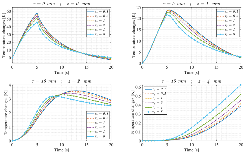
- [§5.4 and §6.4] Section 6.4 states that τ_v 'does not introduce a distinct physical meaning' and is redundant, but Section 5.4 and Figures 13-15 describe distinct, if similar, effects of τ_v compared with τ_θ; reconcile these descriptions so the modeling and the redundancy argument are consistent.
- [Data availability] The paper reports 10-20 repeats per condition but provides no raw data, no per-trial statistics, and no data availability statement; please add representative raw traces and trial statistics or deposit the data in a public repository.
Circularity Check
No significant circularity: the phase-lag estimates are inverse fits or qualitative comparisons against external experiments, not reductions of the model to its own inputs.
full rationale
The paper derives tau_q by an explicit inverse-matching protocol: two laser-energy delivery profiles are simulated under several tau_q values and compared with a measured maximum temperature difference between the two cases (Section 6.1). This is parameter estimation against an external observable, not a quantity fixed by construction. The k* estimate is a null result from experiment (Sensor 1 was never observed to fall below Sensor 2, Figs. 24-25), and tau_v is explicitly marked "assumed value" in Table 3 after showing that k*tau_v is algebraically degenerate with k (Eqs. 13-15). The constitutive equation is cited to the authors' Ref. [30], but the equation is written out and follows standard TPL/Green-Naghdi forms also attributed to Tzou [33] and Podio-Guidugli [34], so the self-citation is not load-bearing. The paper does have non-circular weaknesses: the tau_theta<0.25 entry in Table 3 has no shown experimental derivation; the promised sensitivity study of thermal conductivity (Section 3) is never propagated into re-extraction of tau_q; and a sentence states that phase-lag terms were "considered negligible (i.e., zero) during all the modeling," which contradicts the parameter variations in Sections 5-6. These are evidence-quality and consistency problems, not construction-level circularity.
Assumptions & free parameters
free parameters (4)
- tau_q (heat flux phase lag) =
0.1 s (best match), < 0.5 s range
- tau_theta (temperature gradient phase lag) =
< 0.25 s (proposed range)
- k* (thermal displacement coefficient) =
≈ 0 (negligible)
- tau_v (thermal displacement phase lag) =
0 (assumed)
assumptions (5)
- domain assumption The 2D axisymmetric representation adequately captures the 3D thermal response of the skin sample (Section 2).
- domain assumption Bovine skin is thermally homogeneous with literature values k, c, rho, h (Table 1).
- domain assumption Thermal convection and evaporation heat loss are neglected at the tissue surface except for a constant convection coefficient h (Section 3).
- domain assumption The MLX90614 sensor FOV can be modeled as an arc-shaped area average in the simulation (Section 6).
- domain assumption The TPL constitutive Eq. (3) is the correct model for skin tissue, including the Green-Naghdi thermal displacement term (Section 2).
Cite this review
Pith. "Pith review of Insights into experimental evaluation of the non-fourier heat transfer model in biological tissues." pith.science (2026). https://pith.science/paper/U6AP7RA6
@misc{pith2026250717627,
  author       = {Pith},
  title        = {Pith review of: Insights into experimental evaluation of the non-fourier heat transfer model in biological tissues},
  year         = {2026},
  howpublished = {\url{https://pith.science/paper/U6AP7RA6}},
  note         = {Machine review of arXiv:2507.17627}
}
abstract
A comprehensive understanding of heat transfer mechanisms in biological tissues is essential for the advancement of thermal therapeutic techniques and the development of accurate bioheat transfer models. Conventional models often fail to capture the inherently complex thermal behavior of biological media, necessitating more sophisticated approaches for experimental validation and parameter extraction. In this study, the Two-Dimensional Three-Phase Lag (TPL) heat transfer model, implemented via the finite difference method (FDM), was employed to extract key phase lag parameters characterizing heat conduction in bovine skin tissue. Experimental measurements were obtained using a 450 nm laser source and two non-contact infrared sensors. The influence of four critical parameters was systematically investigated: heat flux phase lag ($\tau_{q}$), temperature gradient phase lag ($\tau_{\theta}$), thermal displacement coefficient ($k^*$), and thermal displacement phase lag ($\tau_{v}$). A carefully designed experimental protocol was used to assess each parameter independently. The results revealed that the extracted phase lag values were substantially lower than those previously reported in the literature. This highlights the importance of high-precision measurements and the need to isolate each parameter during analysis. These findings contribute to the refinement of bioheat transfer models and hold potential for improving the efficacy and safety of clinical thermal therapies.
Figures


Figures from the paper (23 more)






Reference graph
Works this paper leans on
- [1]
-
[2]
Z. Shomali, R. Kovács, P. Ván, I. V. Kudinov, J. Ghazanfarian, Lagging heat models in thermodynamics and bioheat transfer: a critical review, Continuum Mechanics and Thermodynamics 34 (3) (2022) 637–679
work page 2022
-
[3]
Y. Zhang, Generalized dual-phase lag bioheat equations based on nonequilibrium heat transfer in living biological tissues, International Journal of Heat and Mass Transfer 52 (21-22) (2009) 4829–4834
work page 2009
- [4]
-
[5]
E. G. Cravalho, L. R. Fox, J. C. Kan, The application of the bioheat equation to the design of thermal protocols for local hyperthermia, An- nals of the New York Academy of Sciences 335 (1) (1980) 86–97
work page 1980
-
[6]
H.-W. Huang, T.-L. Horng, Bioheat transfer and thermal heating for tumor treatment, in: Heat transfer and fluid flow in biological processes, Elsevier, 2015, pp. 1–42
work page 2015
-
[7]
C. Li, J. Miao, K. Yang, X. Guo, J. Tu, P. Huang, D. Zhang, Fourier and non-fourier bio-heat transfer models to predict ex vivo tempera- ture response to focused ultrasound heating, Journal of Applied Physics 123 (17) (2018)
work page 2018
- [8]
Show all 64 references
-
[9]
de Monte, A
F. de Monte, A. Haji-Sheikh, Bio-heat diffusion under local thermal non-equilibrium conditions using dual-phase lag-based green’s functions, International journal of heat and mass transfer 113 (2017) 1291–1305
2017
-
[10]
J. Zhou, J. Chen, Y. Zhang, Dual-phase lag effects on thermal damage to biological tissues caused by laser irradiation, Computers in biology and medicine 39 (3) (2009) 286–293
2009
-
[11]
M. G. Salem, A. E. Abouelregal, F. Alsharari, H. M. Sedighi, Ther- momechanical response of biological tissues to sudden temperature rise induced by laser beam: insights from three-phase lag theory, Physica Scripta 100 (1) (2024) 015290
2024
-
[12]
Tzou, Lagging behavior in biological systems, in: International Con- ference on Micro/Nanoscale Heat Transfer, Vol
D. Tzou, Lagging behavior in biological systems, in: International Con- ference on Micro/Nanoscale Heat Transfer, Vol. 43918, 2009, pp. 523– 535
2009
-
[13]
Liu, H.-T
K.-C. Liu, H.-T. Chen, Analysis for the dual-phase-lag bio-heat transfer during magnetic hyperthermia treatment, International Journal of Heat and Mass Transfer 52 (5-6) (2009) 1185–1192
2009
-
[14]
Afrin, Y
N. Afrin, Y. Zhang, J. Chen, Thermal lagging in living biological tissue based on nonequilibrium heat transfer between tissue, arterial and ve- nous bloods, International Journal of Heat and Mass Transfer 54 (11-12) (2011) 2419–2426. 49
2011
-
[15]
Liu, H.-T
K.-C. Liu, H.-T. Chen, P.-J. Cheng, Inverse investigation of non-fourier heat conduction in tissue, Journal of thermal biology 62 (2016) 123–128
2016
-
[16]
Lin, C.-Y
S.-M. Lin, C.-Y. Li, Analytical solutions of non-fourier bio-heat conduc- tions for skin subjected to pulsed laser heating, International Journal of Thermal Sciences 110 (2016) 146–158
2016
-
[17]
Dinda, J
A. Dinda, J. Acharya, D. Bhanja, S. Nath, Local thermal non- equilibrium bioheat transfer model for interstitial hyperthermia treat- ment of tumour cell: A numerical approach, Journal of Thermal Biology 110 (2022) 103368
2022
-
[18]
Kumar, K
D. Kumar, K. Rai, Three-phase-lag bioheat transfer model and its val- idation with experimental data, Mechanics Based Design of Structures and Machines 50 (7) (2022) 2493–2507
2022
-
[19]
Singh, P
S. Singh, P. Saccomandi, R. Melnik, Three-phase-lag bio-heat transfer model of cardiac ablation, Fluids 7 (5) (2022) 180
2022
-
[20]
Liu, J.-S
K.-C. Liu, J.-S. Leu, Heat transfer analysis for tissue with surface heat flux based on the non-linearized form of the three-phase-lag model, Jour- nal of Thermal Biology 112 (2023) 103436
2023
-
[21]
Zhang, Y
Q. Zhang, Y. Sun, J. Yang, Thermoelastic responses of biological tissue under thermal shock based on three phase lag model, Case Studies in Thermal Engineering 28 (2021) 101376
2021
-
[22]
Zhang, Y
Q. Zhang, Y. Sun, J. Yang, Bio-heat response of skin tissue based on three-phase-lag model, Scientific Reports 10 (1) (2020) 16421
2020
-
[23]
Zhang, X
L. Zhang, X. Shang, An integral transform solution for bioheat transfer in skin tissue subjected to surface laser irradiation, International Journal of Heat and Mass Transfer 180 (2021) 121706
2021
-
[24]
Gupta, A
P. Gupta, A. Srivastava, Non-fourier transient thermal analysis of bio- logical tissue phantoms subjected to high intensity focused ultrasound, International Journal of Heat and Mass Transfer 136 (2019) 1052–1063
2019
-
[25]
B. Du, G. Xu, D. Xue, J. Wang, Fractional thermal wave bio-heat equa- tion based analysis for living biological tissue with non-fourier neumann boundary condition in laser pulse heating, Optik 247 (2021) 167811. 50
2021
-
[26]
Y. Yin, M. Li, Y. Li, J. Song, Skin pain sensation of epidermal electronic device/skin system considering non-fourier heat conduction, Journal of the Mechanics and Physics of Solids 138 (2020) 103927
2020
-
[27]
Azhdari, G
M. Azhdari, G. Rezazadeh, L. Lambers, T. Ricken, H.-M. Tautenhahn, F. Tautenhahn, S. M. Seyedpour, Refining thermal therapy: Tempera- ture distribution modeling with distinct absorption in multi-layered skin tissue during infrared laser exposure, International Communications i...
2024
-
[28]
Azhdari, G
M. Azhdari, G. Rezazadeh, T. Ricken, R. Pathak, H.-M. Tautenhahn, F. Tautenhahn, S. M. Seyedpour, Temperature distribution in multi- layered skin tissue during laser irradiation considering epidermis sub- layers: Virtual element method approach, Thermal Science and Engi- neeri...
2025
-
[29]
Z. A. Mirza, M. Azhdari, D. Kolomenskiy, G. Rezazadeh, T. Ricken, R. Pathak, H.-M. Tautenhahn, F. Tautenhahn, S. M. Seyedpour, En- hancing laser therapy procedure through surface temperature control in multi-layered skin tissue, Journal of Thermal Biology (2025) 104106
2025
-
[30]
Azhdari, S
M. Azhdari, S. M. Seyedpour, L. Lambers, H.-M. Tautenhahn, F. Taut- enhahn, T. Ricken, G. Rezazadeh, Non-local three phase lag bio ther- mal modeling of skin tissue and experimental evaluation, Interna- tional Communications in Heat and Mass Transfer 149 (2023) 107146. doi:10....
2023
-
[31]
Strąkowska, G
M. Strąkowska, G. De Mey, B. Więcek, Experimental verification of non-fourier heat transfer in a multilayer skin tissue struc-ture by ir tem- perature measurement
-
[32]
D. Y. Tzou, A unified field approach for heat conduction from macro-to micro-scales (1995)
1995
-
[33]
D. Y. Tzou, A unified field approach for heat conduction from macro- to micro-scales, Journal of Heat Transfer 117 (1) (1995) 8–16. doi:10.1115/1.2822329
1995 doi
-
[34]
Podio-Guidugli, A virtual power format for thermomechanics, Con- tinuum Mechanics and Thermodynamics 20 (2009) 479–487
P. Podio-Guidugli, A virtual power format for thermomechanics, Con- tinuum Mechanics and Thermodynamics 20 (2009) 479–487. 51
2009
-
[35]
A. N. Bashkatov, E. A. Genina, V. V. Tuchin, Optical properties of skin, subcutaneous, and muscle tissues: a review, Journal of Innovative Opti- cal Health Sciences 4 (01) (2011) 9–38. doi:10.1142/S1793545811001319
2011 doi
-
[36]
Bhatt, K
M. Bhatt, K. R. Ayyalasomayajula, P. K. Yalavarthy, Generalized beer– lambert model for near-infrared light propagation in thick biological tissues, Journal of Biomedical Optics 21 (7) (2016) 076012–076012. doi:10.1117/1.JBO.21.7.076012
2016 doi
-
[37]
Oshina, J
I. Oshina, J. Spigulis, Beer–lambert law for optical tissue diagnostics: current state of the art and the main limitations, Journal of biomedical optics 26 (10) (2021) 100901–100901. doi:10.1117/1.JBO.26.10.100901
2021 doi
-
[38]
Saemathong, N
J. Saemathong, N. Pannucharoenwong, V. Mongkol, S. Vongpradubchai, P. Rattanadecho, Analyzing two laser thermal energy calculation equa- tions: A comparison of beer-lambert’s law and light transport equation, Engineered Science (2023). doi:10.30919/es912
2023 doi
-
[39]
J. E. Hill, J. D. Leitman, S. JE, Thermal conductivity of various meats, Food technology 21 (8) (1967) 1143
1967
-
[40]
Bhattacharya, R
A. Bhattacharya, R. L. Mahajan, Temperature dependence of ther- mal conductivity of biological tissues, Physiological measurement 24 (3) (2003) 769
2003
-
[41]
J. R. Kanagy, Specific heats of collagen and leather, Journal of Research of the National Bureau of Standards 55 (4) (1955) 191–195
1955
-
[42]
Z. Pan, R. P. Singh, Physical and thermal properties of ground beef during cooking, L WT-Food Science and Technology 34 (7) (2001) 437– 444
2001
-
[43]
J. Xu, A. Psikuta, J. Li, S. Annaheim, R. M. Rossi, Evaluation of the convective heat transfer coefficient of human body and its effect on the human thermoregulation predictions, Building and Environment 196 (2021) 107778
2021
-
[44]
N. N. Mohd Nazali, N. F. Adull Manan, The numerical analysis of hy- perelastic properties in commercial and original aloe vera gels, Journal of Mechanical Engineering 18 (3) (2021) 1–20. 52
2021
-
[45]
P. J. Rodríguez de Rivera, M. Rodríguez de Rivera, F. Socorro, J. A. Calbet, M. Rodríguez de Rivera, A dvantages of in vivo measurement of human skin thermal conductance using a calorimetric sensor, Journal of Thermal Analysis and Calorimetry 147 (18) (2022) 10027–10036
2022
-
[46]
Azhdari, S
M. Azhdari, S. M. Seyedpour, T. Ricken, G. Rezazadeh, On the thermo- vibrational response of multi-layer viscoelastic skin tissue to laser irra- diation, International Journal of Thermal Sciences 187 (2023) 108160
2023
-
[47]
S. M. Seyedpour, M. Azhdari, L. Lambers, T. Ricken, G. Reza- zadeh, One-dimensional thermomechanical bio-heating analy- sis of viscoelastic tissue to laser radiation shapes, Interna- tional Journal of Heat and Mass Transfer 218 (2024) 124747. doi:10.1016/j.ijheatmasstransfer.2...
2024
-
[48]
M. Ciesielski, Application of the alternating direction implicit method for numerical solution of the dual-phase lag equation, Jour- nal of Theoretical and Applied Mechanics 55 (3) (2017) 839–852. doi:10.15632/jtam-pl.55.3.839
2017 doi
-
[49]
Majchrzak, B
E. Majchrzak, B. Mochnacki, Implicit scheme of the finite dif- ference method for 1d dual-phase lag equation, Journal of Ap- plied Mathematics and Computational Mechanics 16 (3) (2017). doi:10.17512/jamcm.2017.3.04
2017 doi
-
[50]
Majchrzak, B
E. Majchrzak, B. Mochnacki, Implicit scheme of the finite differ- ence method for the second-order dual phase lag equation, Jour- nal of Theoretical and Applied Mechanics 56 (2) (2018) 393–402. doi:10.15632/jtam-pl.56.2.393
2018 doi
-
[51]
Majchrzak, B
E. Majchrzak, B. Mochnacki, First and second order dual phase lag equation. numerical solution using the explicit and implicit schemes of the finite difference method, in: MATEC Web of Conferences, Vol. 240, EDP Sciences, 2018, p. 05018. doi:10.1051/matecconf/201824005018
2018
-
[52]
Majchrzak, B
E. Majchrzak, B. Mochnacki, Numerical solutions of the second-order dual-phase-lag equation using the explicit and implicit schemes of the finite difference method, International Journal of Numerical Methods for Heat & Fluid Flow 30 (4) (2020) 2099–2120. doi:10.1108/HFF-11- 20...
2020 doi
-
[53]
S. A. Rukolaine, Unphysical effects of the dual-phase-lag model of heat conduction, International Journal of Heat and Mass Transfer 78 (2014) 58–63
2014
-
[54]
S. A. Rukolaine, Unphysical effects of the dual-phase-lag model of heat conduction: higher-order approximations, International Journal of Thermal Sciences 113 (2017) 83–88
2017
-
[55]
M. A. Ezzat, M. H. Alabdulhadi, Thermomechanical interactions in vis- coelastic skin tissue under different theories, Indian Journal of Physics 97 (1) (2023) 47–60
2023
-
[56]
J. Ma, X. Yang, Y. Sun, J. Yang, Thermal damage in three-dimensional vivo bio-tissues induced by moving heat sources in laser therapy, Scien- tific reports 9 (1) (2019) 10987
2019
-
[57]
X. Li, X. Tian, The thermal injury analysis of skin tissue with a new nonlocal dual phase lag model, Waves in Random and Complex Media (2022) 1–14
2022
-
[58]
Zhang, B
Y. Zhang, B. Chen, D. Li, Non-fourier effect of laser-mediated thermal behaviors in bio-tissues: A numerical study by the dual-phase-lag model, International Journal of Heat and Mass Transfer 108 (2017) 1428–1438
2017
-
[59]
Tiwari, A
R. Tiwari, A. Singhal, R. Kumar, P. Kumar, S. Ghangas, Investigation of memory influences on bio-heat responses of skin tissue due to various thermal conditions, Theory in Biosciences 142 (3) (2023) 275–290
2023
-
[60]
A. Sur, S. Mondal, M. Kanoria, Influence of moving heat source on skin tissue in the context of two-temperature memory-dependent heat transport law, Journal of Thermal Stresses 43 (1) (2020) 55–71
2020
-
[61]
X. Li, X. Tian, Fractional order thermo-viscoelastic theory of biological tissue with dual phase lag heat conduction model, Applied Mathematical Modelling 95 (2021) 612–622
2021
-
[62]
Kumar, R
R. Kumar, R. Tiwari, A. Singhal, S. Mondal, Characterization of ther- mal damage of skin tissue subjected to moving heat source in the purview of dual phase lag theory with memory-dependent derivative, Waves in Random and Complex Media 34 (4) (2024) 3510–3527. 54
2024
-
[63]
A. M. Zenkour, T. Saeed, A. M. Aati, Analyzing the thermoelastic re- sponses of biological tissue exposed to thermal shock utilizing a three- phase lag theory, Journal of Computational Applied Mechanics 55 (2) (2024) 144–164
2024
-
[64]
K. K. Jojare, K. R. Gaikwad, A study of the 3-phase lag model to a two- dimensional isotropic micro-polar thermoelastic medium with memory- dependent properties, Journal of Thermal Stresses 47 (3) (2024) 363– 382. 55
2024
Reviewed August 6, 2026 · model on record in the stance chip above.
Discussion (0). Continue with ORCID to comment.