BCI-Based Assessment of Ocular Response Time Using Dynamic Time Warping Leveraging an RDWT-Driven Deep Neural Framework
Pith reviewed 2026-06-30 20:14 UTC · model grok-4.3
The pith
An RDWT-driven neural network combined with dynamic time warping estimates ocular response times from EEG during VOMS tasks and detects inter-subject differences.
A machine-rendered reading of the paper's core claim, the machinery that carries it, and where it could break.
Core claim
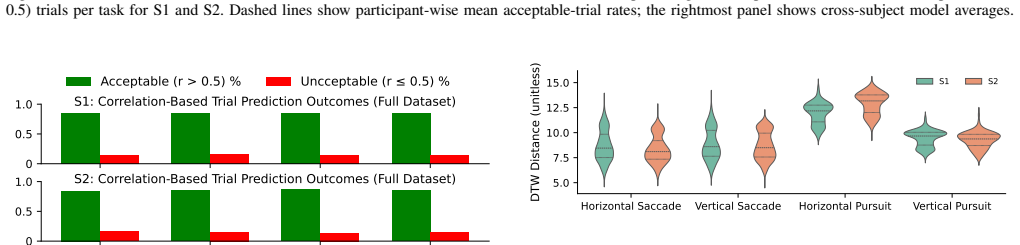
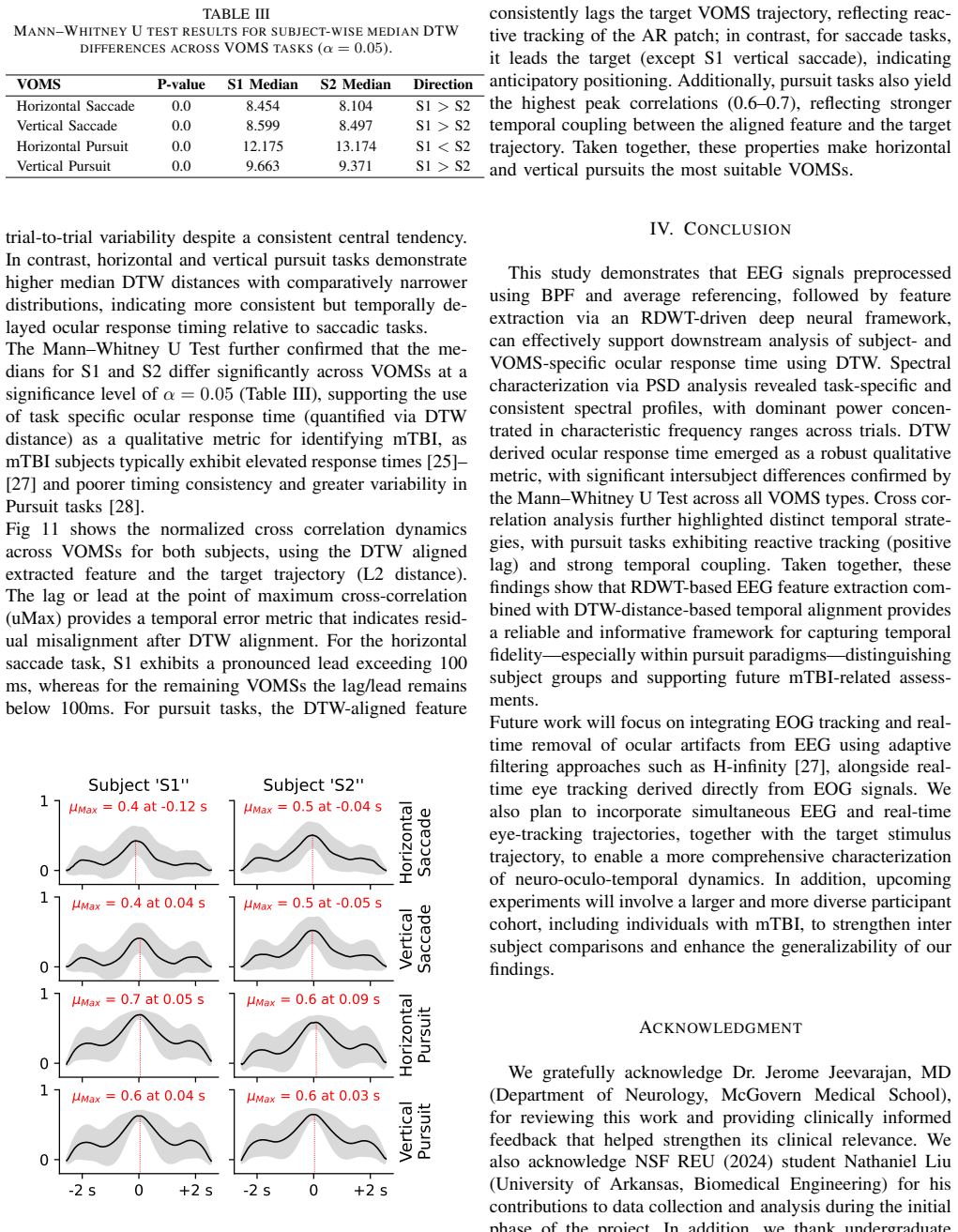
The framework uses RDWT coefficients fed into trainable zero-phase convolutional filters, followed by inverse transform, 2D convolutions, and conv-LSTM decoding to generate predictions from pre-processed EEG. These sliding-window predictions are validated with Pearson correlation of at least 0.5 and then aligned via DTW to estimate response times. Mann-Whitney U tests on the DTW metrics confirm significant inter-subject differences in all VOM tasks, while cross-correlation analysis indicates reactive tracking in pursuit tasks and anticipatory responses in saccades, highlighting the potential of RDWT-based EEG features combined with DTW metrics for multimodal mTBI assessment.
What carries the argument
The RDWT-driven deep neural framework that extracts and denoises EEG features for temporal alignment with dynamic time warping to compute ocular response times.
If this is right
- DTW-derived metrics can distinguish timing differences between subjects in VOMS tasks.
- Pursuit tasks provide particularly useful information for identifying response time variations.
- The combination of RDWT-based EEG features and DTW supports multimodal assessment of mTBI.
- Task-specific temporal patterns emerge, with pursuit showing reactive and saccades anticipatory behavior.
Where Pith is reading between the lines
- This method could enable portable, non-invasive screening tools for mTBI in settings without access to eye-tracking equipment.
- Extending the framework to include direct eye-tracking validation might strengthen the accuracy claims for clinical use.
- Similar approaches could be tested for other conditions involving oculomotor or vestibular dysfunction.
- The sliding-window approach might be adapted for real-time applications in neurorehabilitation.
Load-bearing premise
That neural network predictions of EEG features, validated only by correlation thresholds without simultaneous eye-tracking recordings, accurately reflect true ocular response times.
What would settle it
A study measuring the same subjects' eye movements with both the EEG framework and a gold-standard eye tracker during identical VOMS tasks, then comparing the derived response times.
Figures

read the original abstract
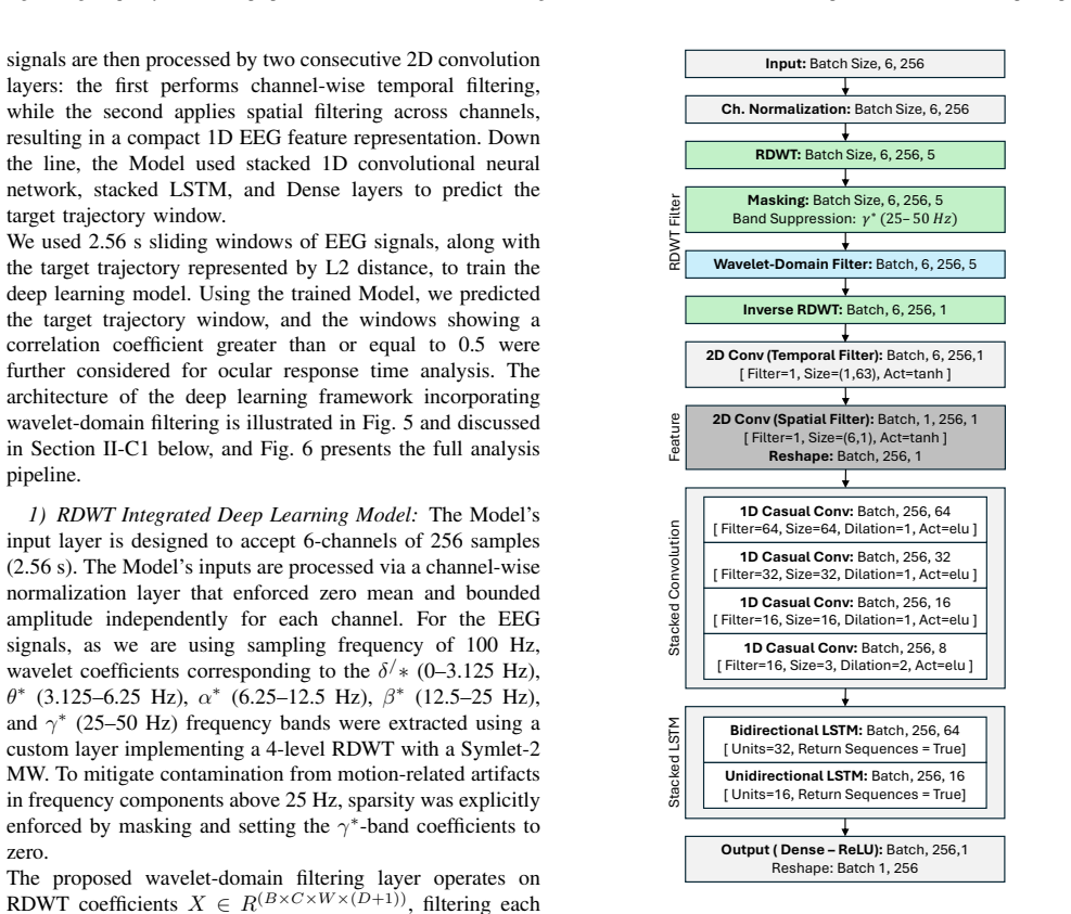
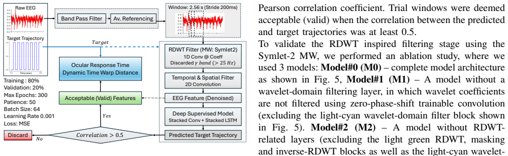
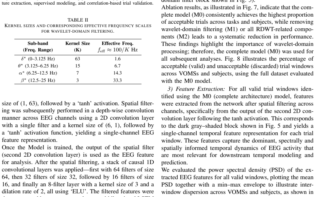
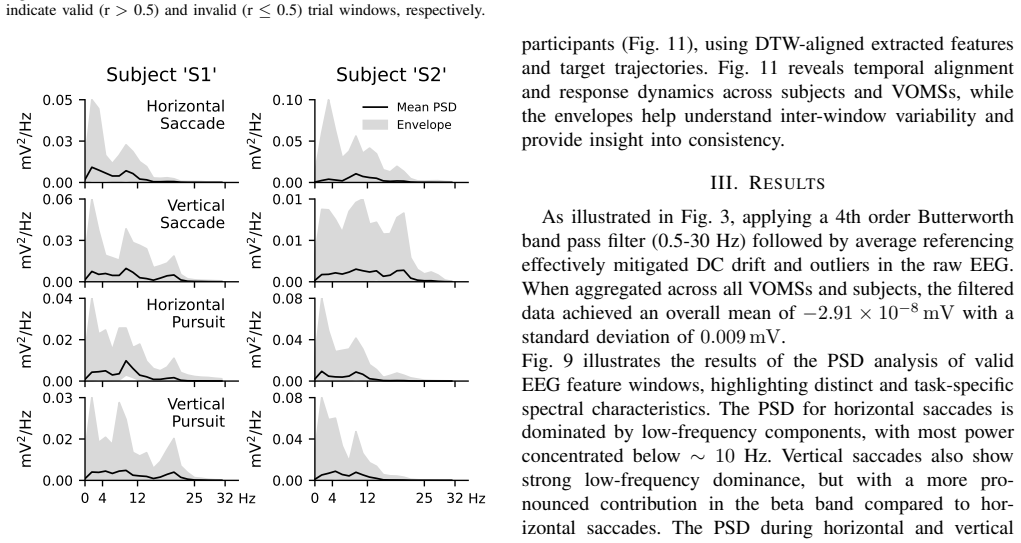
Mild traumatic brain injury (mTBI) is a prevalent condition that remains difficult to diagnose in its early stages. Oculomotor dysfunction is a well-established marker of mTBI, motivating the development of portable tools that capture both eye-movement behavior and underlying neurophysiology. In this work, we present an initial framework that integrates electroencephalogram (EEG) with augmented-reality (AR)-based Vestibular/Ocular Motor Screening (VOMS) tasks to estimate subject-specific ocular response times. Pre-processed EEG signals, obtained through band-pass filtering and average referencing, are analyzed using a Redundant Discrete Wavelet Transform (RDWT)-driven deep neural framework. The RDWT coefficients are subjected to trainable zero-phase convolutional filtering and reconstructed into the time domain via inverse RDWT, followed by channel-wise temporal and spatial filtering using 2D convolution layers and convolutional-LSTM-based decoding. An ablation study demonstrates that wavelet-domain filtering serves as an effective denoising strategy, improving prediction performance. Sliding-window predictions were validated using Pearson correlation (>= 0.5), and Dynamic Time Warping (DTW) was subsequently used to estimate ocular response times. DTW-derived metrics revealed significant inter-subject differences across all VOM tasks, supported by Mann-Whitney U tests. Cross-correlation analysis further revealed task-dependent temporal behaviors: pursuit tasks exhibited reactive tracking, whereas saccades showed anticipatory responses. Overall, the results highlight pursuit tasks as particularly informative for distinguishing timing differences and demonstrate the potential of RDWT-based EEG features combined with DTW metrics for multimodal mTBI assessment.
Editorial analysis
A structured set of objections, weighed in public.
Referee Report
Summary. The manuscript presents a BCI framework that processes EEG during AR-based VOMS tasks via an RDWT-driven DNN (trainable zero-phase convolutional filtering, inverse RDWT, 2D conv + conv-LSTM decoder) to generate sliding-window predictions of ocular response. These predictions are thresholded at Pearson r >= 0.5 and aligned with DTW to extract subject-specific response times; Mann-Whitney U tests on the resulting DTW metrics are reported to show significant inter-subject differences across VOMS tasks, with pursuit tasks highlighted as most informative for mTBI assessment.
Significance. If the DTW-derived latencies can be shown to track true oculomotor timing, the work would offer a portable multimodal signal-processing approach to mTBI screening that fuses neurophysiological features with behavioral timing. The ablation on wavelet-domain filtering and the cross-correlation analysis of task-dependent (reactive vs. anticipatory) behavior are positive elements. However, the absence of any direct validation against simultaneous eye-tracking or clinical timing standards leaves the central quantitative claim unsupported.
major comments (2)
- [Abstract] Abstract (final paragraph) and Results: the only validation reported for the sliding-window DNN outputs is a Pearson correlation threshold of >=0.5; no mean r values, no error distributions, no ablation of the threshold itself, and no comparison to a null or baseline predictor are supplied. Because the DTW step operates directly on these outputs, this threshold alone cannot establish that the extracted response times are accurate rather than artifacts of the model.
- [Abstract] Abstract and Discussion: no simultaneous eye-tracking ground truth, no comparison to clinical VOMS stopwatch timings, and no reported latency error bounds are provided for the DTW-derived ocular response times. Without such anchoring, the reported Mann-Whitney U significance on inter-subject differences cannot be interpreted as reflecting true oculomotor behavior rather than systematic prediction offsets.
minor comments (2)
- [Abstract] The abbreviation 'VOM tasks' appears once; it should be standardized to 'VOMS tasks' throughout for consistency with the method name.
- The ablation study is mentioned but not located in a numbered section or table; a dedicated subsection or table summarizing the performance lift from RDWT filtering would improve traceability.
Simulated Author's Rebuttal
We thank the referee for their constructive feedback on our manuscript. We address each major comment below, agreeing where additional details or clarifications are warranted while noting the preliminary scope of this EEG-focused framework.
read point-by-point responses
-
Referee: [Abstract] Abstract (final paragraph) and Results: the only validation reported for the sliding-window DNN outputs is a Pearson correlation threshold of >=0.5; no mean r values, no error distributions, no ablation of the threshold itself, and no comparison to a null or baseline predictor are supplied. Because the DTW step operates directly on these outputs, this threshold alone cannot establish that the extracted response times are accurate rather than artifacts of the model.
Authors: We agree that the reported validation is limited and that additional metrics are required to substantiate the DNN outputs before DTW application. In the revised manuscript we will add mean Pearson r values with distributions across windows and subjects, an ablation study varying the threshold, and a comparison against a null or baseline predictor to show that the predictions are not artifacts. revision: yes
-
Referee: [Abstract] Abstract and Discussion: no simultaneous eye-tracking ground truth, no comparison to clinical VOMS stopwatch timings, and no reported latency error bounds are provided for the DTW-derived ocular response times. Without such anchoring, the reported Mann-Whitney U significance on inter-subject differences cannot be interpreted as reflecting true oculomotor behavior rather than systematic prediction offsets.
Authors: We acknowledge that the study lacks simultaneous eye-tracking or clinical stopwatch ground truth, as the current work is confined to EEG signal processing during AR-VOMS tasks. The Mann-Whitney results therefore reflect differences in the model's predicted timings rather than validated oculomotor latencies. We will revise the Discussion to explicitly state this limitation and frame the findings as demonstrating the potential of the RDWT-DTW pipeline rather than clinical equivalence. Latency error bounds cannot be computed without ground truth. revision: partial
- Absence of simultaneous eye-tracking ground truth and clinical VOMS timing comparisons, which would require new multimodal data collection not present in the current EEG-only study.
Circularity Check
No significant circularity in derivation chain
full rationale
The provided abstract and context describe a pipeline of RDWT preprocessing, trainable convolutional filtering, 2D conv + conv-LSTM decoding for sliding-window predictions (validated externally via Pearson r >= 0.5 threshold), followed by separate DTW alignment on those outputs to derive response-time metrics, with Mann-Whitney tests applied afterward. No equations, self-definitional loops, fitted-input-as-prediction reductions, or load-bearing self-citations are present in the text. The DTW step operates on independently generated model outputs and task data rather than re-deriving its own inputs by construction. The derivation remains self-contained against external benchmarks and statistical validation steps.
Axiom & Free-Parameter Ledger
axioms (2)
- domain assumption Band-pass filtering and average referencing produce usable EEG for ocular response estimation
- domain assumption Pearson correlation >= 0.5 on sliding windows validates the neural predictions as proxies for ocular timing
Reference graph
Works this paper leans on
-
[1]
(2025) Traumatic Brain Injury & Concussion: Facts about TBI
Centers for Disease Control and Prevention. (2025) Traumatic Brain Injury & Concussion: Facts about TBI. CDC. Accessed:
2025
-
[2]
Available: https://www.cdc.gov/traumatic-brain-injury/ data-research/facts-stats/index.html
[Online]. Available: https://www.cdc.gov/traumatic-brain-injury/ data-research/facts-stats/index.html
-
[3]
DoD TBI Worldwide Num- bers,
Department of Defense, “DoD TBI Worldwide Num- bers,” https://www.health.mil/Military-Health-Topics/ Centers-of-Excellence/Traumatic-Brain-Injury-Center-of-Excellence/ DOD-TBI-Worldwide-Numbers, 2026, Military Health System. Accessed February 20, 2026
2026
-
[4]
Military-related traumatic brain injury and neurodegeneration,
A. C. McKee and M. E. Robinson, “Military-related traumatic brain injury and neurodegeneration,”Alzheimer’s & Dementia: The Journal of the Alzheimer’s Association, vol. 10, no. 3, Suppl, pp. S242–S253,
-
[5]
Available: https://doi.org/10.1016/j.jalz.2014.04.003
[Online]. Available: https://doi.org/10.1016/j.jalz.2014.04.003
-
[6]
T. Covassin, R. J. Elbin, J. L. Stiller-Ostrowski, and A. P. Kontos, “Immediate post-concussion assessment and cognitive testing (impact) practices of sports medicine professionals,”Journal of Athletic Training, vol. 44, no. 6, pp. 639–644, 2009. [Online]. Available: https://doi.org/10.4085/1062-6050-44.6.639
-
[7]
P. Langevin, P. Fr ´emont, P. Fait, and J.-S. Roy, “Responsiveness of the post-concussion symptom scale to monitor clinical recovery after concussion or mild traumatic brain injury,”Orthopaedic Journal of Sports Medicine, vol. 10, no. 10, p. 23259671221127049, 2022. [Online]. Available: https://doi.org/10.1177/23259671221127049
-
[8]
Systematic review of the balance error scoring system,
D. R. Bell, K. M. Guskiewicz, M. A. Clark, and D. A. Padua, “Systematic review of the balance error scoring system,”Sports Health, vol. 3, no. 3, pp. 287–295, 2011. [Online]. Available: https://doi.org/10.1177/1941738111403122
-
[9]
Reliability of computerized neurocognitive tests for concussion assessment: A meta- analysis,
J. L. Farnsworth, L. Dargo, B. G. Ragan, and M. Kang, “Reliability of computerized neurocognitive tests for concussion assessment: A meta- analysis,”Journal of Athletic Training, vol. 52, no. 9, pp. 826–833,
-
[10]
Available: https://doi.org/10.4085/1062-6050-52.6.03
[Online]. Available: https://doi.org/10.4085/1062-6050-52.6.03
-
[11]
Impact test– retest reliability: reliably unreliable?
J. Resch, A. Driscoll, N. McCaffrey, C. Brown, M. S. Ferrara, S. Macciocchi, T. Baumgartner, and K. Walpert, “Impact test– retest reliability: reliably unreliable?”Journal of Athletic Training, vol. 48, no. 4, pp. 506–511, 2013. [Online]. Available: https: //doi.org/10.4085/1062-6050-48.3.09
-
[12]
S. J. Mason, B. S. Davidson, M. Lehto, A. Ledreux, A.-C. Granholm, and K. A. Gorgens, “A cohort study of the temporal stability of impact scores among ncaa division i collegiate athletes: Clinical implications of test-retest reliability for enhancing student athlete safety,”Archives of Clinical Neuropsychology, vol. 35, no. 7, pp. 1131–1144, 2020. [Online...
-
[13]
Eeg artifact removal: State-of-the-art and guidelines,
J. A. Urig ¨uen and B. Garcia-Zapirain, “Eeg artifact removal: State-of-the-art and guidelines,”Journal of Neural Engineering, vol. 12, no. 3, p. 031001, 2015. [Online]. Available: https: //doi.org/10.1088/1741-2560/12/3/031001
-
[14]
Design and validation of a low-cost mobile eeg-based brain–computer interface,
A. Craik, J. J. Gonz ´alez-Espa˜na, A. Alamir, D. Edquilang, S. Wong, L. S ´anchez Rodr´ıguez, J. Feng, G. E. Francisco, and J. L. Contreras- Vidal, “Design and validation of a low-cost mobile eeg-based brain–computer interface,”Sensors, vol. 23, no. 13, p. 5930, 2023. [Online]. Available: https://doi.org/10.3390/s23135930
-
[15]
Sports concussion diagnosis and management,
J. S. Kutcher and C. C. Giza, “Sports concussion diagnosis and management,”Continuum (Minneapolis, Minn.), vol. 20, no. 6, Sports Neurology, pp. 1552–1569, 2014. [Online]. Available: https://doi.org/10.1212/01.CON.0000458974.78766.58
-
[16]
T. T. K. Munia, A. Haider, C. Schneideret al., “A novel eeg based spectral analysis of persistent brain function alteration in athletes with concussion history,”Scientific Reports, vol. 7, p. 17221, 2017. [Online]. Available: https://doi.org/10.1038/s41598-017-17414-x
-
[17]
The role of blood biomarkers for magnetic resonance imaging diagnosis of traumatic brain injury,
J. K. Yue, P. S. Upadhyayula, L. N. Avalos, H. Deng, and K. K. W. Wang, “The role of blood biomarkers for magnetic resonance imaging diagnosis of traumatic brain injury,”Medicina, vol. 56, no. 2, p. 87,
-
[18]
Available: https://doi.org/10.3390/medicina56020087
[Online]. Available: https://doi.org/10.3390/medicina56020087
-
[19]
A. Mucha, M. W. Collins, R. J. Elbin, J. M. Furman, C. Troutman- Enseki, R. M. DeWolf, G. Marchetti, and A. P. Kontos, “A brief vestibular/ocular motor screening (voms) assessment to evaluate concussions: Preliminary findings,”The American Journal of Sports Medicine, vol. 42, no. 10, pp. 2479–2486, 2014. [Online]. Available: https://doi.org/10.1177/036354...
-
[20]
P300 and reaction time in parkinson’s disease,
K. Toda, H. Tachibana, M. Sugita, and K. Konishi, “P300 and reaction time in parkinson’s disease,”Journal of Geriatric Psychiatry and Neurology, vol. 6, no. 3, pp. 131–136, 1993. [Online]. Available: https://doi.org/10.1177/089198879300600301
-
[21]
J. N. van der Geest, C. Kemner, G. Camfferman, M. N. Verbaten, and H. van Engeland, “Eye movements, visual attention, and autism: A saccadic reaction time study using the gap and overlap paradigm,” Biological Psychiatry, vol. 50, no. 8, pp. 614–619, 2001. [Online]. Available: https://doi.org/10.1016/s0006-3223(01)01070-8
-
[22]
The effect of parkinson’s disease on interference control during action selection,
S. A. Wylie, W. P. van den Wildenberg, K. R. Ridderinkhof, T. R. Bashore, V . D. Powell, C. A. Manning, and G. F. Wooten, “The effect of parkinson’s disease on interference control during action selection,” Neuropsychologia, vol. 47, no. 1, pp. 145–157, 2009. [Online]. Available: https://doi.org/10.1016/j.neuropsychologia.2008.08.001
-
[23]
J. Zhang, T. Rittman, C. Nombela, A. Fois, I. Coyle-Gilchrist, R. A. Barker, L. E. Hughes, and J. B. Rowe, “Different decision deficits impair response inhibition in progressive supranuclear palsy and parkinson’s disease,”Brain, vol. 139, no. 1, pp. 161–173, 2016. [Online]. Available: https://doi.org/10.1093/brain/awv331
-
[24]
Seeing beyond words: Visualizing autism spectrum disorder biomarker insights,
X. Xie, R. Zhou, Z. Fang, Y . Zhang, Q. Wang, and X. Liu, “Seeing beyond words: Visualizing autism spectrum disorder biomarker insights,”Heliyon, vol. 10, no. 9, p. e30420, 2024. [Online]. Available: https://doi.org/10.1016/j.heliyon.2024.e30420
-
[25]
K. J. Pope, T. W. Lewis, S. P. Fitzgibbon, A. S. Janani, T. S. Grummett, P. A. H. Williams, M. Battersby, T. Bastiampillai, E. M. Whitham, and J. O. Willoughby, “Managing electromyogram contamination in scalp recordings: An approach identifying reliable beta and gamma eeg features of psychoses or other disorders,”Brain and Behavior, vol. 12, no. 9, p. e27...
-
[26]
Optimal discrete mother wavelet selection for eeg motor imagery decoding: A comparative study,
S. Sarkar and J. L. Contreras-Vidal, “Optimal discrete mother wavelet selection for eeg motor imagery decoding: A comparative study,” inHealth Informatics and Medical Systems and Biomedical Engineering, ser. Communications in Computer and Information Science, A. Alsadoon, F. Shenavarmasouleh, S. Amirian, F. Ghareh Mohammadi, H. R. Arabnia, and L. Deligian...
-
[27]
Computing and visualizing dynamic time warping alignments in r: The dtw package,
T. Giorgino, “Computing and visualizing dynamic time warping alignments in r: The dtw package,”Journal of Statistical Software, vol. 31, no. 7, p. 1–24, 2009. [Online]. Available: https://www. jstatsoft.org/index.php/jss/article/view/v031i07
2009
-
[28]
Combining eeg and eye movement recording in free viewing: Pitfalls and possibilities,
A. R. Nikolaev, R. N. Meghanathan, and C. van Leeuwen, “Combining eeg and eye movement recording in free viewing: Pitfalls and possibilities,”Brain and Cognition, vol. 107, pp. 55–83, 2016. [Online]. Available: https://doi.org/10.1016/j.bandc.2016.06.004
-
[29]
Brain function associated with reaction time after sport-related concussion,
N. W. Churchill, M. G. Hutchison, S. J. Graham, and T. A. Schweizer, “Brain function associated with reaction time after sport-related concussion,”Brain Imaging and Behavior, vol. 15, no. 3, pp. 1508–1517, 2021. [Online]. Available: https://doi.org/10.1007/ s11682-020-00349-9
2021
-
[30]
B. A. Christensen, B. Clark, A. M. Muir, W. D. Allen, E. M. Corbin, T. Jaggi, N. Alder, A. Clawson, T. J. Farrer, E. D. Bigler, and M. J. Larson, “Interhemispheric transfer time and concussion in adolescents: A longitudinal study using response time and event-related potential measures,”Frontiers in Human Neuroscience, vol. 17, p. 1161156, 2023. [Online]....
-
[31]
Saccadic eye movements in mild traumatic brain injury: a pilot study,
S. J. Mullen, Y . H. Y ¨ucel, M. Cusimano, T. A. Schweizer, A. Oentoro, and N. Gupta, “Saccadic eye movements in mild traumatic brain injury: a pilot study,”The Canadian Journal of Neurological Sciences / Le Journal Canadien des Sciences Neurologiques, vol. 41, no. 1, pp. 58–65, 2014. [Online]. Available: https://doi.org/10.1017/S0317167100016279
-
[32]
J. L. Stubbs, S. L. Corrow, B. R. Kiang, J. C. Corrow, H. L. Pearce, A. Y . Cheng, J. J. S. Barton, and W. J. Panenka, “Working memory load improves diagnostic performance of smooth pursuit eye movement in mild traumatic brain injury patients with protracted recovery,”Scientific Reports, vol. 9, no. 1, p. 291, 2019. [Online]. Available: https://doi.org/10...
discussion (0)
Sign in with ORCID, Apple, or X to comment. Anyone can read and Pith papers without signing in.