REVIEW 3 major objections 5 minor 84 references
Active Matter Invasion
T0 review · 3 major / 5 minor · reviewed 2026-08-14 · deepseek-v4-flash
Pith's one-line read A growing active fluid invading a narrow capillary switches among three distinct invasion patterns—flat coherent front, S-shaped wavy front, and detaching clusters—depending only on a single dimensionless activity number.
desk verdict A competent and largely convincing numerical study mapping three invasion regimes for growing active nematics in a capillary; the main weakness is an unsupported claim about robustness to the growth implementation. read the letter →
The pith
A machine-rendered reading of the paper's core claim, the machinery that carries it, and where it could break.
The reading
What carries the argument
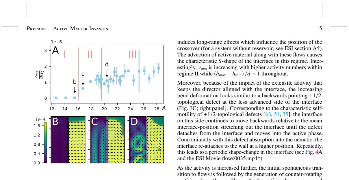
The central object is a two-phase active nematohydrodynamics model: a scalar phase field φ marks active versus passive fluid, a nematic tensor Q_αβ tracks orientational order, and an active stress −ζφQ_αβ injects energy at the scale set by the activity ζ. The organizing dimensionless group is the activity number A = d/√(K_Q/ζ), the capillary width over the active length scale, which collapses data from different channel widths onto one phase map. The argument works by identifying two observables—interface deformation (h_max − h_min)/d and number of detached clusters N_c—and showing that both jump at A ≈ 16 and A ≈ 20, with the mechanisms being spontaneous flow onset and activity-versus-surface-tension pinch-off respectively.
What would settle it
Run the same two-phase active-nematic equations with a different growth source term, such as spatially uniform proliferation instead of random local events, and check whether the two crossovers in (h_max − h_min)/d and cluster count N_c stay at A ≈ 16 and A ≈ 20; a shift or disappearance would show the regime classification depends on the growth implementation. Alternatively, measure interface flatness and detached cluster count in an expanding bacterial or epithelial monolayer in a channel while varying available chemical energy, and see whether the same two abrupt changes appear.
Extended reading notes
Core claim
The paper claims that a growing active nematic invading a fluid-filled capillary from a reservoir passes through three qualitatively distinct invasion regimes as the dimensionless activity number A = d/√(K_Q/ζ) increases: a flat-interface, flow-free regime (A ≲ 16) where invasion is purely growth and diffusion controlled; an S-shaped-interface regime (roughly 16 ≲ A ≲ 20) driven by spontaneous flow generation that advects material, with periodic flipping of the front between capillary walls as vortices form; and a regime (A ≳ 20) where active clusters pinch off from the main body and penetrate 0.5 to 1 capillary widths deeper, even though total active material in the capillary is similar to regime II. The first crossover is traced to the well-known hydrodynamic instability to spontaneous flow in confined active nematics; the second is attributed to active stresses overcoming surface tension at the interface, aided by bulk dynamics and +1/2 topological defects.
Load-bearing premise
The random local growth events that feed the active phase are assumed not to change the invasion regime; the paper compares growth versus no growth and reservoir versus no reservoir, but never varies how growth is implemented, so the regime boundaries could in principle be artifacts of that particular source term.
Editorial extensions
If this is right
- The activity number A, rather than activity, channel width, or elasticity separately, sets the invasion mode: the same crossovers appear for capillaries of different widths when plotted against A.
- Before the first crossover, invasion is slow and nearly independent of activity because transport is purely diffusive and growth-driven, with no spontaneous flows in the capillary.
- After the first crossover, activity-induced flows advect active material into the capillary and the invasion index rises approximately linearly with A.
- After the second crossover, detached clusters add little to the total amount of active material in the channel but extend maximum reach by up to one capillary width.
- Within regime II, the front's S-shape switches from one wall to the other when the most-forward vortex reverses its rotation, giving periodic front oscillations.
Reading between the lines
- If the three-regime classification holds generally, invasive efficiency in confined geometries could be regulated by biochemical energy supply alone: a colony could switch from coherent to cluster-shedding invasion by tuning its activity, suggesting a physical control point for slowing or aiding spread.
- The ESI result that removing the reservoir shifts the second crossover to higher A implies that upstream geometry and growth pressure participate in setting cluster detachment thresholds; an experimental study varying reservoir size while holding capillary activity fixed would test this long-range influence.
- A natural extension is to give the activity coefficient a curvature dependence at the interface, mimicking leader cells; one prediction of the model framework is that such a term would move the first crossover or alter the S-shape switching frequency, which could be checked in particle-based simulations.
- Cluster detachment as a way to reach deeper suggests a generic physical rationale for the advantage of disseminating small groups during collective invasion, independent of specific biochemical signalling.
Signed reviews
Editorial analysis
A structured set of objections, weighed in public.
Referee Report
Summary. The paper uses two-dimensional hybrid lattice Boltzmann simulations of a two-phase active nematohydrodynamic model to study the invasion of a growing active nematic from a reservoir into a narrower capillary filled with isotropic fluid. Growth is implemented as stochastic local increases of the phase field in the reservoir with logistic saturation. Varying the dimensionless activity number A = d/sqrt(KQ/zeta) reveals three qualitatively distinct invasion regimes: a flat-interface coherent regime (I), an S-shaped interface with spontaneous flows and vortex-induced switching (II), and a regime in which active clusters detach from the main body (III). The authors characterize the two crossovers using the interface height difference (hmax - hmin)/d and the number of detached clusters Nc, connect the first crossover to the spontaneous flow transition in confined active nematics, and propose that the second crossover arises from active stresses overcoming surface tension. They further show that cluster detachment lets the active material protrude about 0.5 to 1 capillary-width deeper into the channel.
Significance. If the classification is robust, the paper provides a useful organizing framework for how growth, activity, and confinement combine to produce distinct modes of collective invasion, with potential relevance to biofilm expansion and collective cell migration. The strengths of the manuscript are the fully specified governing equations and parameters, well-defined observables with error bars from repeated initial conditions, and explicit falsifiable predictions (the two crossover thresholds in A and the cluster-penetration depth). The ESI adds useful controls, including no-growth and no-reservoir comparisons and a check of two channel widths. The authors are also honest about the uncertainty in the second-crossover mechanism. The central risk is the untested sensitivity of the regime structure to the details of the stochastic growth protocol, which is the ingredient that makes the regimes exist.
major comments (3)
- [Section 2.2 and ESI A] The claim that 'the details of this implementation or even the geometry of a reservoir are not important for the qualitative dynamics in the capillary (see ESI section A)' is not supported by the evidence presented. ESI A tests only two binary variations: removing growth entirely and removing the reservoir. It does not vary the stochastic growth parameters r, tau_g, r_g, alpha, and phi_c, nor does it test growth placed inside the capillary. Since growth is the component that makes the three invasion regimes appear, a hidden sensitivity of the crossover positions or of cluster detachment to these parameters would directly undermine the central classification. I therefore request either additional simulations varying the growth protocol (at least for a representative set of parameters and for growth distributed in the capillary) or a reformulation of the claim to state that the classification is demonstrated for the specific growth implementation used.
- [Section 3.2.2 and Figure 4C] The mechanistic explanation for the crossover from regime II to III is that active stresses overcome the stabilizing effect of surface tension, leading to pinch-off of clusters. However, the text itself admits that 'it is not possible to decide to what degree this effect is purely interfacial and whether dynamics in the bulk are important.' Because the paper explicitly claims to characterize 'the mechanical mechanisms underlying the crossovers,' a quantitative test is needed. For example, varying the surface-tension-related coefficients D or K_phi at fixed A and measuring Nc and the threshold would distinguish interfacial from bulk contributions. Without such a test, the proposed mechanism remains a plausible hypothesis rather than a demonstrated result; the existence of the regimes is still supported by the observables, but the explanatory part of the central claim is not yet established.
- [Section 3.1 and Figure 2] The text describes the two transitions as 'well-defined crossovers,' but the data points are spaced by increments of order unity in A (e.g., A = 15.5, 16.7, 17.9, 19.2), so the sharpness of the transitions is not resolved. A denser sampling of A near the claimed thresholds, together with a quantitative criterion for locating the crossover (for instance, the value of A where (hmax - hmin)/d first exceeds a threshold or where Nc becomes non-zero), would strengthen the claim that the regimes are separated by well-defined crossovers rather than by gradual changes over a finite range.
minor comments (5)
- [Section 2.2] The stochastic growth algorithm is described verbally; please specify the time cadence of growth attempts (e.g., whether each lattice site in the reservoir is considered once per lattice-Boltzmann time step or once per tau_g) so that the implementation is unambiguous and reproducible.
- [ESI A and Supplementary Figure 1] The statement that 'growth is an essential factor to create the phenomena reported in the main text' appears to be in tension with the note in the caption of Supplementary Figure 1 that 'clusters are also present for a system lacking growth, but it takes longer for them to appear.' Please clarify in what sense growth is essential (e.g., for the S-shaped interface and the early appearance of clusters) and reconcile these two statements.
- [Figure 3A] The axis label in Figure 3A uses units of d over the active time scale tau_zeta, but the main text says 'rms-velocity v_rms in units of d' without specifying the time normalization until the caption; please make the axis label and text consistent.
- [ESI B and Supplementary Figure 2] For the channel-width comparison, please state explicitly whether the same activity coefficients zeta were used for both d values (with A varying through d) or whether zeta was adjusted; this affects how the collapse onto A in Supplementary Figure 2 should be interpreted.
- [References] References [10] and [19] appear to be the same reference (Conrad and Poling-Skutvik, Annu. Rev. Chem. Biomol. Eng. 9:175–200, 2018) cited twice with slightly different formatting; please merge them into a single entry.
Circularity Check
No significant circularity: the three-regime classification is read directly from simulation observables, the spontaneous-flow benchmark is an external literature result, and no fitted parameter is renamed as a prediction.
full rationale
The paper's central claim is a classification of invasion into three regimes as the activity number A is varied. The crossover observables (hmax-hmin)/d and Nc are direct measurements from the simulations, and the crossover positions are read off the data; they are not fitted parameters and are not constructed from the classification itself. The dimensionless activity number A = d/sqrt(KQ/zeta) is a standard combination of model parameters defined independently of the output regimes, so no self-definitional loop is present. The I-to-II crossover is explained by reference to the well-established spontaneous flow transition in confined active nematics [49,50]; although some cited authors overlap with the present paper, that transition is an external, previously established result and the paper explicitly notes that growth and reservoir geometry shift its location, showing it is not being asserted by construction. The II-to-III crossover is described mechanistically as a competition between active stresses and surface tension, with the paper honestly stating that 'it is not possible to decide to what degree this effect is purely interfacial and whether dynamics in the bulk are important'; this is a hedged interpretation, not a circular reduction. One robustness gap should be flagged: Section 2.2 claims that 'the details of this implementation or even the geometry of a reservoir are not important for the qualitative dynamics in the capillary (see ESI section A)', but ESI section A only tests growth versus no-growth and reservoir versus no-reservoir; it does not vary the stochastic growth parameters r, tau_g, r_g, alpha, phi_c, or the location of growth. This is a missing-support issue for the insensitivity claim, not a circularity issue, because the regime structure is not definitionally tied to those implementation details and the claim remains empirically testable. No equation in the paper reduces to its own input, no fitted quantity is renamed as a prediction, and the central qualitative result is a direct simulation outcome rather than an output forced by a self-citation chain.
Assumptions & free parameters
free parameters (4)
- Kφ =
0.08
- D =
0.08
- CLQ =
0.15
- growth parameters (r, τg, rg, α, φc) =
r=0.001, τg=10000, rg=5, α=0.01, φc=1.2
assumptions (4)
- domain assumption Continuum active nematohydrodynamics (Eqs. 1-16) captures the essential physics of growing biological active matter in confinement.
- ad hoc to paper Growth can be represented by random local mass sources with logistic saturation (Section 2.2).
- domain assumption The activity number A = d / sqrt(KQ/ζ) is the relevant dimensionless control parameter.
- domain assumption No-slip boundaries and von Neumann conditions for φ and Q (no anchoring) in the capillary.
Cite this review
Pith. "Pith review of Active Matter Invasion." pith.science (2026). https://pith.science/paper/UQRJGN5O
@misc{pith2026190800768,
  author       = {Pith},
  title        = {Pith review of: Active Matter Invasion},
  year         = {2026},
  howpublished = {\url{https://pith.science/paper/UQRJGN5O}},
  note         = {Machine review of arXiv:1908.00768}
}
read the original abstract
Biological active materials such as bacterial biofilms and eukaryotic cells thrive in confined micro-spaces. Here, we show through numerical simulations that confinement can serve as a mechanical guidance to achieve distinct modes of collective invasion when combined with growth dynamics and the intrinsic activity of biological materials. We assess the dynamics of the growing interface and classify these collective modes of invasion based on the activity of the constituent particles of the growing matter. While at small and moderate activities the active material grows as a coherent unit, we find that blobs of active material collectively detach from the cohort above a well-defined activity threshold. We further characterise the mechanical mechanisms underlying the crossovers between different modes of invasion and quantify their impact on the overall invasion speed.
Figures


Figures from the paper (2 more)


Reference graph
Works this paper leans on
-
[1]
A. G. Clark and D. M. Vignjevic. Modes of cancer cell invasion and the role of the microenvironment. Curr. Opin. Cell Biol., 36:13–22, 2015
2015
-
[2]
Cancer cells in the tumor core exhibit spatially coordinated migration patterns
Ralitza Staneva, Fatima El Marjou, Jorge Barbazan, De- nis Krndija, Sophie Richon, Andrew Clark, and Dani- jela Matic Vignjevic. Cancer cells in the tumor core exhibit spatially coordinated migration patterns. J. Cell Sci., 132(6):jcs.220277, 2019. ISSN 0021-9533. doi: 10.1242/jcs.220277. URL http://jcs.biologists. org/content/early/2019/02/11/jcs.220277
-
[3]
Collective motion of bacteria in two dimensions
Yilin Wu. Collective motion of bacteria in two dimensions. Quant. Biol., 3:199–205, 2015
2015
-
[4]
Re- cent advances in studying single bacteria and biofilm me- chanics
Catherine Even, Christian Marlière, Jean-Marc Ghigo, Jean-Marc Allain, Alba Marcellan, and Eric Raspaud. Re- cent advances in studying single bacteria and biofilm me- chanics. Adv. Colloid Interface Sci., 247:573–588, 2017
2017
-
[5]
Emergence of three-dimensional order and structure in growing biofilms
Raimo Hartmann, Praveen K Singh, Philip Pearce, Rachel Mok, Boya Song, Francisco Díaz-Pascual, Jörn Dunkel, and Knut Drescher. Emergence of three-dimensional order and structure in growing biofilms. Nat. Phys., 15:251–256, 2018
2018
-
[6]
Biomass accumulation and control strategies in gas biofiltration
Chunping Yang, Hong Chen, Guangming Zeng, Guan- long Yu, and Shenglian Luo. Biomass accumulation and control strategies in gas biofiltration. Biotech- nol. Adv. , 28(4):531 – 540, 2010. ISSN 0734-
work page 2010
-
[7]
Gritsenko, Olga Ilina, and Peter Friedl
Pavlo G. Gritsenko, Olga Ilina, and Peter Friedl. In- terstitial guidance of cancer invasion. The Journal of Pathology, 226(2):185–199, 2011. doi: 10.1002 /path
work page 2011
-
[8]
In- travital third harmonic generation microscopy of collective melanoma cell invasion
Bettina Weigelin, Gert-Jan Bakker, and Peter Friedl. In- travital third harmonic generation microscopy of collective melanoma cell invasion. IntraVital, 1(1):32–43, 2012. doi: 10.4161/intv.21223. URL https://doi.org/10.4161/ intv.21223
Show all 84 references
-
[9]
William Costerton, and Paul Stoodley
Luanne Hall-Stoodley, J. William Costerton, and Paul Stoodley. Bacterial biofilms: from the natural environ- ment to infectious diseases. Nat. Rev. Microbiol., 2:95– 108, Feb 2004. URL http://dx.doi.org/10.1038/ nrmicro821. Review Article
2004
-
[10]
Confined flow: Consequences and implications for bacteria and biofilms
Jacinta C Conrad and Ryan Poling-Skutvik. Confined flow: Consequences and implications for bacteria and biofilms. Annu. Rev. Chem. Biomol. Eng., 9:175–200, 2018
2018
-
[11]
Kabla, Chwee Teck Lim, and Benoît Ladoux
Sri Ram Krishna Vedula, Man Chun Leong, Tan Lei Lai, Pascal Hersen, Alexandre J. Kabla, Chwee Teck Lim, and Benoît Ladoux. Emerging modes of collective cell migration induced by geometrical constraints. Proc. Natl. Acad. Sci. U. S. A. , 109(32):12974–12979, 2012. ISSN 0027-842...
2012
-
[12]
Anna Kristina Marel, Matthias Zorn, Christoph Klingner, Roland Wedlich-Söldner, Erwin Frey, and Joachim O. Rädler. Flow and di ffusion in channel-guided cell mi- gration. Biophys. J., 107(5):1054–1064, 2014. ISSN 15420086. doi: 10.1016 /j.bpj.2014.07.017
2014
-
[13]
Alignment of cell division axes in directed epithelial cell migration
Anna-Kristina Marel, Nils Podewitz, Matthias Zorn, Joachim Oskar Rädler, and Jens Elgeti. Alignment of cell division axes in directed epithelial cell migration. New J. Phys., 16(11):115005, 2014. URL http://stacks. iop.org/1367-2630/16/i=11/a=115005
2014
-
[14]
Dean, Reza Riahi, and Pak Kin Wong
Yongliang Yang, Nima Jamilpour, Baoyin Yao, Zachary S. Dean, Reza Riahi, and Pak Kin Wong. Probing leader cells in endothelial collective migration by plasma lithography geometric confinement. Sci. Rep., 6:srep22707, Mar 2016. URL http://dx.doi.org/10.1038/srep22707. Arti- cle
2016 doi
-
[15]
Fardin, and Benoît Ladoux
Estelle Gauquelin, Sham Tlili, Cyprien Gay, Grégoire Peyret, René-Marc Mège, Marc A. Fardin, and Benoît Ladoux. Influence of proliferation on the motions of ep- ithelial monolayers invading adherent strips. Soft Matter, 15:2798–2810, 2019. doi: 10.1039 /C9SM00105K. URL http://d...
2019 doi
-
[16]
Topological defects in con- fined populations of spindle-shaped cells
Guillaume Duclos, Christoph Erlenkämper, Jean-François Joanny, and Pascal Silberzan. Topological defects in con- fined populations of spindle-shaped cells. Nat. Phys., 13: 58–62, Sep 2017. URL http://dx.doi.org/10.1038/ nphys3876
2017
-
[17]
Duclos, C
G. Duclos, C. Blanch-Mercader, V . Yashunsky, G. Sal- breux, J.-F. Joanny, J. Prost, and P. Silberzan. Spon- taneous shear flow in confined cellular nematics. Nat. Phys., 14(7):728–732, 2018. ISSN 1745-2481. doi: 10.1038/s41567-018-0099-7. URL https://doi.org/ 10.1038/s41567-018-0099-7
2018 doi
-
[18]
Directed collec- tive motion of bacteria under channel confinement
H Wioland, E Lushi, and R E Goldstein. Directed collec- tive motion of bacteria under channel confinement. New J. Phys., 18(7):075002, 2016. URL http://stacks.iop. org/1367-2630/18/i=7/a=075002
2016
-
[19]
Conrad and Ryan Poling-Skutvik
Jacinta C. Conrad and Ryan Poling-Skutvik. Confined flow: Consequences and implications for bacteria and biofilms. Annu. Rev. Chem. Biomol. Eng., 9(1):175–200,
-
[20]
Spatial confinement of active microtubule networks induces large-scale rotational cytoplasmic flow
Kazuya Suzuki, Makito Miyazaki, Jun Takagi, Takeshi Itabashi, and Shin’ichi Ishiwata. Spatial confinement of active microtubule networks induces large-scale rotational cytoplasmic flow. Proc. Natl. Acad. Sci. U. S. A., 114(11): 2922–2927, 2017. ISSN 0027-8424. doi: 10.1073 /pnas...
2017
-
[21]
Kun-Ta Wu, Jean Bernard Hishamunda, Daniel T. N. Chen, Stephen J. DeCamp, Ya-Wen Chang, Alberto Fernández-Nieves, Seth Fraden, and Zvonimir Dogic. Transition from turbulent to coherent flows in confined three-dimensional active fluids. Science, 355(6331): eaal1979, 2017. ISSN 003...
2017
-
[22]
Woodhouse, Jörn Dunkel, John O
Hugo Wioland, Francis G. Woodhouse, Jörn Dunkel, John O. Kessler, and Raymond E. Goldstein. Confine- ment stabilizes a bacterial suspension into a spiral vor- tex. Phys. Rev. Lett. , 110:268102, Jun 2013. doi: 10.1103/PhysRevLett.110.268102. URL https://link. aps.org/doi/10.110...
2013 doi
-
[23]
Reconfigurable flows and defect landscape of confined active nematics
Jérôme Hardoüin, Rian Hughes, Amin Doostmohammadi, Justine Laurent, Teresa Lopez-Leon, Julia M Yeomans, Jordi Ignés-Mullol, and Francesc Sagués. Reconfigurable flows and defect landscape of confined active nematics. page arXiv:1903.01787, 2019
1903 arXiv
-
[24]
Norton, Michael P
Achini Opathalage, Michael M. Norton, Michael P. N. Juniper, Blake Langeslay, S. Ali Aghvami, Seth Fraden, and Zvonimir Dogic. Self-organized dynamics and the transition to turbulence of confined active nematics. Proc. Natl. Acad. Sci. U. S. A., 116(11):4788–4797, 2019. ISSN 00...
2019
-
[25]
E ffective diffusivity of microswimmers in a crowded environment
Marvin Brun-Cosme-Bruny, Eric Bertin, Benoît Coasne, Philippe Peyla, and Salima Rafaï. E ffective diffusivity of microswimmers in a crowded environment. J. Chem. Phys., 150(10):104901, 2019. doi: 10.1063 /1.5081507. URL https://doi.org/10.1063/1.5081507
2019 doi
-
[26]
Transport and dispersion of active parti- cles in periodic porous media
Roberto Alonso-Matilla, Brato Chakrabarti, and David Saintillan. Transport and dispersion of active parti- cles in periodic porous media. Phys. Rev. Fluids , 4: 043101, Apr 2019. doi: 10.1103 /PhysRevFluids.4. 043101. URL https://link.aps.org/doi/10.1103/ PhysRevFluids.4.043101
2019
-
[27]
Wensink, Jörn Dunkel, Sebastian Heiden- reich, Knut Drescher, Raymond E
Henricus H. Wensink, Jörn Dunkel, Sebastian Heiden- reich, Knut Drescher, Raymond E. Goldstein, Hartmut Löwen, and Julia M. Yeomans. Meso-scale turbulence in living fluids. Proc. Natl. Acad. Sci. U. S. A., 109(36): 14308–14313, 2012. ISSN 0027-8424. doi: 10.1073 /pnas. 12020321...
2012
-
[28]
Thampi, Ramin Golestanian, and Julia M
Sumesh P. Thampi, Ramin Golestanian, and Julia M. Yeomans. V orticity, defects and correlations in ac- tive turbulence. Philos. Trans. R. Soc., A , 372 (2029):20130366, 2014. doi: 10.1098 /rsta.2013.0366. URL https://royalsocietypublishing.org/doi/ abs/10.1098/rsta.2013.0366
2014
-
[29]
Geometry and topology of turbulence in active nematics
Luca Giomi. Geometry and topology of turbulence in active nematics. Phys. Rev. X, 5:031003, Jul 2015. doi: 10.1103/PhysRevX.5.031003. URL https://link.aps. org/doi/10.1103/PhysRevX.5.031003
2015 doi
-
[30]
Martin James, Wouter J. T. Bos, and Michael Wilczek. Turbulence and turbulent pattern formation in a min- imal model for active fluids. Phys. Rev. Fluids , 3: 061101, Jun 2018. doi: 10.1103 /PhysRevFluids.3. 061101. URL https://link.aps.org/doi/10.1103/ PhysRevFluids.3.061101
2018
-
[31]
New class of turbulence in active fluids
Vasil Bratanov, Frank Jenko, and Erwin Frey. New class of turbulence in active fluids. Proc. Natl. Acad. Sci. U. S. A., 112(49):15048–15053, 2015. ISSN 0027-8424. doi: 10. 1073/pnas.1509304112. URL https://www.pnas.org/ content/112/49/15048
2015
-
[32]
Elgeti, M
J. Elgeti, M. E. Cates, and D. Marenduzzo. Defect hy- drodynamics in 2d polar active fluids. Soft Matter , 7: 3177–3185, 2011. doi: 10.1039 /C0SM01097A. URL http://dx.doi.org/10.1039/C0SM01097A
2011 doi
-
[33]
Bowick, Xu Ma, and M
Luca Giomi, Mark J. Bowick, Xu Ma, and M. Cristina Marchetti. Defect annihilation and proliferation in active nematics. Phys. Rev. Lett., 110:228101, May 2013. doi: 10.1103/PhysRevLett.110.228101. URL https://link. aps.org/doi/10.1103/PhysRevLett.110.228101
2013 doi
-
[34]
Defect dynam- ics in active nematics
Luca Giomi, Mark J Bowick, Prashant Mishra, Rastko Sknepnek, and M Cristina Marchetti. Defect dynam- ics in active nematics. Philos. Trans. R. Soc., A , 372 (2029):20130365, 2014. doi: 10.1098 /rsta.2013.0365. URL https://royalsocietypublishing.org/doi/ abs/10.1098/rsta.2013.0365
2014
-
[35]
Yeomans, and Benoit Ladoux
Thuan Beng Saw, Amin Doostmohammadi, Vincent Nier, Leyla Kocgozlu, Sumesh Thampi, Yusuke Toyama, Philippe Marcq, Chwee Teck Lim, Julia M. Yeomans, and Benoit Ladoux. Topological defects in epithelia govern cell death and extrusion. Nature, 544:212–216, Apr 2017. URL http://dx....
2017 doi
-
[36]
Modeling collective cell migration in geometric confine- ment
Victoria Tarle, Estelle Gauquelin, S R K Vedula, Joseph D’Alessandro, C T Lim, Benoit Ladoux, and Nir S Gov. Modeling collective cell migration in geometric confine- ment. Phys. Biol. , 14(3):035001, 2017. URL http: //stacks.iop.org/1478-3975/14/i=3/a=035001
2017
-
[37]
Blow, Sumesh P
Matthew L. Blow, Sumesh P. Thampi, and Julia M. Yeo- mans. Biphasic, lyotropic, active nematics.Phys. Rev. Lett., 113:248303, Dec 2014. doi: 10.1103 /PhysRevLett.113. 248303. URL http://link.aps.org/doi/10.1103/ PhysRevLett.113.248303
2014
-
[38]
Marenduzzo, E
D. Marenduzzo, E. Orlandini, M. E. Cates, and J. M. Yeo- mans. Steady-state hydrodynamic instabilities of active liquid crystals: Hybrid lattice boltzmann simulations. Phys. Rev. E, 76:031921, Sep 2007. doi: 10.1103 /PhysRevE.76. 031921. URL https://link.aps.org/doi/10.1103/ P...
2007
-
[39]
Aditi Simha and Sriram Ramaswamy
R. Aditi Simha and Sriram Ramaswamy. Hydrody- namic fluctuations and instabilities in ordered suspen- sions of self-propelled particles. Phys. Rev. Lett. , 89:058101, Jul 2002. doi: 10.1103 /PhysRevLett.89. 058101. URL https://link.aps.org/doi/10.1103/ PhysRevLett.89.058101
2002
-
[40]
Thampi, Ramin Golestanian, and Julia M
Sumesh P. Thampi, Ramin Golestanian, and Julia M. Yeo- mans. Velocity correlations in an active nematic.Phys. Rev. Lett., 111:118101, Sep 2013. doi: 10.1103 /PhysRevLett. 111.118101. URL https://link.aps.org/doi/10. 1103/PhysRevLett.111.118101
2013
-
[41]
DeCamp, Gabriel S
Stephen J. DeCamp, Gabriel S. Redner, Aparna Baskaran, Michael F. Hagan, and Zvonimir Dogic. Orientational order of motile defects in active nematics. Nat. Mater., 14: 1110–1115, Aug 2015. URL http://dx.doi.org/10. 1038/nmat4387
2015
-
[42]
Liverpool
Dario Cortese, Jens Eggers, and Tanniemola B. Liverpool. Pair creation, motion, and annihilation of topological de- fects in two-dimensional nematic liquid crystals. Phys. Rev. E, 97:022704, Feb 2018. doi: 10.1103 /PhysRevE.97. Preprint – Active Matter Inv asion 10 022704. URL...
2018
-
[43]
Yeomans, and Francesc Sagués
Amin Doostmohammadi, Jordi Ignés-Mullol, Julia M. Yeomans, and Francesc Sagués. Active nematics. Nat. Commun., 9(1):3246, 2018. ISSN 2041-1723. doi: 10.1038/s41467-018-05666-8. URL https://doi.org/ 10.1038/s41467-018-05666-8
2018 doi
-
[44]
Tim Sanchez, Daniel T. N. Chen, Stephen J. DeCamp, Michael Heymann, and Zvonimir Dogic. Spontaneous motion in hierarchically assembled active matter. Nature, 491:431–434, Nov 2012. URL http://dx.doi.org/10. 1038/nature11591
2012
-
[45]
Dynamics of anisotropic tissue growth
Thomas Bittig, Ortrud Wartlick, Anna Kicheva, Mar- cos González-Gaitán, and Frank Jülicher. Dynamics of anisotropic tissue growth. New J. Phys., 10(6):063001,
-
[46]
Duclos, S
G. Duclos, S. Garcia, H. G. Yevick, and P. Silberzan. Perfect nematic order in confined monolayers of spindle- shaped cells. Soft Matter , 10:2346–2353, 2014. doi: 10.1039/C3SM52323C. URL http://dx.doi.org/10. 1039/C3SM52323C
2014 doi
-
[47]
Thampi, Thuan B
Amin Doostmohammadi, Sumesh P. Thampi, Thuan B. Saw, Chwee T. Lim, Benoit Ladoux, and Julia M. Yeomans. Celebrating soft matter’s 10th anniversary: Cell division: a source of active stress in cellular monolayers. Soft Matter, 11:7328–7336, 2015. doi: 10.1039 /C5SM01382H. URL h...
2015 doi
-
[48]
Tsimring
Dmitri V olfson, Scott Cookson, Je ff Hasty, and Lev S. Tsimring. Biomechanical ordering of dense cell popula- tions. Proc. Natl. Acad. Sci. U. S. A. , 105(40):15346– 15351, 2008. ISSN 0027-8424. doi: 10.1073 /pnas. 0706805105. URL http://www.pnas.org/content/ 105/40/15346
2008
-
[49]
V oituriez, J
R. V oituriez, J. F. Joanny, and J. Prost. Spontaneous flow transition in active polar gels. EPL, 70(3):404,
-
[50]
S. A. Edwards and J. M. Yeomans. Spontaneous flow states in active nematics: A unified picture. EPL, 85(1):18008,
-
[51]
Shendruk, Amin Doostmohammadi, Kristian Thi- jssen, and Julia M
Tyler N. Shendruk, Amin Doostmohammadi, Kristian Thi- jssen, and Julia M. Yeomans. Dancing disclinations in con- fined active nematics. Soft Matter, 13:3853–3862, 2017. doi: 10.1039/C6SM02310J. URL http://dx.doi.org/ 10.1039/C6SM02310J
2017 doi
-
[52]
Shendruk, Kristian Thijssen, and Julia M
Amin Doostmohammadi, Tyler N. Shendruk, Kristian Thijssen, and Julia M. Yeomans. Onset of meso- scale turbulence in active nematics. Nat. Commun. , 8:15326, 2017. URL http://dx.doi.org/10.1038/ ncomms15326. Article
2017
-
[53]
Norton, Arvind Baskaran, Achini Opatha- lage, Blake Langeslay, Seth Fraden, Aparna Baskaran, and Michael F
Michael M. Norton, Arvind Baskaran, Achini Opatha- lage, Blake Langeslay, Seth Fraden, Aparna Baskaran, and Michael F. Hagan. Insensitivity of active nematic liquid crystal dynamics to topological constraints. Phys. Rev. E, 97:012702, Jan 2018. doi: 10.1103 /PhysRevE.97. 01270...
2018
-
[54]
de Gennes and J
Pierre G. de Gennes and J. Prost. The Physics of Liquid Crystals. Oxford University Press, Oxford, 1995
1995
-
[55]
Chaikin and Taylor C
Paul M. Chaikin and Taylor C. Lubensky. Principles of Condensed Matter Physics. Cambridge University Press, Cambridge, 2000
2000
-
[56]
Orlandini, M
E. Orlandini, M. R. Swift, and J. M. Yeomans. A lattice boltzmann model of binary-fluid mixtures. EPL, 32(6):463,
-
[57]
R. G. Larson. The structure and rheology of complex fluids. Oxford University Press, New York (N.Y .), 1999
1999
-
[58]
The mechanics and statistics of active matter
Sriram Ramaswamy. The mechanics and statistics of active matter. Annu. Rev. Condens. Matter Phys. , 1:323–345, 2010
2010
-
[59]
Rossen, Jens M
Ninna S. Rossen, Jens M. Tarp, Joachim Mathiesen, Mo- gens H. Jensen, and Lene B. Oddershede. Long-range ordered vorticity patterns in living tissue induced by cell division. Nat. Commun., 5:5720, Dec 2014. URL http://dx.doi.org/10.1038/ncomms6720. Article
2014 doi
-
[60]
Stabilization of active matter by flow-vortex lattices and defect ordering
Amin Doostmohammadi, Michael F Adamer, Sumesh P Thampi, and Julia M Yeomans. Stabilization of active matter by flow-vortex lattices and defect ordering. Nat. Commun., 7:10557, 2016
2016
-
[61]
Hemingway, Prashant Mishra, M
Ewan J. Hemingway, Prashant Mishra, M. Cristina Marchetti, and Suzanne M. Fielding. Correlation lengths in hydrodynamic models of active nematics. Soft Matter, 12:7943–7952, 2016. doi: 10.1039 /C6SM00812G. URL http://dx.doi.org/10.1039/C6SM00812G
2016 doi
-
[62]
Taming active turbulence with patterned soft interfaces
Pau Guillamat, Jordi Ignés-Mullol, and Francesc Sagués. Taming active turbulence with patterned soft interfaces. Nat. Commun., 8:564, 2017
2017
-
[63]
Phase separation and emergent structures in an active nematic fluid
Elias Putzig and Aparna Baskaran. Phase separation and emergent structures in an active nematic fluid. Phys. Rev. E, 90:042304, Oct 2014. doi: 10.1103 /PhysRevE.90. 042304. URL https://link.aps.org/doi/10.1103/ PhysRevE.90.042304
2014
-
[64]
Geometric control of active collective motion
Maxime Theillard, Roberto Alonso-Matilla, and David Saintillan. Geometric control of active collective motion. Soft Matter, 13:363–375, 2017
2017
-
[65]
Thampi, Amin Doostmohammadi, Ramin Golestanian, and Julia M
Sumesh P. Thampi, Amin Doostmohammadi, Ramin Golestanian, and Julia M. Yeomans. Intrinsic free energy in active nematics. EPL, 112(2):28004, 2015. URL http: //stacks.iop.org/0295-5075/112/i=2/a=28004
2015
-
[66]
He Li, Xia-qing Shi, Mingji Huang, Xiao Chen, Minfeng Xiao, Chenli Liu, Hugues Chaté, and H. P. Zhang. Data- driven quantitative modeling of bacterial active nematics. Proc. Natl. Acad. Sci. U. S. A. , 116(3):777–785, 2019. ISSN 0027-8424. doi: 10.1073 /pnas.1812570116. URL ht...
2019
-
[67]
Nematic liquid crystals formed by living amoeboid cells
H Gruler, U Dewald, and M Eberhardt. Nematic liquid crystals formed by living amoeboid cells. Euro. Phys. J. B, 11:187–192, 1999
1999
-
[68]
Turbulent dynamics of epithelial cell cultures
C Blanch-Mercader, V Yashunsky, S Garcia, G Duclos, L Giomi, and P Silberzan. Turbulent dynamics of epithelial cell cultures. Phys. Rev. Lett., 120:208101, 2018. Preprint – Active Matter Inv asion 11
2018
-
[69]
Topological defects control collective dynam- ics in neural progenitor cell cultures
Kyogo Kawaguchi, Ryoichiro Kageyama, and Masaki Sano. Topological defects control collective dynam- ics in neural progenitor cell cultures. Nature, 545:327– 331, Apr 2017. URL http://dx.doi.org/10.1038/ nature22321
2017
-
[70]
Biological tissues as active nematic liq- uid crystals
Thuan Beng Saw, Wang Xi, Benoit Ladoux, and Chwee Teck Lim. Biological tissues as active nematic liq- uid crystals. Adv. Mater., 30(47):1802579, 2018. doi: 10. 1002/adma.201802579. URL https://onlinelibrary. wiley.com/doi/abs/10.1002/adma.201802579
2018 doi
-
[71]
Computational model for cell morphodynamics
Danying Shao, Wouter-Jan Rappel, and Herbert Levine. Computational model for cell morphodynamics. Phys. Rev. Lett., 105:108104, Sep 2010. doi: 10.1103 /PhysRevLett. 105.108104. URL https://link.aps.org/doi/10. 1103/PhysRevLett.105.108104
2010
-
[72]
Igor S. Aranson. Physical Models of Cell Motility. Springer International Publishing, Switzerland, 2016
2016
-
[73]
Yeomans, and Amin Doost- mohammadi
Romain Mueller, Julia M. Yeomans, and Amin Doost- mohammadi. Emergence of active nematic behavior in monolayers of isotropic cells. Phys. Rev. Lett. , 122: 048004, Feb 2019. doi: 10.1103 /PhysRevLett.122. 048004. URL https://link.aps.org/doi/10.1103/ PhysRevLett.122.048004
2019
-
[74]
Bridging the gap between single cell migra- tion and collective dynamics
Florian Thueroff, Andriy Goychuk, Matthias Reiter, and Erwin Frey. Bridging the gap between single cell migra- tion and collective dynamics. page bioRxiv/548677, 2019. doi: 10.1101 /548677. URL https://www.biorxiv. org/content/early/2019/02/13/548677
2019
-
[75]
Collective migration of an epithelial monolayer in response to a model wound
Mathieu Poujade, Erwan Grasland-Mongrain, A Hertzog, J Jouanneau, Philippe Chavrier, Benoît Ladoux, Axel Buguin, and Pascal Silberzan. Collective migration of an epithelial monolayer in response to a model wound. Proc. Natl. Acad. Sci. U. S. A., 104:15988–15993, 2007
2007
-
[76]
Physical forces during collective cell migration
Xavier Trepat, Michael R Wasserman, Thomas E Angelini, Emil Millet, David A Weitz, James P Butler, and Jeffrey J Fredberg. Physical forces during collective cell migration. Nat. Phys., 5(6):426, 2009
2009
-
[77]
Physical model of the dynamic instability in an expanding cell culture
Shirley Mark, Roie Shlomovitz, Nir S Gov, Mathieu Pou- jade, Erwan Grasland-Mongrain, and Pascal Silberzan. Physical model of the dynamic instability in an expanding cell culture. Biophys. J., 98:361–370, 2010. Preprint – Active Matter Inv asion 12 SupplementaryMaterials A R o...
2010
-
[1995]
URL http://stacks.iop.org/0295-5075/32/ i=6/a=001
-
[2005]
URL http://stacks.iop.org/0295-5075/70/ i=3/a=404
-
[2008]
URL http://stacks.iop.org/1367-2630/10/ i=6/a=063001
-
[2009]
URL http://stacks.iop.org/0295-5075/85/ i=1/a=18008
-
[2018]
URL https://doi.org/10.1146/ annurev-chembioeng-060817-084006
doi: 10.1146 /annurev-chembioeng-060817-084006. URL https://doi.org/10.1146/ annurev-chembioeng-060817-084006 . PMID: 29561646
-
[3031]
URL https://onlinelibrary.wiley.com/ doi/abs/10.1002/path.3031
-
[9750]
doi: https: //doi.org/10.1016/j.biotechadv.2010. 04.002. URL http://www.sciencedirect.com/ science/article/pii/S0734975010000418
2010 doi
Reviewed August 14, 2026 · model on record in the stance chip above.
Discussion (0). Continue with ORCID to comment.