REVIEW 2 major objections 4 minor 27 references
Site selective synthesis of in situ Ni filled multiwalled carbon nanotubes using Ni(salen) as a catalyst source
T0 review · 2 major / 4 minor · reviewed 2026-08-14 · deepseek-v4-flash
Pith's one-line read This paper reports that a photoresist loaded with Ni(salen) can be patterned by ordinary lithography and, in a single atmospheric-pressure CVD step, grow nickel-filled multiwalled carbon nanotubes only on the patterned regions.
desk verdict The Ni(salen)/photoresist route to site-selective Ni-filled CNT growth is a real, modest synthesis advance, but the coercivity claim is oversold by one unsubtracted SQUID curve on the whole as-grown film. read the letter →
The pith
A machine-rendered reading of the paper's core claim, the machinery that carries it, and where it could break.
The reading
What carries the argument
The load-bearing object is the modified photoresist, 'Mod-PR': a conventional positive photoresist (HPR 504) in which 0.2 M of Ni(salen), $N,N'$-bis(salicylidene)ethylenediiminato nickel(II), is dispersed. It does two jobs at once. Lithographically, exposure and development leave catalyst patterns wherever they are drawn; chemically, the 900 °C anneal decomposes the complex into 10–100 nm Ni particles. During growth, the tubes advance by a tip-growth mechanism, and the paper invokes capillary action of liquid-like Ni particles at the nucleation stage to explain how nickel is drawn into the tube cavity, producing the partial filling.
What would settle it
Measure the hysteresis loop of the same as-grown Ni-filled film after selectively removing the catalyst particles at the nanotube tips (for example by mild chemical etching that leaves the tubes intact) and compare it with the as-grown loop: if the 94 Oe coercivity at 5 K drops toward the bulk nickel value, then the nanowire filling is not what produces the reported magnetic enhancement.
Extended reading notes
Core claim
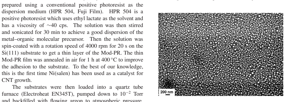
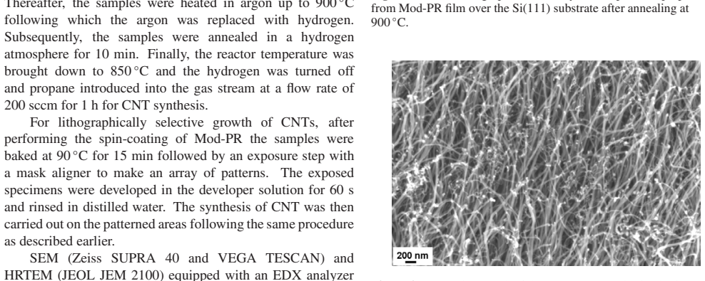
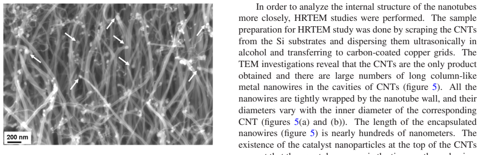
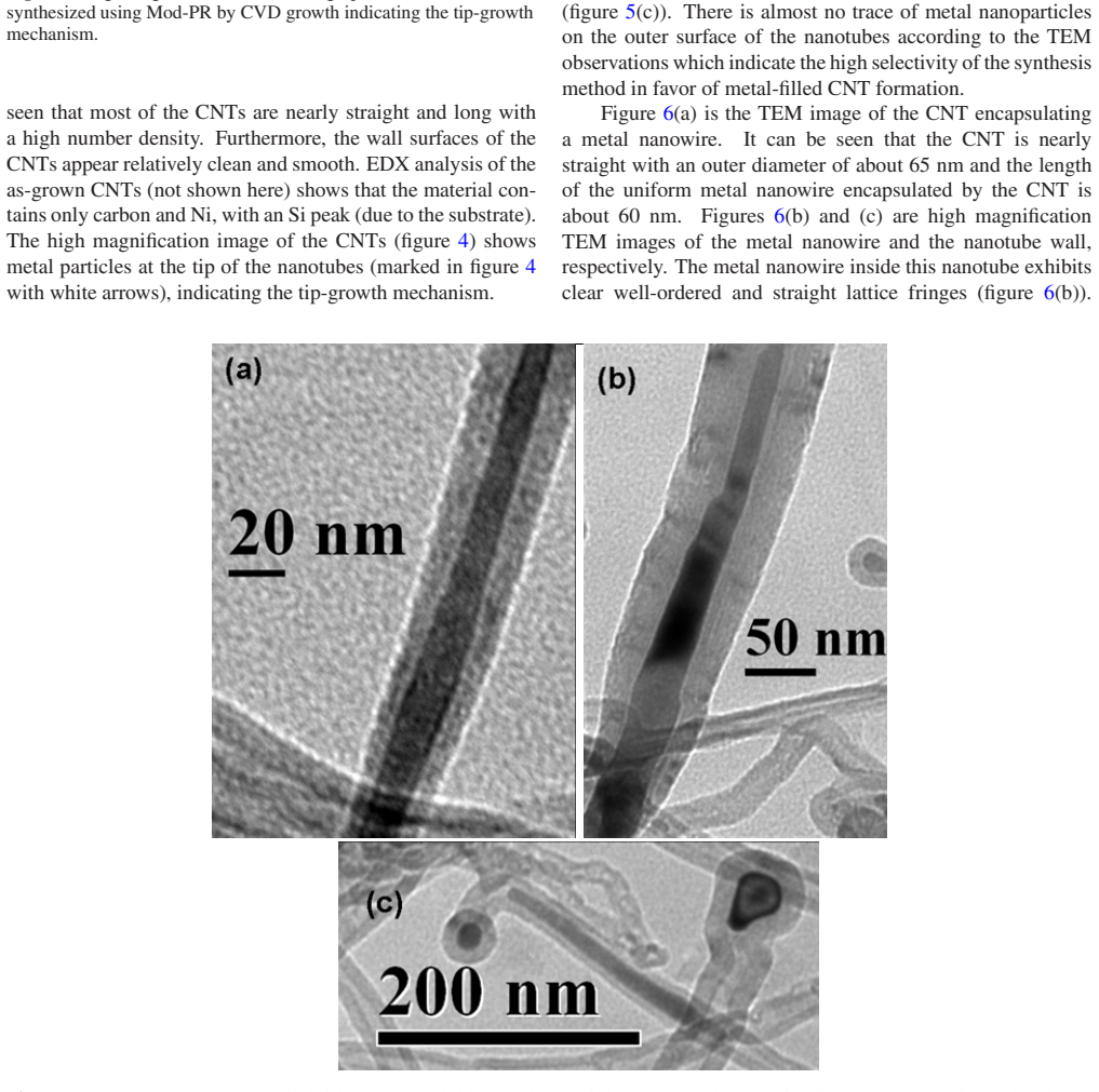
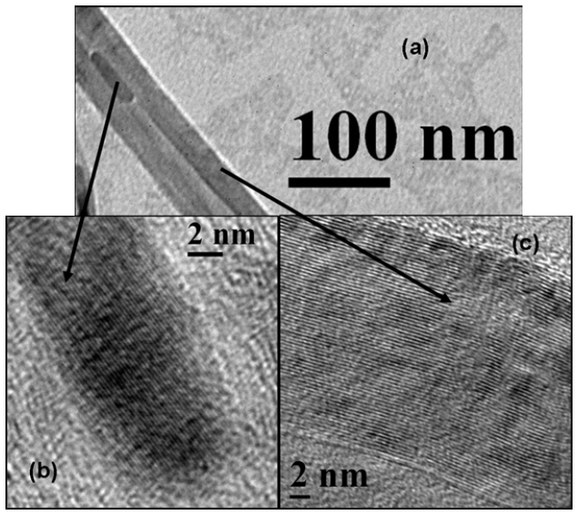
The central claim is that simply dissolving Ni(salen) in a standard positive photoresist (HPR 504) turns the photoresist itself into a catalyst source that both defines where nanotubes grow and supplies the metal that fills them. After spin-coating and a 400 °C anneal, the modified resist forms uniformly distributed Ni catalyst particles 10–100 nm in size. Hydrogen annealing and propane CVD at 850 °C then produce long, clean multiwalled nanotubes whose tips carry catalyst particles, indicating tip growth, and whose cavities contain continuous nickel nanowires with 0.21 nm lattice fringes matching the Ni(111) planes. The paper states that this is the first use of Ni(salen) as a carbon-nanotube catalyst, and that the method is scalable and site-selective.
Load-bearing premise
The coercivity claim assumes the SQUID signal is dominated by the nickel nanowires inside the nanotubes; the measurement was made on the whole as-grown film, which also contains nickel catalyst particles at the nanotube tips, and no control or subtraction separates the two contributions.
Editorial extensions
If this is right
- Standard photolithography alone can place Ni-filled carbon nanotubes at predefined locations, removing a barrier to integrating filled nanotubes into devices.
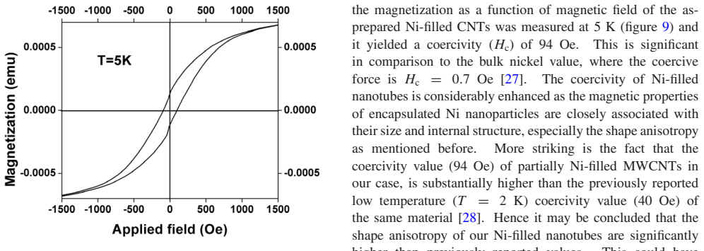
- The as-grown film is ferromagnetic at 5 K with a coercivity of 94 Oe, about 130 times the bulk nickel value of 0.7 Oe and higher than the 40 Oe earlier reported for the same material at 2 K.
- Atmospheric-pressure CVD with propane yields well-graphitized multiwalled nanotubes with $I_D/I_G = 0.32$, clean outer surfaces, and no metal residue outside the tubes.
- Because the nickel filling is formed in situ rather than by a separate infiltration step, the entire fabrication is a single growth run.
Reading between the lines
- I infer that the photoresist-mixing strategy should extend to other metal–salen or metal–organic precursors whose decomposition temperatures match the CVD anneal, giving site-selective iron- or cobalt-filled nanotubes for other magnetic or catalytic uses; the paper only demonstrates nickel.
- A testable extension is to tune the filling fraction through growth time, temperature, and propane flow, since the paper explains filling as capillary uptake during nucleation but does not report such a sweep.
- Because the SQUID measurement was made on the whole as-grown film, the natural control is to measure the film after selectively removing the tip catalyst particles; the paper does not include that control, so the 94 Oe value is best read as a film-level observation rather than a nanowire-only property.
Signed reviews
Editorial analysis
A structured set of objections, weighed in public.
Referee Report
Summary. The manuscript reports the growth of Ni-filled multiwalled carbon nanotubes by atmospheric-pressure chemical vapor deposition at 850 °C using propane on Si, with a modified photoresist containing Ni(salen) as the catalyst source. The authors claim that this simple photolithographic route enables scalable, site-selective growth of high-quality, partially Ni-filled MWCNTs. Evidence includes SEM showing uniform CNT growth and tip-growth morphology, HRTEM showing Ni nanowires encapsulated in the tubes with a 0.21 nm (111) Ni lattice spacing, EDX confirming Ni filling, and Raman spectroscopy with an ID/IG ratio of 0.32. Magnetic measurements by SQUID at 5 K are reported to show ferromagnetic behavior with a coercivity of 94 Oe, which the authors compare to bulk Ni and to a previously reported Ni-filled CNT value.
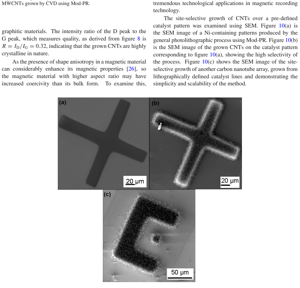
Significance. If the claims are supported, the photoresist-based patterning method would be a practically useful advance for integrating Ni-filled CNTs into devices, since it avoids separate catalyst deposition and achieves in situ filling. The TEM and EDX evidence for encapsulated Ni nanowires is direct and convincing for the synthesis claim, and the site-selective growth shown in Figure 10 is a clear strength. The Raman data support the high-quality claim. However, the magnetic characterization, which is a headline quantitative result, is not yet sufficiently controlled to support the stated ferromagnetic and shape-anisotropy conclusions. The paper is suitable for an applied-physics journal if this load-bearing gap is addressed.
major comments (2)
- [§3, Figure 9] The coercivity of 94 Oe is measured on the whole as-grown film, which contains Ni catalyst particles at the nanotube tips (Figures 4 and 5(c)) and may contain residual surface Ni particles from the annealed Mod-PR film (Figure 2). No control measurement on a sample without filling, no subtraction of the exposed-Ni contribution, and no acid-wash or selective-etch experiment are reported. Therefore the hysteresis loop cannot be attributed specifically to the encapsulated Ni nanowires, and the comparison to bulk Ni and to ref [28] does not establish the claimed shape-anisotropy enhancement. The authors should either isolate the filling contribution or temper the magnetic claim accordingly.
- [§3, coercivity comparison paragraph] The comparison with ref [28] compares a coercivity measured at 5 K with a value reported at 2 K. Since coercivity is strongly temperature-dependent in ferromagnetic nanomaterials, the temperature difference alone prevents a meaningful quantitative comparison. The authors should either remeasure at the same temperature, report the temperature dependence, or discuss the expected effect of the temperature difference. The same paragraph also quotes bulk Ni Hc = 0.7 Oe without specifying the measurement temperature, which should be provided.
minor comments (4)
- [§3, EDX analysis] The text states that EDX analysis of the as-grown CNTs 'is not shown here'; including this spectrum, or a representative one, in the main text or supporting information would strengthen the compositional claim.
- [Figure 9] The magnetization curve in Figure 9 lacks visible axis labels and units, and the sample mass is not reported. Since the manuscript presents quantitative magnetic data, the authors should provide calibrated axes and report the magnetization normalization (per gram or per volume of Ni).
- [§3, growth model] The growth model for partial Ni filling is referenced to ref [25] but not summarized. For self-containedness, please add one or two sentences describing the capillary-action mechanism and how it applies to the present experimental conditions.
- [Figure 10] The site-selectivity claim would be easier to evaluate if the SEM images in Figure 10 included scale bars and if the text quantified the contrast between patterned and unpatterned regions (e.g., presence or absence of CNTs outside the catalyst patterns).
Circularity Check
No significant circularity: the paper reports direct experimental observations with no fitted-input or self-citation chain that reduces a 'prediction' to its inputs.
full rationale
This is an experimental synthesis-and-characterization paper. The central claims—site-selective growth, Ni-filled MWCNTs, high quality evidenced by Raman, and ferromagnetic behavior with measured coercivity—are supported by direct SEM, TEM, EDX, Raman, and SQUID measurements. There is no derivation chain in which an output quantity is constructed to equal an input quantity. The only self-citation is reference [25], invoked for the growth model: 'The details of the growth model are discussed elsewhere [25].' That citation explains a proposed mechanism for partial Ni filling, but it is not load-bearing for the reported observations; the filling is directly imaged by TEM and identified by HRTEM lattice spacing and EDX, and the growth model is not used to generate or predict the measured coercivity or the Raman ID/IG ratio. The coercivity comparison to bulk Ni (0.7 Oe, ref [27]) and to a prior Ni-filled nanotube value (40 Oe at 2 K, ref [28]) is an external benchmark, not an input to the experiment. No fitted parameter is renamed as a prediction, and no uniqueness or existence theorem from prior work by these authors is invoked to force a conclusion. Consequently, the circularity burden is minimal; the finding is no significant circularity.
Assumptions & free parameters
assumptions (4)
- domain assumption Ni(salen) mixed in photoresist decomposes during the 900 C anneal into metallic Ni nanoparticles that catalyze CNT growth.
- domain assumption Propane is the carbon source and the carbonized photoresist neither contributes carbon nor poisons growth.
- domain assumption The bulk SQUID magnetization and coercivity are attributed to encapsulated Ni nanowires rather than to catalyst particles at tips or on the substrate.
- domain assumption A single Raman spectrum and selected TEM images represent the quality and filling state of the whole sample.
Cite this review
Pith. "Pith review of Site selective synthesis of in situ Ni filled multiwalled carbon nanotubes using Ni(salen) as a catalyst source." pith.science (2026). https://pith.science/paper/UVQY63HA
@misc{pith2026190804516,
  author       = {Pith},
  title        = {Pith review of: Site selective synthesis of in situ Ni filled multiwalled carbon nanotubes using Ni(salen) as a catalyst source},
  year         = {2026},
  howpublished = {\url{https://pith.science/paper/UVQY63HA}},
  note         = {Machine review of arXiv:1908.04516}
}
read the original abstract
The synthesis of Ni filled multiwalled carbon nanotubes was performed by atmospheric pressure chemical vapor deposition with propane on Si at 850 C using a simple mixture of Ni(salen), and a conventional photoresist. Analysis of the carbon nanotubes using scanning electron microscopy together with high resolution transmission electron microscopy show that the nanotubes have grown by a tip growth mechanism and exhibit a multi walled structure with partial Ni filling. The high quality of the Ni filled nanotubes is evidenced by Raman spectroscopy. The magnetic properties of Ni filled nanotubes were analyzed using a superconducting quantum interference device which revealed their ferromagnetic behavior with large coercivity. A scalable as well as site selective growth of high quality Ni filled carbon nanotubes is achieved by a simple photolithographic method.
Figures

Figures from the paper (6 more)

Reference graph
Works this paper leans on
-
[28]
Tyagi P K et al 2004 Thin Solid Films 469 127 6
work page 2004
-
[25]
Sengupta J and Jacob C 2010 J. Nanosci. Nanotechnol. 10 3064
work page 2010
-
[1]
Winkler A, M¨uhl T, Menzel S, Koseva R K, Hampel S, Leonhardt A and B¨uchner B 2006 J. Appl. Phys. 99 104905
work page 2006
-
[2]
Kuo C T, Lin C H and Lo A Y 2003 Diamond Relat. Mater. 12 799
work page 2003
-
[3]
Vyalikh A et al 2008 Nanomedicine 3 321
work page 2008
-
[4]
Popov A M, Lozovik Y E, Fiorito S and Yahia L 2007 Int. J. Nanomed. 2 361
work page 2007
-
[5]
Narayanan T N, Sunny V, Shaijumon M M, Ajayan P M and Anantharaman M R 2009 Electrochem. Solid-State Lett. 12 K21
work page 2009
-
[6]
Moench I, Meye A, Leonhardt A, Kraemer K, Kozhuharova R, Gemming T, Wirth M P and Buechner B 2005 J. Magn. Magn. Mater.290 276
work page 2005
Show all 27 references
-
[7]
Schneider C M et al 2004 Diamond Relat. Mater. 13 215
2004
-
[8]
Watts P C P, Hsu W K, Randall D P, Kotzeva V and Chen G Z 2002 Chem. Mater. 14 4505
2002
-
[9]
93 145901
Svensson K, Olin H and Olsson E 2004 P h y s .R e v .L e t t . 93 145901
2004
-
[10]
Dong L, Tao X, Zhang L, Zhang X and Nelson B J 2007 Nano Lett. 7 58
2007
-
[11]
Che G, Lakshmi B B, Martin C R and Fisher E R 1999 Langmuir 15 750
1999
-
[12]
Krupskaya Y, Mahn C, Parame swaran A, Taylor A, Kraemer K, Hampel S, Leonhardt A, Ritschel M, Buechner B and Klingeler R 2009 J. Magn. Magn. Mater.321 4067
2009
-
[13]
Karmakar S, Sharma S M, Mukadam M D, Yusuf S M and Sood A K 2005 J. Appl. Phys. 97 054306
2005
-
[14]
Sloan J, Kirkland A I, Hutchison J L and Green M L H 2002 Chem. Commun. 13 1319
2002
-
[15]
Ajayan P M and Iijima S 1993 Nature 361 333
1993
-
[16]
Tsang S C, Chen Y K, Harris P J F and Green M L H 1994 Nature 372 159
1994
-
[17]
Loiseau A and Pascard H 1996 Chem. Phys. Lett. 256 246
1996
-
[18]
Bao J, Tie C, Xu Z, Suo Z, Zhou Q and Hong J 2002 Adv. Mater. 14 1483
2002
-
[19]
Hsu W K et al 1999 Chem. Phys. Lett. 301 159
1999
-
[20]
Leonhardt A, Ritschel M, Kozhuharova R, Graff A, Muhl T, Huhle R, Monch I, Elefant D and Schneider C M 2003 Diamond Relat. Mater. 12 790
2003
-
[21]
Status Solidi a 203 1064
Deck C P and Vecchio K 2005 Carbon 43 2608 [22] M¨uller C, Golberg D, Leonhardt A, Hampel S and B¨uchner B 2006 Phys. Status Solidi a 203 1064
2005
-
[23]
Zhang X, Cao A, Wei B, Li Y, Wei J, Xu C and Wu D 2002 Chem. Phys. Lett. 362 285
2002
-
[24]
Ajayan P M 1999 Chem. Rev. 99 1787
1999
-
[26]
Pelecky D L L and Rieke R D 1996 Chem. Mater. 8 1770
1996
-
[27]
Chikazumi S 1964 Physics of Magnetism (New York: Wiley)
1964
Reviewed August 14, 2026 · model on record in the stance chip above.
Discussion (0). Continue with ORCID to comment.