REVIEW 3 major objections 7 minor 23 references
Heartbeat Classification in Wearables Using Multi-layer Perceptron and Time-Frequency Joint Distribution of ECG
T0 review · 3 major / 7 minor · reviewed 2026-08-14 · deepseek-v4-flash
Pith's one-line read A sparse time-frequency representation of each heartbeat, fed to a multi-layer perceptron, classifies ECG beats with 95.7% average accuracy and cuts false negatives to 3.7%.
desk verdict A beat-level split inflates the headline accuracy; the underlying sparse STFT idea is worth a second look with proper patient-disjoint evaluation. read the letter →
The pith
A machine-rendered reading of the paper's core claim, the machinery that carries it, and where it could break.
The reading
What carries the argument
The load-bearing object is the ECG sparse distributed signature S = [f{R(X)} f{I(X)} P], where X is the short-time Fourier transform of the QRS complex, f is a transformation applied to the real and imaginary parts, and P is a padded zero region. Each signature is an 82x82 matrix with sparsity below 1%, so the positions of high-magnitude entries encode the beat's identity. The signature is fed to a multi-layer perceptron with 6,724 input neurons, and the paper argues that this representation, not any hand-coded feature set, is what drives the accuracy and noise tolerance.
What would settle it
Retrain the same signature-plus-MLP pipeline on some of the 48 patients in the labeled ECG database and test on the remaining patients, reporting per-patient accuracy; if patient-exclusive accuracy falls materially below 95.7% or false negatives rise above 3.7%, the claimed improvement depends on same-patient beat overlap rather than on the signature.
Extended reading notes
Core claim
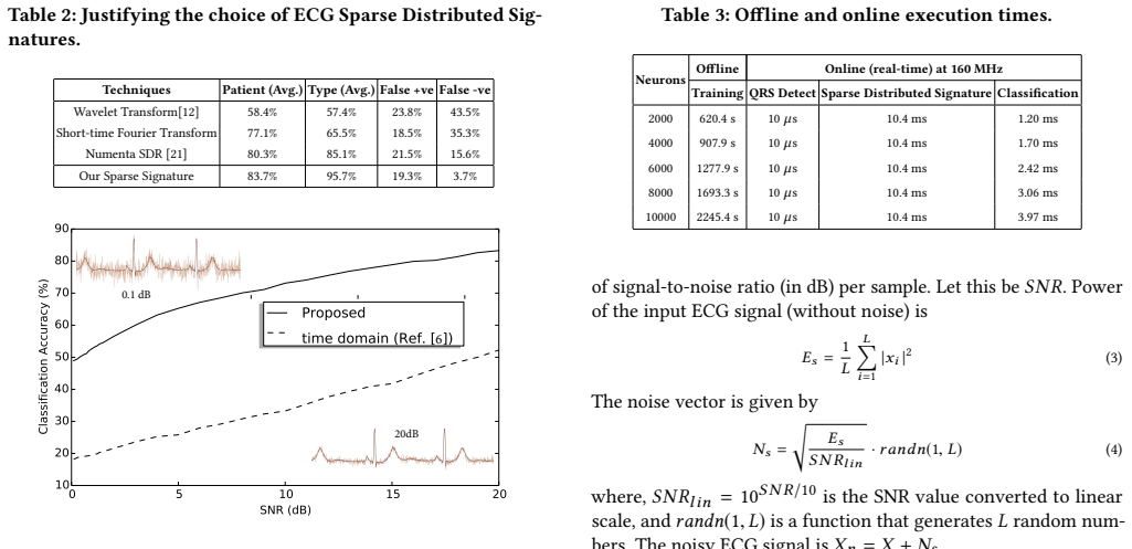
The central claim is that a sparse distributed signature formed from the real and imaginary parts of the short-time Fourier transform of a QRS complex, combined into an 82x82 matrix with fewer than 1% nonzero entries, carries enough information for a multi-layer perceptron to separate the 23 heartbeat classes. With this representation, the classifier reaches 80.7% to 100% accuracy per class, averaging 95.7%, and reduces false negatives to 3.7%, about 89% lower than wavelet and STFT representations tested with the same classifier. The authors further report that the signature keeps classification accuracy high under additive white Gaussian noise, and that the online pipeline of QRS detection, signature conversion, and classification finishes in milliseconds, supporting real-time wearable use.
Load-bearing premise
The reported accuracy assumes that randomly splitting beats from the same 48 patients into training and test sets predicts how the classifier will perform on a new person's ECG; if beats from the same patient are too similar, the test accuracy is inflated.
Editorial extensions
If this is right
- The online pipeline, at roughly 10.4 ms for signature conversion and 1.2 to 4.0 ms for classification at 160 MHz, is fast enough for real-time wearable arrhythmia monitoring.
- Training the same classifier on the five clinically grouped heartbeat classes instead of all 23 improves per-class accuracy by 0.4 to 5.1 percentage points, so the method can be specialized to specific clinical questions.
- The false-negative rate of 3.7% means fewer missed arrhythmic beats than the wavelet, STFT, and sparse representations compared in the paper, which is the failure mode that matters most for screening.
- Under additive white Gaussian noise from 0 to 20 dB, the proposed signatures hold classification accuracy substantially above a time-domain baseline, suggesting resilience to acquisition noise.
Reading between the lines
- Because the train and test beats are drawn from the same 48 patients, the reported 95.7% average likely overstates performance on a brand-new person; a held-out-patient evaluation would be the natural next test.
- The signature's reliance on the positions of high-value entries suggests the same construction could be applied to other transient biomedical signals, such as EEG or EMG, where non-stationarity is also the norm.
- The deliberately padded zero block in the signature leaves room to fuse auxiliary sensor streams, for example accelerometer-based motion data, without changing the classifier, which could address the motion artifacts the paper mentions only briefly.
Signed reviews
Editorial analysis
A structured set of objections, weighed in public.
Referee Report
Summary. The manuscript proposes a two-phase pipeline for heartbeat classification in wearable settings. In the offline phase, QRS complexes from the MIT-BIH arrhythmia database are transformed into sparse 82×82 signatures built from the real and imaginary parts of the short-time Fourier transform, and a multi-layer perceptron (MLP) is trained on these signatures; in the online phase, the Pan-Tompkins algorithm detects QRS peaks in live ECG and the trained MLP classifies each beat. The paper reports an average classification accuracy of 95.7% across 23 heartbeat classes (claimed 22% higher than a time-domain baseline), a false-negative rate of 3.7% for the proposed signatures versus 15.6%–43.5% for three alternative representations, improved robustness to additive white Gaussian noise, and online execution times compatible with real-time use. The central results are presented in Section 5, with accuracy curves in Figure 6, representation comparisons in Table 2, noise analysis in Section 5.3, and timing measurements in Table 3.
Significance. The paper's contribution, if its numbers hold, is a compact sparse time-frequency signature that lets a simple MLP classify ECG beats with high accuracy and low false negatives, plus a plausible real-time implementation story. The signature construction is deterministic — computed from the STFT and a threshold, not fitted to labels — and the controlled comparison across four input representations in Table 2 is a reasonable experimental design. However, the significance is limited by two factors. The reported 95.7% accuracy is produced under a beat-level split in which training and test beats come from the same patients, so it does not support the wearable-use-case claim of generalization to new patients; and the experiment is described with too few details to be reproduced or independently verified. If a patient-disjoint re-evaluation confirms the advantage, the sparse signature representation would be a useful addition to the ECG-classification literature.
major comments (3)
- [§5, Evaluation protocol] The central claim of 95.7% average accuracy rests on a beat-level random split: Section 5 states that 6500 labeled QRS complexes are 'selected randomly from 120K QRS complexes for 48 patients' and that the test set is the remaining beats 'represented from all 48 patients'. Because MIT-BIH contains many beats per patient and beats from the same patient share morphology and recording conditions, this protocol allows the MLP to learn patient-specific patterns during training and then be evaluated on other beats from the very same patients. For the stated wearable deployment scenario, where the classifier must generalize to unseen patients, this does not establish the result; the 'Patient (Avg.)' column in Table 2 averages over the same 48 patients under the same split and does not address inter-patient generalization. The evaluation should be redone with a patient-disjoint split (e.g., the DS1/DS2 split commonly used in the literature and consistent with ANSI/AAMI EC57), reporting accuracy, sensitivity, and positive predictive value on held-out patients, and the corresponding claims in the abstract, Sections 5.1 and 5.2 must then be revised.
- [§5 and §4.1, Reproducibility] The implementation details needed to reproduce the headline result are missing. The STFT parameters (analysis window, length, stride) are not reported — the paper only mentions 'specgram of python-matplotlib' — and the sparsity threshold that turns the STFT into a sparse signature is not quantified (Section 4.1 says only 'values above a certain threshold (sparsity < 1%)'). The MLP configuration is likewise unspecified: Section 5 says a 'design space exploration' determined the hidden layers and neurons, but the final architecture is never given, and Table 3 lists 2000–10000 neurons without identifying which setting produced the 95.7% accuracy. The paper should report the exact STFT settings, the threshold, the final MLP architecture, and the training hyperparameters (learning rate, epochs, regularization), and should state whether the design-space exploration used the test set, which would introduce additional leakage.
- [§5.2, Baseline comparisons] The claimed relative improvements — 22% over the time-domain baseline, 89% lower false negatives than the wavelet representation, 91% lower than the STFT-only representation — are only interpretable if the four representations in Table 2 are compared under identical conditions. The patient-average accuracy of 58.4% reported for the wavelet representation is far below typical results for wavelet-based ECG classifiers reported elsewhere, which raises the question of whether the baselines were run with the same pre-processing, the same split, and the same (unspecified) MLP configuration. The authors should specify the common experimental setup, provide confusion matrices for the four representations, and confirm that the false-positive and false-negative definitions in Section 5.2 match those used for the baselines; without this, the abstract's 3.7% false-negative claim is not verifiable.
minor comments (7)
- [Keywords] The keyword list (Spiking neural network, global synapse, particle swarm optimization, CxQuad, spike disorder count, inter-spike distortion) does not correspond to any content in the manuscript; it should be replaced with terms such as ECG, heartbeat classification, short-time Fourier transform, sparse distributed representation, and multi-layer perceptron.
- [§4.1, Eq. (2)] The padded zero component P in the signature definition carries no information; the authors should either justify its inclusion or explicitly describe it as a placeholder, and clarify whether its constant zeros affect the stated sparsity of fewer than 1% nonzero entries.
- [§5.3, Noise tolerance] Figure 7 shows the noise-tolerance curve for a single patient (id 106), yet the text claims an 'average 133%' improvement over the time-domain baseline for the SNR range 0–20 dB; the average should be supported by reporting the distribution across patients rather than a statement that a similar trend was observed.
- [§5.3, Motion artifacts] The motion-artifact sentence reports 'comparable accuracy' for motion-compensated ECG compared with a wavelet-ICA approach but provides no numbers or details; either quantitative results should be added or the claim removed.
- [Table 1] The mapping from the 23 MIT-BIH classes to the five classes used in the 'training with 5 classes' column is not described; the authors should state the mapping explicitly so that Table 1 is reproducible.
- [Abstract and §5.1] The abstract states 'an improvement of 22% over state-of-the-art approaches', while Section 5.1 specifies the improvement against a single time-domain baseline [6]; the phrasing should be made precise to avoid overclaiming.
- [References and typos] Reference [20] misattributes the 1986 backpropagation paper to Williams and Hinton; the correct first author is Rumelhart. In Section 5, 'Hashwell CPU' should read 'Haswell CPU', and the name 'Pan & Tompkin's Algorithm' in Section 3 should be 'Pan & Tompkins'.
Circularity Check
Mild selection-on-test bias: the network architecture and sparse-signature choice are selected using the test set, so the reported 95.7% accuracy is a selected maximum rather than an independent prediction.
-
fitted input called prediction
[Section 3 (Proposed Approach) and Section 5 (Results and Discussions), incl. Table 2]
"Once trained, the neural network is validated using a test set. A design space exploration is then performed to determine dimensions of the neural network (hidden layers, number of neurons, etc). ... The test set is composed of all 120K QRS complexes (except the ones used for the training set). ... Table 2 reports results justifying our choice of ECG sparse distributed signatures for classification."
The reported test accuracy is obtained on the same test set that was explicitly used for model selection. Section 3 says the network is 'validated using a test set' and then a 'design space exploration' sets the hidden layers and neuron counts. Section 5 confirms that the test set contains all non-training beats. Thus the architecture is chosen by maximizing accuracy on this very test set, and the same test set then produces the headline 95.7% figure. The reported accuracy is therefore a selected maximum, statistically forced by the selection procedure, and not an independent estimate of predictive performance. Table 2 similarly 'justifies' the choice of representation using the same test results, so the representation choice is also selected on the test set.
full rationale
The core derivation is not circular. The sparse signature is produced deterministically by the short-time Fourier transform (Eq. 1), the concatenation of real and imaginary components with padding (Eq. 2), and thresholding; no label information enters the representation construction. The MLP is a standard supervised classifier trained by back-propagation on labeled beats, and the test beats are disjoint from the training beats. There is no load-bearing self-citation chain, no uniqueness theorem imported from the authors, and no ansatz smuggled in via a citation. The one genuine circularity concern is the use of the same test set for model selection: Section 3 states that after validation on a test set, a design space exploration fixes the network dimensions, and Section 5 reports accuracy on the non-training beats. Consequently, the headline 95.7% figure is a maximum over architectures chosen on that test set, not an unbiased estimate. A separate validation limitation, not itself circularity, is that the split is beat-level within the same 48 patients, so the result does not demonstrate generalization to new wearable users. Overall, no derivation reduces to its input by construction, so the circularity score is low.
Assumptions & free parameters
free parameters (5)
- STFT analysis window and stride =
not reported (uses matplotlib specgram defaults)
- Signature matrix size =
82x82
- Sparsity threshold =
not reported (sparsity < 1%)
- MLP architecture =
not reported (hidden layers and neurons chosen by design space exploration)
- Number of training beats =
6500
assumptions (4)
- domain assumption MIT-BIH annotations are ground truth for heartbeat classes.
- domain assumption The ANSI/AAMI five-class grouping is sufficient for arrhythmia.
- domain assumption Pan-Tompkins algorithm detects QRS complexes correctly on the test data.
- standard math The short-time Fourier transform, as defined in Eq. 1, is standard and exact.
Cite this review
Pith. "Pith review of Heartbeat Classification in Wearables Using Multi-layer Perceptron and Time-Frequency Joint Distribution of ECG." pith.science (2026). https://pith.science/paper/VUGZPRMT
@misc{pith2026190806865,
  author       = {Pith},
  title        = {Pith review of: Heartbeat Classification in Wearables Using Multi-layer Perceptron and Time-Frequency Joint Distribution of ECG},
  year         = {2026},
  howpublished = {\url{https://pith.science/paper/VUGZPRMT}},
  note         = {Machine review of arXiv:1908.06865}
}
read the original abstract
Heartbeat classification using electrocardiogram (ECG) data is a vital assistive technology for wearable health solutions. We propose heartbeat feature classification based on a novel sparse representation using time-frequency joint distribution of ECG. Fundamental to this is a multi-layer perceptron, which incorporates these signatures to detect cardiac arrhythmia. This approach is validated with ECG data from MIT-BIH arrhythmia database. Results show that our approach has an average 95.7% accuracy, an improvement of 22% over state-of-the-art approaches. Additionally, ECG sparse distributed representations generates only 3.7% false negatives, reduction of 89% with respect to existing ECG signal classification techniques.
Figures


Figures from the paper (3 more)


Reference graph
Works this paper leans on
-
[6]
V. Queiroz, E. Luz, G. Moreira, Á. Guarda, and D. Menotti, “Automatic cardiac arrhythmia detection and classification using vectorcardiograms and complex networks, ” inEMBC. IEEE, 2015
work page 2015
-
[1]
The impact of the MIT-BIH arrhythmia database,
G. B. Moody and R. G. Mark, “The impact of the MIT-BIH arrhythmia database, ” IEEE Engineering in Medicine and Biology Magazine , 2001
work page 2001
-
[2]
A. for the Advancement of Medical Instrumentation et al., “Testing and reporting performance results of cardiac rhythm and st segment measurement algorithms. american national standards institute, inc.(ansi), ” Inc.(ANSI), ANSI/AAMI/ISO EC57, 2008
work page 2008
-
[3]
Automatic classification of heartbeats using ECG morphology and heartbeat interval features,
P. De Chazal, M. O’Dwyer, and R. B. Reilly, “Automatic classification of heartbeats using ECG morphology and heartbeat interval features, ”IEEE Transactions on Biomedical Engineering, 2004
work page 2004
-
[4]
Heartbeat classification using feature selec- tion driven by database generalization criteria,
M. Llamedo and J. P. Martínez, “Heartbeat classification using feature selec- tion driven by database generalization criteria, ”IEEE Transactions on Biomedical Engineering, 2011
work page 2011
-
[5]
Arrhythmia detection using amplitude difference features based on random forest,
J. Park, S. Lee, and K. Kang, “Arrhythmia detection using amplitude difference features based on random forest, ” inIEEE EMBC, 2015
work page 2015
-
[7]
Heartbeat classification using disease-specific feature selection,
Z. Zhang, J. Dong, X. Luo, K.-S. Choi, and X. Wu, “Heartbeat classification using disease-specific feature selection, ”Elsevier Computers in biology and medicine , 2014
work page 2014
-
[8]
Deep learning ap- proach for active classification of electrocardiogram signals,
M. Al Rahhal, Y. Bazi, H. AlHichri, N. Alajlan, F. Melganiet al., “Deep learning ap- proach for active classification of electrocardiogram signals, ”Elsevier Information Sciences, 2016
work page 2016
Show all 23 references
-
[9]
ECG classification using wavelet packet entropy and random forests,
T. Li and M. Zhou, “ECG classification using wavelet packet entropy and random forests, ”MDPI Entropy, 2016
2016
-
[10]
PcHD: Personalized classification of heartbeat types using a decision tree,
J. Park and K. Kang, “PcHD: Personalized classification of heartbeat types using a decision tree, ”Elsevier Computers in biology and medicine , 2014
2014
-
[11]
Computer aided diagnosis of atrial arrhythmia using dimensionality reduction methods on transform domain representation,
R. J. Martis, U. R. Acharya, H. Adeli, H. Prasad, J. H. Tan et al. , “Computer aided diagnosis of atrial arrhythmia using dimensionality reduction methods on transform domain representation, ”Elsevier Biomedical Signal Processing and Control, 2014
2014
-
[12]
ECG signals classification based on discrete wavelet transform, time domain and frequency domain features,
S. A. Shufni and M. Y. Mashor, “ECG signals classification based on discrete wavelet transform, time domain and frequency domain features, ” inIEEE Confer- ence on Biomedical Engineering (ICoBE) , 2015
2015
-
[13]
A real-time QRS detection algorithm,
J. Pan and W. J. Tompkins, “A real-time QRS detection algorithm, ”IEEE transac- tions on biomedical engineering , 1985
1985
-
[14]
Theano: Deep learning on GPUs with python,
J. Bergstra et al., “Theano: Deep learning on GPUs with python, ” inNIPS BigLearn- ing Workshop, 2011
2011
-
[15]
Neuromorphic architectures for spiking deep neural networks,
G. Indiveri, F. Corradi, and N. Qiao, “Neuromorphic architectures for spiking deep neural networks, ” inIEEE IEDM. IEEE, 2015, pp. 4–2
2015
-
[16]
28.4 a battery-powered efficient multi- sensor acquisition system with simultaneous ECG, BIO-Z, GSR, and PPG,
M. Konijnenburg, S. Stanzione et al., “28.4 a battery-powered efficient multi- sensor acquisition system with simultaneous ECG, BIO-Z, GSR, and PPG, ” in IEEE ISSCC, 2016
2016
-
[17]
Real-time ECG monitoring and arrhythmia detection using Android-based mobile devices,
S. Gradl, P. Kugler, C. Lohmüller, and B. Eskofier, “Real-time ECG monitoring and arrhythmia detection using Android-based mobile devices, ” inIEEE EMBC, 2012
2012
-
[18]
The fractional fourier transform and time-frequency representa- tions,
L. B. Almeida, “The fractional fourier transform and time-frequency representa- tions, ”IEEE Transactions on signal processing , 1994
1994
-
[19]
Stable recovery of sparse overcom- plete representations in the presence of noise,
D. L. Donoho, M. Elad, and V. N. Temlyakov, “Stable recovery of sparse overcom- plete representations in the presence of noise, ”IEEE Transactions on information theory, 2006
2006
-
[20]
Learning representations by back-propagating errors,
D. Williams and G. Hinton, “Learning representations by back-propagating errors, ” Nature, 1986
1986
-
[21]
Encoding data for HTM systems,
S. Purdy, “Encoding data for HTM systems, ”arXiv preprint arXiv:1602.05925, 2016
2016 arXiv
-
[22]
Rate-adaptive compressed-sensing and sparsity variance of biomedical signals,
V. Behravan, N. E. Glover, R. Farry, P. Y. Chiang, and M. Shoaib, “Rate-adaptive compressed-sensing and sparsity variance of biomedical signals, ” inIEEE BSN, 2015
2015
-
[23]
Ecg artifact removal from surface emg signal using an automated method based on wavelet-ica,
S. Abbaspour, M. Lindén, and H. Gholamhosseini, “Ecg artifact removal from surface emg signal using an automated method based on wavelet-ica, ” inInterna- tional conference on wearable micro and nanotechnologies for personalized health. Sweden: Västerås, 2015, pp. 91–7. 6
2015
Reviewed August 14, 2026 · model on record in the stance chip above.
Discussion (0). Continue with ORCID to comment.