REVIEW 4 major objections 5 minor 12 references
Deep Brain Net: An Optimized Deep Learning Model for Brain tumor Detection in MRI Images Using EfficientNetB0 and ResNet50 with Transfer Learning
T0 review · 4 major / 5 minor · reviewed 2026-08-06 · deepseek-v4-flash
Pith's one-line read DeepBrainNet fuses EfficientNetB0 and ResNet50 to classify four brain MRI tumor categories, claiming 88% accuracy, 88.75% weighted F1, and a 98.17% macro AUC.
desk verdict A standard two-backbone transfer-learning application whose headline accuracy is unsupported by an internally contradictory dataset description and inconsistent metric reporting. read the letter →
The pith
A machine-rendered reading of the paper's core claim, the machinery that carries it, and where it could break.
The reading
What carries the argument
The machine doing the work is a parallel hybrid: an EfficientNetB0 branch, whose mobile inverted bottleneck (MBConv) blocks use depthwise separable convolutions to cut parameters and computational cost, joined with a ResNet50 branch that is initialized from ImageNet weights and fine-tuned, so residual connections keep gradients flowing during transfer learning. Around this sit a preprocessing chain (resizing, CLAHE contrast enhancement, histogram equalization, margin cropping), fuzzy C-means feature selection that retains features with high membership across clusters, and training-time augmentation with rotations, flips, zooms, shears, brightness changes, and shifts. Early stopping on validation loss and dynamic learning-rate reduction stabilize training.
What would settle it
Re-run the architecture on the same 7,023-image data with a strictly disjoint test split and report test accuracy and weighted F1; if test accuracy falls below 88% by more than split sampling error, the central claim is not supported. A cheaper control is an ablation training EfficientNetB0 and ResNet50 alone under identical settings: the hybrid must beat its better single branch, otherwise the fusion is not what carries the result.
Extended reading notes
Core claim
The central claim of the paper is that a single deep network can get the best of both efficiency and depth by fusing EfficientNetB0's mobile inverted bottleneck blocks with a ResNet50 branch that carries over ImageNet features. Trained with on-the-fly augmentation, dropout, early stopping, and fuzzy C-means feature selection, the hybrid reports 88% overall accuracy on a 7,023-image four-class MRI dataset. The class-level report shows the strongest separation for the no-tumor class (precision 0.946) and the weakest for meningioma (F1 0.808). The paper presents these numbers as outperforming prior deep-learning classifiers on the same benchmark and as evidence that an architecture tuned for efficiency on natural images transfers well to medical imaging.
Load-bearing premise
The claim depends on treating the validation set, which was also used for early stopping and model selection, as a measure of generalization; if that set is not a cleanly separated test set, the 88% accuracy figure is not a supported estimate of performance on new scans.
Editorial extensions
If this is right
- If the reported accuracy holds on a clean test set, DeepBrainNet could serve as a first-pass four-class triage tool in settings where an MRI radiologist is not immediately available.
- A macro AUC near 0.98 means the model ranks classes well even where hard labels are missed, so threshold tuning could trade sensitivity against specificity per tumor type.
- The roughly 24 million trainable parameters and the EfficientNetB0 branch's depthwise separable convolutions keep the model light enough for deployment on clinical workstations rather than large GPU clusters.
- Per-class results point to meningioma as the most error-prone category, so future data collection or class weighting should target that class first.
Reading between the lines
- Because no separate held-out test set is described, the 88% figure should be read as a validation-based estimate; a clean test split may land lower.
- The paper never compares the hybrid against its own two backbones run alone, so the claimed fusion advantage is not yet isolated; an ablation with identical training would settle it.
- Tumor and no-tumor images come from different source repositories, so some of the discriminative signal may be source-specific rather than tumor-specific; testing on an external dataset with matched acquisition is the cleanest check.
- The comparison table's baselines [14] and [15] have no corresponding entries in the reference list, so the claim that DeepBrainNet beats those systems cannot be independently verified from this manuscript alone.
Editorial analysis
A structured set of objections, weighed in public.
Referee Report
Summary. The paper proposes DeepBrainNet, a hybrid deep learning model combining EfficientNetB0 and ResNet50 with transfer learning for four-class brain tumor classification from MRI images. The authors report an overall accuracy of 88%, a weighted F1-score of 88.75%, and a macro AUC-ROC of 98.17% on a publicly available dataset, and claim that this consistently outperforms existing state-of-the-art methods. The manuscript describes dataset collection, preprocessing, fuzzy C-means feature selection, model architecture, training details, and a comparative analysis.
Significance. If the reported results were reproducible and properly validated, the model would be a modest but potentially useful engineering contribution to automated brain tumor classification. The paper's strengths are that it uses publicly available data, provides training details, includes a confusion matrix and ROC curves, and compares with two prior systems. However, the contribution is incremental—a standard late-fusion ensemble of two pretrained CNNs—and the experimental validation as presented is not rigorous enough to support the central claims of consistent superiority and clinical potential.
major comments (4)
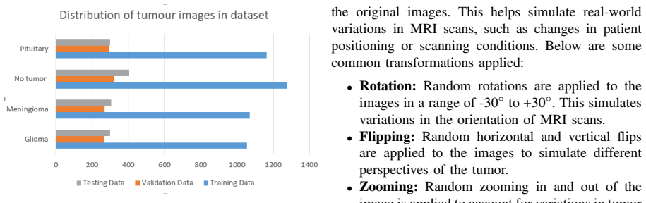
- [§III-A (Dataset)] The dataset description is internally contradictory. The section first states that the study uses 7023 images from figshare, SARTAJ, and Br35H, with no-tumor images taken from Br35H. It then states that SARTAJ images were removed and 'The final dataset used for this study consists of images from figshare.' If the figshare collection contains only the three tumor classes (as is standard for this dataset), then a figshare-only dataset cannot yield the no-tumor results reported in Table III and the four-class confusion matrix in Figure 11. If Br35H no-tumor images were retained, then the final dataset is not figshare-only and the exact per-class counts and total are not specified. Either way, the input dataset for the experiments is not uniquely defined, which makes the reported 88% accuracy unreproducible.
- [§V (Results) and Abstract/Table IV] The central accuracy claim is reported inconsistently and without a defined evaluation split. The Abstract states 88% accuracy, Table IV lists 89% for DeepBrainNet, and Section V reports a validation accuracy of 93.2% (and later 94.7%) for the same model. The manuscript never defines a held-out test set; the models are selected by early stopping on validation loss, so the reported metrics appear to be validation-set metrics that are optimistically biased. Without a clearly separated test partition, the claim that the model 'generalizes well to unseen data' is unsupported.
- [§III-D (Feature Selection)] Fuzzy C-means (FCM) feature selection is described in detail and Algorithm 1 is provided, but FCM never appears in the results section, no ablation study is performed, and there is no explanation of how the selected features are integrated into the classification pipeline. As written, the feature-selection component has no observable effect on the empirical results, so its role in the claimed performance is unclear.
- [Table IV (Comparative Analysis)] The comparison in Table IV is not adequately grounded. The table claims 'same dataset used in this study,' but given the ambiguity in §III-A, it is unclear which dataset and split were used for the cited baselines [14] and [15]. No details are provided on the preprocessing, augmentation, or evaluation protocol for the baselines, and no error bars or multiple-run statistics are given. The claim that DeepBrainNet 'consistently outperforms' prior work is therefore not supported by the reported evidence.
minor comments (5)
- [§IV-A] The description of EfficientNetB0 incorrectly states that it combines EfficientNet and VNet for segmentation; this paper addresses classification, and the quoted text appears to have been reused from a segmentation study.
- [Equation (1)] The variables in the resizing equation are inconsistently named: 'Iresized = f(I original, Dnew)' but the definition list mentions 'Original image' rather than 'I original'.
- [Algorithm 1] The membership update equation in Algorithm 1 has a typesetting error: 'uij = db(xi, vj)Pc k=1 db(xi, vk)' is missing the division symbol, making the formula unreadable.
- [Reference [4]] Reference [4] cites 'arXiv:2304.100394', which appears to contain an extra digit; the correct ID appears to be arXiv:2304.10039.
- [Figure 3] The bar chart in Figure 3 is not annotated with axis labels or class counts, making it difficult to verify the class distribution described in the text.
Circularity Check
No significant circularity: the paper reports an empirical training-and-evaluation outcome with no derivation chain that reduces to its own inputs.
full rationale
This paper makes no formal derivation claim. Its central result is an empirical measurement: a hybrid EfficientNetB0/ResNet50 model trained with transfer learning and evaluated on brain MRI data, yielding 88% accuracy, an 88.75% weighted F1-score, and a 98.17% macro AUC-ROC. The preprocessing equations (resizing, normalization, CLAHE, histogram equalization) and the fuzzy C-means algorithm are standard operations, not derived results, and none of them is defined in terms of the final accuracy or F1-score. The evaluation metrics such as precision, recall, and F1 are textbook definitions applied to the model's predictions, not quantities that were fitted to produce the reported values. There are no load-bearing self-citations: the cited works are external and, while some are the sources of dataset descriptions and algorithm conventions, the reported performance numbers are not obtained from those citations. No uniqueness theorem from the authors is invoked, and no ansatz is smuggled in via self-citation. The paper does contain a serious internal inconsistency in the dataset description: Section III-A says the final dataset consists of images from figshare, while also stating that no-tumor images were taken from Br35H, which would be needed for the four-class results in Table III and Figure 11. That is a reproducibility and correctness risk, not a circularity of the kind this axis targets. Similarly, the absence of a clearly defined held-out test set and the possibility of validation-based early stopping inflating reported metrics are empirical validity concerns, not circular reasoning. Because the central claim is an experimental outcome with no derivation chain that reduces to its inputs, the appropriate circularity score is 0.
Assumptions & free parameters
free parameters (2)
- Trained ResNet50 and EfficientNetB0 weights including fusion head =
~24M trainable parameters; final weights not released
- Training hyperparameters: epochs 40, batch size 32, input 224x224, augmentation settings =
As stated in Section V; learning rate and dropout rates not specified
assumptions (3)
- domain assumption The merged figshare, SARTAJ, and Br35H dataset has accurate labels after removal of misclassified SARTAJ glioma images.
- domain assumption The validation split is representative and does not leak between training and validation.
- domain assumption ImageNet-pretrained features transfer to MRI brain images.
Cite this review
Pith. "Pith review of Deep Brain Net: An Optimized Deep Learning Model for Brain tumor Detection in MRI Images Using EfficientNetB0 and ResNet50 with Transfer Learning." pith.science (2026). https://pith.science/paper/WGX55CXO
@misc{pith2026250707011,
  author       = {Pith},
  title        = {Pith review of: Deep Brain Net: An Optimized Deep Learning Model for Brain tumor Detection in MRI Images Using EfficientNetB0 and ResNet50 with Transfer Learning},
  year         = {2026},
  howpublished = {\url{https://pith.science/paper/WGX55CXO}},
  note         = {Machine review of arXiv:2507.07011}
}
read the original abstract
In recent years, deep learning has shown great promise in the automated detection and classification of brain tumors from MRI images. However, achieving high accuracy and computational efficiency remains a challenge. In this research, we propose Deep Brain Net, a novel deep learning system designed to optimize performance in the detection of brain tumors. The model integrates the strengths of two advanced neural network architectures which are EfficientNetB0 and ResNet50, combined with transfer learning to improve generalization and reduce training time. The EfficientNetB0 architecture enhances model efficiency by utilizing mobile inverted bottleneck blocks, which incorporate depth wise separable convolutions. This design significantly reduces the number of parameters and computational cost while preserving the ability of models to learn complex feature representations. The ResNet50 architecture, pre trained on large scale datasets like ImageNet, is fine tuned for brain tumor classification. Its use of residual connections allows for training deeper networks by mitigating the vanishing gradient problem and avoiding performance degradation. The integration of these components ensures that the proposed system is both computationally efficient and highly accurate. Extensive experiments performed on publicly available MRI datasets demonstrate that Deep Brain Net consistently outperforms existing state of the art methods in terms of classification accuracy, precision, recall, and computational efficiency. The result is an accuracy of 88 percent, a weighted F1 score of 88.75 percent, and a macro AUC ROC score of 98.17 percent which demonstrates the robustness and clinical potential of Deep Brain Net in assisting radiologists with brain tumor diagnosis.
Figures


Figures from the paper (9 more)


Reference graph
Works this paper leans on
-
[1]
MRI brain tumour detection us- ing deep learning and machine learning ap- proaches
S. Anantharajan, S. Gunasekaran, and T. Sub- ramanian. “MRI brain tumour detection us- ing deep learning and machine learning ap- proaches”. In:Measurement: Sensors31.1 (2024), p. 101026.DOI: 10.1016/j.measen.2024. 101026
-
[2]
Employing deep learn- ing and transfer learning for accurate brain tumour detection
S. K. Mathivanan et al. “Employing deep learn- ing and transfer learning for accurate brain tumour detection”. In:Scientific Reports14.2 (2024), p. 7232.DOI: 10 . 1038 / s41598 - 024 - 57970-7
work page 2024
-
[3]
Brain tumour detection from images and compar- ison with transfer learning methods and 3- layer CNN
M. Z. Khaliki and M. S. Bas ¸arslan. “Brain tumour detection from images and compar- ison with transfer learning methods and 3- layer CNN”. In:Scientific Reports14.3 (2024), p. 2664.DOI: 10.1038/s41598-024-52823-9
-
[4]
Brain tumor multi classification and segmentation in MRI images using deep learning
B. Amin et al. “Brain tumour multi clas- sification and segmentation in MRI images using deep learning”. In:arXiv preprint arXiv:2304.100394 (2023).DOI: 10 . 48550 / arXiv.2304.10039
work page Pith review arXiv doi:10.48550/arxiv.2304.10039 2023
-
[5]
E. Albalawi et al. “Enhancing brain tumour classification in MRI scans with a multi-layer customized convolutional neural network ap- proach”. In:Frontiers in Computational Neuro- science18.5 (2024), p. 1418546.DOI: 10.3389/ fncom.2024.1418546
-
[6]
S. Natha et al. “Automated brain tumour iden- tification in biomedical radiology images: A multi-model ensemble deep learning approach”. In:Applied Sciences14.6 (2024), p. 2210.DOI: 10.3390/app14052210
-
[7]
Ali M. Reza. “Realization of the contrast limited adaptive histogram equalization (CLAHE) for real-time image enhancement”. In:Journal of VLSI signal processing systems for signal, im- age and video technology38.7 (2004), pp. 35– 44.DOI: 10.1023/B:VLSI.0000028532.53893. 82
-
[8]
A review of histogram equalization techniques in image enhancement application
W. A. Mustafa and M. M. M. Abdul Kader. “A review of histogram equalization techniques in image enhancement application”. In:Journal of Physics: Conference Series. V ol. 1019. 8. IOP Publishing. 2018, p. 012026
work page 2018
Show all 12 references
-
[9]
Comparison of Fuzzy C-Means, Fuzzy Kernel C-Means, and Fuzzy Kernel Robust C-Means to Classify Thalassemia Data
Zuherman Rustam et al. “Comparison of Fuzzy C-Means, Fuzzy Kernel C-Means, and Fuzzy Kernel Robust C-Means to Classify Thalassemia Data”. In:International Journal on Advanced Science, Engineering and Information Technol- ogy9.9 (2019), p. 1205.DOI: 10.18517/ijaseit. 9.4.9580
2019 doi
-
[10]
Un- supervised Multiview Fuzzy C-Means Cluster- ing Algorithm
I. Hussain, K. P. Sinaga, and M.-S. Yang. “Un- supervised Multiview Fuzzy C-Means Cluster- ing Algorithm”. In:Electronics12.10 (2023), p. 4467.DOI: 10.3390/electronics12214467
2023 doi
-
[11]
Efficientnet: Rethinking model scaling for convolutional neu- ral networks
Mingxing Tan and Quoc V . Le. “Efficientnet: Rethinking model scaling for convolutional neu- ral networks”. In:International conference on machine learning. 11. PMLR. 2019, pp. 6105– 6114
2019
-
[12]
Potato Leaf Disease De- tection and Classification Using VGG16
V . L. Adluri et al. “Potato Leaf Disease De- tection and Classification Using VGG16”. In: Proceedings of International Joint Conference on Advances in Computational Intelligence. 12. Springer Nature Singapore. 2024, pp. 147–158. DOI: 10.1007/978-981-97-0180-3 13
2024 doi
Reviewed August 6, 2026 · model on record in the stance chip above.
Discussion (0). Sign in to comment.