REVIEW 4 major objections 6 minor 64 references
Image-based physical characterization of magnetotactic bacteria from an environmental sample
T0 review · 4 major / 6 minor · reviewed 2026-08-07 · deepseek-v4-flash
Pith's one-line read The paper claims that automated measurements of cell size, swimming speed, and magnetic moment can separate coexisting magnetotactic bacterial populations in an uncultured river sample, and it demonstrates this on a newly discovered…
desk verdict A useful open-source MTB imaging workflow and a new field site, packaged inside a paper whose 'three separate species' claim outruns what AIC on a single sample can support. read the letter →
The pith
A machine-rendered reading of the paper's core claim, the machinery that carries it, and where it could break.
The reading
What carries the argument
The load-bearing object is the automated U-turn method, which fits each recorded turning trajectory to the theoretical shape $y = -\frac{L}{\pi}\ln(\sec(\pi x/L))$, where $L$ is the asymptotic U-turn width. From that width the cell magnetic moment follows as $m = \pi\alpha v/(HL)$, with rotational drag coefficient $\alpha = 8\pi^2\eta R^3$, field strength $H$, swimming speed $v$, and effective radius $R$. This turns a standard alternating-magnetic-field experiment into a per-cell moment measurement. On the velocimetry side, a static field is reversed to draw long swimming tracks from cells concentrated at a capillary wall, and automated multiple-hypothesis tracking reconstructs them. Population counts are then chosen by AIC-constrained multi-Gaussian fits over velocity-radius or moment-radius histograms, with only statistically significant components retained.
What would settle it
Isolate, sort, or micromanipulate individual bacteria from each of the three fitted magnetic-moment populations and sequence single-cell 16S rRNA genes; if cells within one fitted cluster do not share a consistent genotype, or if a fresh sample from the same site yields a different cluster count on the same day, the population-resolution claim would be refuted. A cheaper partial check is to compare TEM cell-size and magnetosome-presence counts against the model's predicted clusters and its critical-radius intercept.
Extended reading notes
Core claim
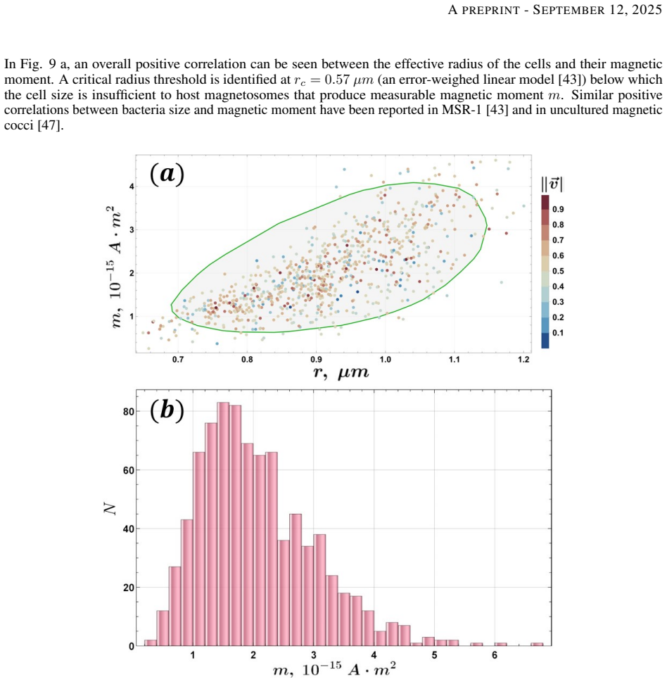
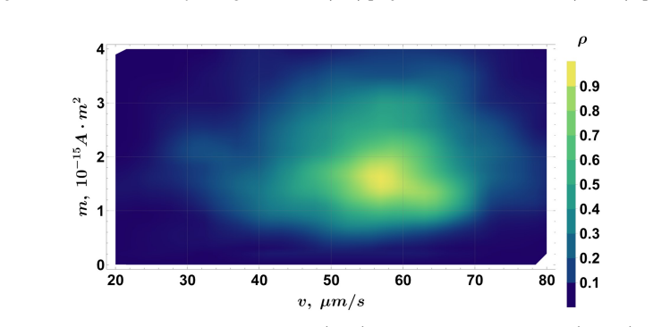
The central claim is that integrating automated velocimetry and magnetic moment analysis provides a powerful and accessible way to differentiate magnetotactic bacteria populations in complex environmental samples. On a sample from the Ogre River, the U-turn analysis of 846 individual tracks gives a most probable magnetic moment of $m = 1.5\times10^{-15}\,\mathrm{A\,m^2}$, a mean of $\langle m\rangle = 2.06\times10^{-15}\,\mathrm{A\,m^2}$, and a critical radius $r_c = 0.57\,\mu\mathrm{m}$ below which cells show no measurable moment. The same data, fit in the $(m, r)$ plane with an AIC-selected three-population Gaussian mixture, separate three magnetic groups, one of which shows a negative radius-moment correlation that the paper notes is biologically unusual. Velocimetry alone finds four populations on the first day and two on the second, and the comparison shows that one velocimetry population is non-magnetic. The paper's conclusion is that these physical fingerprints resolve MTB populations that genetic and morphological methods cannot easily separate in an uncultured mixture.
Load-bearing premise
The fitted statistical clusters in size, velocity, and magnetic moment correspond to real biological groups such as species or cell types, rather than artifacts of the Gaussian fitting, ongoing sample decay, or contamination by non-magnetic cells.
Editorial extensions
If this is right
- MTB diversity in an environmental sample can be assessed with an optical microscope, a permanent magnet, and the open-source code, without cultivation, TEM, or sequencing.
- Velocimetry and magnetic-moment analysis can be combined to flag non-magnetic bacteria that would otherwise be misread as separate magnetotactic populations.
- Velocity distributions measured on consecutive days provide a quantitative readout of how fast distinct MTB populations decline under ex situ storage.
- The critical radius near $0.57\,\mu\mathrm{m}$ gives a concrete size cutoff below which cells in this environment are unlikely to carry a detectable magnetosome chain.
- Three magnetic populations with distinct $(m,r)$ correlations imply that behavioral fingerprinting can distinguish species-level groups within a mixed wild sample.
Reading between the lines
- The authors do not map their fitted clusters onto individual genotypes; a natural extension would be single-cell sorting of the three magnetic populations followed by single-cell 16S rRNA sequencing to test whether the clusters correspond one-to-one with species.
- The unusual population with negative $m(r)$ correlation could be a growth-stage artifact or a genuinely distinct magnetosome arrangement; imaging sorted cells from that cluster with TEM would settle which.
- Because the static-field velocimetry needs only a permanent magnet, the pipeline could be deployed as a field-screen for MTB-rich sites, measuring population diversity on site before any enrichment is attempted.
- Repeating the two-day measurement at more frequent intervals would let the method estimate population-specific survival half-lives, which would inform how quickly samples must be processed after collection.
Editorial analysis
A structured set of objections, weighed in public.
Referee Report
Summary. The manuscript reports a new magnetotactic bacteria (MTB)-rich site in the Ogre River, Latvia, and presents two open-source image-based analysis pipelines: a static-field velocimetry method for cell velocity and effective radius, and an alternating-field U-turn method for estimating single-cell magnetic moments. Using AIC-constrained multi-Gaussian fits on velocity-radius and moment-radius data, the authors claim to resolve four velocity populations on Day 1 (two on Day 2) and three magnetic-moment populations, which they interpret as three separate MTB species, together with one non-magnetic population. The physical methods are combined with 16S rRNA metagenomics and TEM to characterize the sample, and the paper concludes that the workflows provide a powerful approach for differentiating MTB populations in complex environmental samples.
Significance. If the central claim is sustained, the paper would provide a valuable, cultivation-independent, low-equipment toolset for rapid field screening of MTB populations, and it contributes open-source code for tracking, U-turn analysis, and magnetic-moment retrieval. The new MTB-rich field site is also a useful empirical contribution. The strengths are the automated tracking pipeline, the explicit magnetic-moment statistics from 846 trajectories, and the authors' candid discussion of the biological ambiguity of the fitted populations. However, the key scientific claim that the statistically identified Gaussian components correspond to real biological populations is not independently validated, and the negative-correlation component in the (r, m) fit is presented with an inadequate plausibility check. These issues directly affect the paper's central demonstration that the methods can differentiate MTB populations.
major comments (4)
- [Methods, 'MTB population identification'; Results, 'Magnetic moment-based characterization'] The central claim that three MTB populations (and by extension, 'three separate species') are resolved rests on AIC-constrained Gaussian mixture components, but AIC only selects the best model within a candidate family; it does not establish that the components correspond to real clusters. The manuscript provides no independent validation: no linkage of clusters to 16S OTUs, no single-cell isolation or staining, no replicate sampling, and no comparison to a null model. The variational Gaussian mixture check in Appendix A is not independent because it assumes the same Gaussian cluster structure on the same features. Consequently, the 'three separate species' interpretation in the Conclusions is not supported by the evidence presented.
- [Results, 'Magnetic moment-based characterization' (Table 3, row 2; Fig. 10)] The negative-correlation component (rho = -0.26 +/- 0.02) is described by the authors as biologically unusual and previously unreported. Their dismissal of aggregates because effective radii of 0.86-0.93 um are 'too small to accommodate multiple cells' is not convincing: a doublet of two cells with radii near 0.6 um would have an area-based effective radius of roughly 0.85 um, so aggregates are not excluded by the stated dimensions. Furthermore, the three components overlap heavily (mean radii separated by less than 0.15 um, sigma_2 between 0.03 and 0.10 um), so skewness, boundary effects, or the chosen number of components could produce an apparent negative correlation. The paper should demonstrate robustness through bootstrapped fits, skewness-aware models, or independent biophysical validation before treating this component as a real biological population.
- [Introduction (Section 1) vs. Methods, 'Magnetic moment calculation'] The Introduction lists 'an improved Bean model, integrated with an automated U-turn analysis framework' as a central methodological component, but the Methods section describes only the standard U-turn shape analysis from Ref. [43] (Eqs. 1 and 2) and never defines or mentions a 'Bean model' or any improvement to it. As written, the claimed novelty of this methodological component cannot be evaluated. The authors should either present the model and its validation or remove the claim from the Introduction.
- [Methods, 'MTB population identification'; Tables 2 and 3] The manuscript does not report sample sizes for the velocimetry datasets, the AIC values or Delta-AIC for the selected models, or a quantitative definition of the significance criterion based on 'contributions to the PDF integral volume reconstruction.' Without these details, the reader cannot assess whether the four-population (Day 1) and three-population (m) fits are meaningfully better than simpler alternatives. Please provide these values, along with a bootstrap stability analysis or equivalent, for the reported population counts.
minor comments (6)
- [Throughout] The name 'Aikake' should be spelled 'Akaike' (Introduction and Velocimetry-based characterization).
- [Results, 'Morphological and genetic diversity'] The word 'coboochtoedral' appears to be a typo for 'cubooctahedral', and 'bilopotrichous' should likely be 'lophotrichous'.
- [Table 1 and 16S rRNA sequencing section] '16-s rRNA' and '16-s' should be written as '16S rRNA' and '16S'.
- [16S rRNA gene sequencing data analysis] 'Miliseq' should be 'MiSeq'.
- [Figure 8 caption] The caption contains the typo 'Velocitmetry'; it should be 'Velocimetry'.
- [Results, 'Velocimetry-based characterization'] Please report the number of cells (N) used for the velocity histograms in Figures 4 and 5, as this is essential for interpreting the fitted population fractions.
Circularity Check
No circular derivation: populations are fit from independent single-cell measurements; self-citations to prior U-turn methodology are reuse, not load-bearing circularity.
full rationale
Walking the derivation chain: (i) cell radii and velocities are direct image-analysis observables from reconstructed trajectories; (ii) magnetic moments are obtained by fitting U-turn trajectories to the theoretical shape in Eq. (1) and then computing m from Eq. (2), a physical relation developed in the authors' prior work [43]; (iii) the population split is produced by AIC-constrained multi-Gaussian fitting of the (r,v) and (r,m) data, and the negative-correlation component is explicitly not a fitting constraint; (iv) the 'three separate species' statement is an interpretation of the AIC fit, not an input to it. None of these steps sets the conclusion equal to an input: the measured trajectories, radii, velocities, and magnetic moments are independent observables, and the AIC procedure selects a model rather than imposing the claimed population count. The reuse of Eqs. (1)-(2) and the rc = 0.57 um threshold from [43] is methodological reuse of independently developed, code-released prior work, not a self-justifying premise; the threshold is an external reference from MSR-1 data and is not used to construct the fitted populations. The Appendix A variational-GMM check is a same-data internal consistency analysis and therefore is not independent validation, but that is a statistical caveat, not circularity. The paper itself acknowledges that 'AIC provides a statistically robust framework for model selection' yet 'does not impose biophysical constraints' and that different individuals may be recorded in different datasets; these are honest limitations. The skeptical concern that AIC-selected Gaussian components may not correspond to biologically real species is a validity/identifiability risk, not a reduction of the output to the input. No circular step can be quoted with a specific equation-to-equation reduction, so the analysis is self-contained against the measured data; the score reflects only minor non-load-bearing self-citation.
Assumptions & free parameters
free parameters (3)
- Critical radius threshold rc =
0.57 µm
- Multi-Gaussian population parameters =
Populations in Tables 2 and 3 (means, variances, correlations, amplitudes)
- PDF significance threshold =
Not stated numerically
assumptions (5)
- domain assumption The U-turn shape function (Eq. 1) and magnetic moment formula m = παv/(HL) from arXiv:2501.09869 are valid for cells in this environmental sample.
- domain assumption Effective spherical approximation for rotational drag, α = 8π²ηR³, with R from trajectory-averaged equivalent circle radius.
- domain assumption Medium viscosity η = 0.90·10^-3 Pa·s and field B = 2.55·10^-4 T are accurate and constant.
- standard math AIC-constrained Gaussian mixture modeling identifies biologically real populations.
- ad hoc to paper Each fitted population corresponds to a distinct species or cell type.
Cite this review
Pith. "Pith review of Image-based physical characterization of magnetotactic bacteria from an environmental sample." pith.science (2026). https://pith.science/paper/WQCHB2LB
@misc{pith2026250604011,
  author       = {Pith},
  title        = {Pith review of: Image-based physical characterization of magnetotactic bacteria from an environmental sample},
  year         = {2026},
  howpublished = {\url{https://pith.science/paper/WQCHB2LB}},
  note         = {Machine review of arXiv:2506.04011}
}
read the original abstract
Magnetotactic bacteria (MTB) are a diverse group of microorganisms that are able to biomineralize magnetic nanoparticles. Most MTB remain uncultured, making population-level characterization from natural environments difficult. We report the discovery of a new and diverse MTB-rich site in the Ogre River, Latvia, and present an integrated approach combining 16S rRNA sequencing, transmission electron microscopy, and novel open-source, automated image-based physical methods to characterize bacteria populations within environmental samples. We introduce a pipeline for cell velocimetry using a static magnetic field and a method to classify cell populations based on their magnetic moment using a modified U-turn method where cell behavior is studied in an alternating magnetic field. This study demonstrates that our physical analysis methods provide a powerful, fast, and robust toolset for MTB population analysis in complex environmental samples.
Figures


Figures from the paper (12 more)




Reference graph
Works this paper leans on
-
[43]
Mara Smite et al. “Explicit and fully automatic analysis of magnetotactic bacteria motion reveals the magnitude and length scaling of magnetic moments”. In: (2025). arXiv: 2501.09869 [physics.bio-ph].URL: https: //arxiv.org/abs/2501.09869
work page Pith review arXiv 2025
-
[1]
Richard Blakemore. “Magnetotactic Bacteria”. In:Science190.4212 (1975), pp. 377–379.DOI: 10.1126/ science . 170679. eprint: https : / / www . science . org / doi / pdf / 10 . 1126 / science . 170679.URL: https://www.science.org/doi/abs/10.1126/science.170679
-
[2]
Ecology, Diversity, and Evolution of Magnetotactic Bacteria
Christopher T. Lefèvre and Dennis A. Bazylinski. “Ecology, Diversity, and Evolution of Magnetotactic Bacteria”. en. In:Microbiology and Molecular Biology Reviews77.3 (Sept. 2013), pp. 497–526.ISSN: 1092-2172, 1098- 5557.DOI: 10.1128/MMBR.00021-13 .URL: https://journals.asm.org/doi/10.1128/MMBR.00021- 13(visited on 03/31/2023)
-
[3]
Swimming with magnets: From biological organisms to synthetic devices
Stefan Klumpp et al. “Swimming with magnets: From biological organisms to synthetic devices”. en. In: Physics Reports789 (Jan. 2019), pp. 1–54.ISSN: 03701573.DOI: 10.1016/j.physrep.2018.10.007.URL: https://linkinghub.elsevier.com/retrieve/pii/S0370157318302862(visited on 08/10/2023)
-
[4]
George A. Schaible et al. “Multicellular magnetotactic bacteria are genetically heterogeneous consortia with metabolically differentiated cells”. In:PLOS Biology22.7 (July 2024), pp. 1–26.DOI: 10.1371/journal. pbio.3002638.URL:https://doi.org/10.1371/journal.pbio.3002638
doi:10.1371/journal 2024
-
[5]
Magnetosome formation in prokaryotes
Dennis A. Bazylinski and Richard B. Frankel. “Magnetosome formation in prokaryotes”. en. In:Nature Reviews Microbiology2.3 (Mar. 2004), pp. 217–230.ISSN: 1740-1526, 1740-1534.DOI: 10.1038/nrmicro842.URL: https://www.nature.com/articles/nrmicro842(visited on 01/10/2024)
-
[6]
A Compass To Boost Navigation: Cell Biology of Bacterial Magnetotaxis
Frank D. Müller, Dirk Schüler, and Daniel Pfeiffer. “A Compass To Boost Navigation: Cell Biology of Bacterial Magnetotaxis”. en. In:Journal of Bacteriology202.21 (Oct. 2020). Ed. by William Margolin.ISSN: 0021-9193, 1098-5530.DOI: 10.1128/JB.00398-20 .URL: https://journals.asm.org/doi/10.1128/JB.00398- 20(visited on 09/03/2024)
-
[7]
Spatial arrangement of chains of magnetosomes in magnetotactic bacteria
Marianne Hanzlik, Michael Winklhofer, and Nikolai Petersen. “Spatial arrangement of chains of magnetosomes in magnetotactic bacteria”. en. In:Earth and Planetary Science Letters145.1-4 (Dec. 1996), pp. 125–134. ISSN: 0012821X.DOI: 10.1016/S0012-821X(96)00191-4 .URL: https://linkinghub.elsevier.com/ retrieve/pii/S0012821X96001914(visited on 06/04/2024)
Show all 64 references
-
[8]
Biomineralization and Magnetism of Uncultured Magnetotactic Coccus Strain THC-1 With Non-chained Magnetosomal Magnetite Nanoparticles
Jinhua Li et al. “Biomineralization and Magnetism of Uncultured Magnetotactic Coccus Strain THC-1 With Non-chained Magnetosomal Magnetite Nanoparticles”. In:Journal of Geophysical Research: Solid Earth125.12 (2020). e2020JB020853 2020JB020853, e2020JB020853.DOI: https://doi.or...
2020 doi
-
[9]
Misalignment between the magnetic dipole moment and the cell axis in the magnetotactic bacteriumMagnetospirillum magneticumAMB-1
Lucas Le Nagard et al. “Misalignment between the magnetic dipole moment and the cell axis in the magnetotactic bacteriumMagnetospirillum magneticumAMB-1”. In:Physical Biology16.6 (Sept. 2019), p. 066008.ISSN: 1478-3975.DOI: 10.1088/1478- 3975/ab2858 .URL: https://iopscience.io...
2019 doi
-
[10]
Configuration of the magnetosome chain: a natural magnetic nanoarchitecture
Inaki Orue et al. “Configuration of the magnetosome chain: a natural magnetic nanoarchitecture”. In:Nanoscale 10 (Feb. 2018), pp. 7407–7419.DOI:10.1039/C7NR08493E
2018 doi
-
[11]
Magnetotactic bacteria and magnetofossils: ecology, evolution and environmental implications
Pranami Goswami et al. “Magnetotactic bacteria and magnetofossils: ecology, evolution and environmental implications”. en. In:npj Biofilms and Microbiomes8.1 (June 2022), p. 43.ISSN: 2055-5008.DOI: 10.1038/ s41522-022-00304-0 .URL: https://www.nature.com/articles/s41522-022-00...
2022
-
[12]
Physiological magnetic field strengths help magnetotactic bacteria navigate in simulated sediments
Agnese Codutti et al. “Physiological magnetic field strengths help magnetotactic bacteria navigate in simulated sediments”. In:eLife13 (May 2025).DOI:10.7554/eLife.98001.3
2025 doi
-
[13]
Dynamics of Magnetotactic Bacteria in a Rotating Magnetic Field
Kaspars ¯Erglis et al. “Dynamics of Magnetotactic Bacteria in a Rotating Magnetic Field”. en. In:Biophysical Journal93.4 (Aug. 2007), pp. 1402–1412.ISSN: 00063495.DOI: 10.1529/biophysj.107.107474 .URL: https://linkinghub.elsevier.com/retrieve/pii/S000634950771398X(visited on 0...
2007 doi
-
[14]
2024.DOI: 10.13140/RG.2.2
Mihails Birjukovs et al.Magnetic control of magnetotactic bacteria swarms. 2024.DOI: 10.13140/RG.2.2. 33910.00325. arXiv:2404.18941 [physics.bio-ph].URL:https://arxiv.org/abs/2404.18941
2024
-
[15]
Hydrodynamic Interactions, Hidden Order, and Emergent Collective Behavior in an Active Bacterial Suspension
C. J. Pierce et al. “Hydrodynamic Interactions, Hidden Order, and Emergent Collective Behavior in an Active Bacterial Suspension”. In:Phys. Rev. Lett.121 (18 Nov. 2018), p. 188001.DOI: 10.1103/PhysRevLett.121. 188001.URL:https://link.aps.org/doi/10.1103/PhysRevLett.121.188001
2018 doi
-
[16]
Tunable self-assembly of magnetotactic bacteria: Role of hydrodynamics and mag- netism
Christopher Pierce et al. “Tunable self-assembly of magnetotactic bacteria: Role of hydrodynamics and mag- netism”. In:AIP Advances10 (Jan. 2020), p. 015335.DOI:10.1063/1.5129925
2020 doi
-
[17]
Tuning bacterial hydrodynamics with magnetic fields
Christopher Pierce et al. “Tuning bacterial hydrodynamics with magnetic fields”. In:Physical Review E95 (June 2017), p. 062612.DOI:10.1103/PhysRevE.95.062612. 17 APREPRINT- SEPTEMBER12, 2025
2017 doi
-
[18]
Applications of Magnetotactic Bacteria, Magnetosomes and Magnetosome Crystals in Biotechnology and Nanotechnology: Mini-Review
Gabriele Vargas et al. “Applications of Magnetotactic Bacteria, Magnetosomes and Magnetosome Crystals in Biotechnology and Nanotechnology: Mini-Review”. en. In:Molecules23.10 (Sept. 2018), p. 2438.ISSN: 1420-3049.DOI: 10.3390/molecules23102438.URL: http://www.mdpi.com/1420-304...
2018 doi
-
[19]
Biomedical applications of magnetosomes: State of the art and perspectives
Gang Ren et al. “Biomedical applications of magnetosomes: State of the art and perspectives”. en. In:Bioactive Materials28 (Oct. 2023), pp. 27–49.ISSN: 2452199X.DOI: 10.1016/j.bioactmat.2023.04.025 .URL: https://linkinghub.elsevier.com/retrieve/pii/S2452199X23001433(visited on...
2023 doi
-
[20]
Magnetotactic bacteria for cancer therapy
M. L. Fdez-Gubieda et al. “Magnetotactic bacteria for cancer therapy”. en. In:Journal of Applied Physics 128.7 (Aug. 2020), p. 070902.ISSN: 0021-8979, 1089-7550.DOI: 10 . 1063 / 5 . 0018036.URL: https : //pubs.aip.org/jap/article/128/7/070902/347601/Magnetotactic- bacteria- fo...
2020
-
[21]
Heating Efficiency of Different Magnetotactic Bacterial Species: Influence of Mag- netosome Morphology and Chain Arrangement
Danny Villanueva et al. “Heating Efficiency of Different Magnetotactic Bacterial Species: Influence of Mag- netosome Morphology and Chain Arrangement”. en. In:ACS Applied Materials & Interfaces16.49 (Dec. 2024), pp. 67216–67224.ISSN: 1944-8244, 1944-8252.DOI: 10 . 1021 / acsam...
2024 doi
-
[22]
Gwisai et al.Magnetic torque-driven living microrobots for enhanced tumor infiltration
T. Gwisai et al.Magnetic torque-driven living microrobots for enhanced tumor infiltration. en. preprint. Bio- engineering, Jan. 2022.DOI: 10.1101/2022.01.03.473989.URL: http://biorxiv.org/lookup/doi/10. 1101/2022.01.03.473989(visited on 06/12/2023)
2022 doi
-
[23]
Spatially selective delivery of living magnetic microrobots through torque-focusing
Nima Mirkhani et al. “Spatially selective delivery of living magnetic microrobots through torque-focusing”. en. In: Nature Communications15.1 (Mar. 2024), p. 2160.ISSN: 2041-1723.DOI: 10.1038/s41467-024-46407-4 . URL:https://www.nature.com/articles/s41467-024-46407-4(visited o...
2024 doi
-
[24]
Engineering Magnetotactic Bacteria as Medical Microrobots
Jiaqi Wang et al. “Engineering Magnetotactic Bacteria as Medical Microrobots”. In:Advanced Materials (2025).DOI: 10 . 1002 / adma . 202416966 .URL: https : / / www . scopus . com / inward / record . uri ? eid = 2 - s2 . 0 - 105002720634 & doi = 10 . 1002 % 2fadma . 202416966 &...
2025
-
[25]
Magnetotactic bacteria: Characteristics and environmental applications
Xinjie Wang et al. “Magnetotactic bacteria: Characteristics and environmental applications”. en. In:Frontiers of Environmental Science & Engineering14.4 (Aug. 2020), p. 56.ISSN: 2095-2201, 2095-221X.DOI: 10.1007/ s11783- 020- 1235- z.URL: http://link.springer.com/10.1007/s1178...
2020 doi
-
[26]
Large-Scale Cultivation of Magnetotactic Bacteria and the Optimism for Sustainable and Cheap Approaches in Nanotechnology
Anderson de Souza Cabral et al. “Large-Scale Cultivation of Magnetotactic Bacteria and the Optimism for Sustainable and Cheap Approaches in Nanotechnology”. In:Marine Drugs21.2 (2023).DOI: 10 . 3390 / md21020060.URL: https://www.scopus.com/inward/record.uri?eid=2-s2.0-85148873...
2023
-
[27]
Intracellular Structures of Prokaryotes: Inclusions, Compartments and Assemblages
J.M. Shively et al. “Intracellular Structures of Prokaryotes: Inclusions, Compartments and Assemblages”. In: Encyclopedia of Microbiology (Third Edition). Ed. by Moselio Schaechter. Third Edition. Oxford: Academic Press, 2009, pp. 404–424.ISBN: 978-0-12-373944-5.DOI: https://d...
2009 doi
-
[28]
Magnetotactic Bacteria and Magnetosomes
Damien Faivre and Dirk Schüler. “Magnetotactic Bacteria and Magnetosomes”. en. In:Chemical Reviews 108.11 (Nov. 2008), pp. 4875–4898.ISSN: 0009-2665, 1520-6890.DOI: 10.1021/cr078258w.URL: https: //pubs.acs.org/doi/10.1021/cr078258w(visited on 06/05/2024)
2008 doi
-
[29]
Single-cell analysis reveals a novel uncultivated magnetotactic bacterium within the candidate division OP3
Sebastian Kolinko et al. “Single-cell analysis reveals a novel uncultivated magnetotactic bacterium within the candidate division OP3”. en. In:Environmental Microbiology14.7 (July 2012), pp. 1709–1721.ISSN: 1462-2912, 1462-2920.DOI: 10.1111/j.1462-2920.2011.02609.x.URL: https:...
2012
-
[30]
Diversity of Magneto-Aerotactic Behaviors and Oxygen Sensing Mechanisms in Cultured Magnetotactic Bacteria
Christopher. Lefèvre et al. “Diversity of Magneto-Aerotactic Behaviors and Oxygen Sensing Mechanisms in Cultured Magnetotactic Bacteria”. en. In:Biophysical Journal107.2 (July 2014), pp. 527–538.ISSN: 00063495. DOI: 10 . 1016 / j . bpj . 2014 . 05 . 043.URL: https : / / linkin...
2014
-
[31]
Short-term effects of temperature on the abundance and diversity of magnetotactic cocci
Wei Lin, Yinzhao Wang, and Yongxin Pan. “Short-term effects of temperature on the abundance and diversity of magnetotactic cocci”. In:MicrobiologyOpen1.1 (2012), pp. 53–63.DOI: 10.1002/mbo3.7.URL: https: //www.scopus.com/inward/record.uri?eid=2- s2.0- 84872545017&doi=10.1002%2...
2012 doi
-
[32]
Growing Magnetotactic Bacteria of the Genus Magnetospirillum: Strains MSR-1, AMB-1 and MS-1
Lucas Le Nagard et al. “Growing Magnetotactic Bacteria of the Genus Magnetospirillum: Strains MSR-1, AMB-1 and MS-1”. en. In:Journal of Visualized Experiments140 (Oct. 2018), p. 58536.ISSN: 1940-087X.DOI: 10.3791/58536 .URL: https://www.jove.com/video/58536/growing- magnetotac...
2018 doi
-
[33]
Ecology and Spatial Distribution of Magnetotactic Bacteria in Araguaia River Floodplain
Igor Taveira et al. “Ecology and Spatial Distribution of Magnetotactic Bacteria in Araguaia River Floodplain”. In:Environmental Microbiology Reports17.1 (2025).DOI: 10 . 1111 / 1758 - 2229 . 70073.URL: https : / / www . scopus . com / inward / record . uri ? eid = 2 - s2 . 0 -...
2025
-
[34]
Culture-independent characterization of a novel, uncultivated magnetotactic member of theNitrospiraephylum
Christopher T. Lefèvre et al. “Culture-independent characterization of a novel, uncultivated magnetotactic member of theNitrospiraephylum”. en. In:Environmental Microbiology13.2 (Feb. 2011), pp. 538–549.ISSN: 1462-2912, 1462-2920.DOI: 10.1111/j.1462- 2920.2010.02361.x .URL: ht...
2011
-
[35]
MORPHOLOGICAL CHARACTERIZATION OF BIOGENIC MAGNETITE NANOPARTICLES WITH DISTINCT SHAPES
Regina Kövér, Peter Pekker, and Mihály Pósfai. “MORPHOLOGICAL CHARACTERIZATION OF BIOGENIC MAGNETITE NANOPARTICLES WITH DISTINCT SHAPES”. In: Sept. 2024
2024
-
[36]
Distribution and diversity of magnetotactic bacteria in sediments of the Yellow Sea continental shelf
Cong Xu et al. “Distribution and diversity of magnetotactic bacteria in sediments of the Yellow Sea continental shelf”. en. In:Journal of Soils and Sediments18.7 (July 2018), pp. 2634–2646.ISSN: 1439-0108, 1614-7480.DOI: 10.1007/s11368- 018- 1912- 8 .URL: http://link.springer....
2018 doi
-
[37]
Combined Approach for Characterization of Uncultivated Magnetotactic Bacteria from Various Aquatic Environments
Christine B. Flies, Jörg Peplies, and Dirk Schüler. “Combined Approach for Characterization of Uncultivated Magnetotactic Bacteria from Various Aquatic Environments”. en. In:Applied and Environmental Microbiology 71.5 (May 2005), pp. 2723–2731.ISSN: 0099-2240, 1098-5336.DOI: 1...
2005
-
[38]
Bazylinski et al
Dennis A. Bazylinski et al. “Magnetococcus marinus gen. nov., sp. nov., a marine, magnetotactic bacterium that represents a novel lineage (Magnetococcaceae fam. nov., Magnetococcales ord. nov.) at the base of the Alphaproteobacteria”. en. In:International Journal of Systematic...
2013 doi
-
[39]
Ecology, physiology, and phylogeny of deep subsurface Sphingomonas sp
J K Fredrickson et al. “Ecology, physiology, and phylogeny of deep subsurface Sphingomonas sp”. en. In:Journal of Industrial Microbiology and Biotechnology23.4-5 (Oct. 1999), pp. 273–283.ISSN: 1367-5435, 1476-5535. DOI: 10.1038/sj.jim.2900741 .URL: https://academic.oup.com/jim...
1999 doi
-
[40]
Sphingomonas and Related Genera
David L. Balkwill, J. K. Fredrickson, and M. F. Romine. “Sphingomonas and Related Genera”. In:The Prokary- otes: Volume 7: Proteobacteria: Delta, Epsilon Subclass. Ed. by Martin Dworkin et al. New York, NY: Springer New York, 2006, pp. 605–629.ISBN: 978-0-387-30747-3.DOI: 10 ....
2006 doi
-
[41]
N 2 -dependent growth and nitrogenase activity in the metal-metabolizing bacteria, GeobacterandMagnetospirillumspecies
Dennis A. Bazylinski et al. “N 2 -dependent growth and nitrogenase activity in the metal-metabolizing bacteria, GeobacterandMagnetospirillumspecies”. en. In:Environmental Microbiology2.3 (June 2000), pp. 266–273. ISSN: 1462-2912, 1462-2920.DOI: 10.1046/j.1462-2920.2000.00096.x...
-
[42]
A New Look At The Statistical Model Identification
Hirotugu Akaike. “A New Look At The Statistical Model Identification”. In:Automatic Control, IEEE Transac- tions on19 (Jan. 1975), pp. 716–723.DOI:10.1109/TAC.1974.1100705
1975
-
[44]
Measurement of the magnetic moment of single Magnetospirillum gryphiswaldense cells by magnetic tweezers
C. Zahn et al. “Measurement of the magnetic moment of single Magnetospirillum gryphiswaldense cells by magnetic tweezers”. en. In:Scientific Reports7.1 (Dec. 2017), p. 3558.ISSN: 2045-2322.DOI: 10.1038/s41598- 017-03756-z.URL:http://www.nature.com/articles/s41598-017-03756-z(v...
2017 doi
-
[45]
Magnetic response of Magnetospirillum gryphiswaldense observed inside a microfluidic channel
M.P. Pichel et al. “Magnetic response of Magnetospirillum gryphiswaldense observed inside a microfluidic channel”. In:Journal of Magnetism and Magnetic Materials460 (2018), pp. 340–353.ISSN: 0304-8853.DOI: 10.1016/j.jmmm.2018.04.004
2018 doi
-
[46]
Interplay of surface interaction and magnetic torque in single-cell motion of magnetotactic bacteria in microfluidic confinement
Agnese Codutti et al. “Interplay of surface interaction and magnetic torque in single-cell motion of magnetotactic bacteria in microfluidic confinement”. en. In:eLife11 (July 2022), e71527.ISSN: 2050-084X.DOI: 10.7554/ eLife.71527.URL:https://elifesciences.org/articles/71527(v...
2022
-
[47]
U-turn trajectories of magnetotactic cocci allow the study of the correlation between their magnetic moment, volume and velocity
Daniel Acosta-Avalos et al. “U-turn trajectories of magnetotactic cocci allow the study of the correlation between their magnetic moment, volume and velocity”. In:European Biophysics Journal48.6 (June 2019), pp. 513–521. ISSN: 1432-1017.DOI: 10.1007/s00249-019-01375-2 .URL: ht...
2019 doi
-
[48]
Collective magnetotaxis of microbial holobionts is optimized by the three-dimensional organization and magnetic properties of ectosymbionts
Daniel M. Chevrier et al. “Collective magnetotaxis of microbial holobionts is optimized by the three-dimensional organization and magnetic properties of ectosymbionts”. en. In:Proceedings of the National Academy of Sciences 120.10 (Mar. 2023), e2216975120.ISSN: 0027-8424, 1091...
2023 doi
-
[49]
Quantile tomography: Using quantiles with multivariate data
Linglong Kong and Ivan Mizera. “Quantile tomography: Using quantiles with multivariate data”. In:Statistica Sinica22 (June 2008).DOI:https://arxiv.org/abs/0805.0056
2008 arXiv
-
[50]
2014.URL: https : / / mathematicaforprediction
Anton Antonov.Directional quantile envelopes. 2014.URL: https : / / mathematicaforprediction . wordpress.com/2014/11/03/directional-quantile-envelopes/
2014
-
[51]
Similarity measurement using polygon curve representation and Fourier descriptors for shape-based vertebral image retrieval
Lee Dah-Jye, Sameer Antani, and L. Long. “Similarity measurement using polygon curve representation and Fourier descriptors for shape-based vertebral image retrieval”. In:Proceedings of SPIE - The International Society for Optical Engineering5032 (Dec. 2003).DOI:10.1117/12.481912
2003 doi
-
[52]
MHT-X: offline multiple hypothesis tracking with algorithm X
P¯eteris Zvejnieks et al. “MHT-X: offline multiple hypothesis tracking with algorithm X”. In:Experiments in Fluids63 (Mar. 2022).DOI:10.1007/s00348-022-03399-5
2022 doi
-
[53]
Particle tracking velocimetry in liquid gallium flow around a cylindrical obstacle
Mihails Birjukovs et al. “Particle tracking velocimetry in liquid gallium flow around a cylindrical obstacle”. In:Experiments in Fluids63 (June 2022).DOI: 10.1007/s00348- 022- 03445- 2 .URL: https://link. springer.com/article/10.1007/s00348-022-03445-2
2022 doi
-
[54]
Particle tracking velocimetry and trajectory curvature statistics for particle-laden liquid metal flow in the wake of a cylindrical obstacle
Mihails Birjukovs et al. “Particle tracking velocimetry and trajectory curvature statistics for particle-laden liquid metal flow in the wake of a cylindrical obstacle”. In:Experiments in Fluids65 (Apr. 2024).DOI: 10.1007/ s00348-024-03793-1
2024
-
[55]
Motion of Magnetotactic Microorganisms
Darci Motta S. Esquivel and Henrique G. P. Lins De Barros. “Motion of Magnetotactic Microorganisms”. en. In:Journal of Experimental Biology121.1 (Mar. 1986), pp. 153–163.ISSN: 0022-0949, 1477-9145.DOI: 10.1242/jeb.121.1.153 .URL: https://journals.biologists.com/jeb/article/121...
1986 doi
-
[56]
An improved dual-indexing approach for multiplexed 16S rRNA gene sequencing on the Illumina MiSeq platform
Douglas W Fadrosh et al. “An improved dual-indexing approach for multiplexed 16S rRNA gene sequencing on the Illumina MiSeq platform”. en. In:Microbiome2.1 (Dec. 2014), p. 6.ISSN: 2049-2618.DOI: 10.1186/2049- 2618-2-6.URL: https://microbiomejournal.biomedcentral.com/articles/1...
2014 doi
-
[57]
Trimmomatic: a flexible trimmer for Illumina sequence data
Anthony M. Bolger, Marc Lohse, and Bjoern Usadel. “Trimmomatic: a flexible trimmer for Illumina sequence data”. en. In:Bioinformatics30.15 (Aug. 2014), pp. 2114–2120.ISSN: 1367-4811, 1367-4803.DOI:10.1093/ bioinformatics / btu170.URL: https : / / academic . oup . com / bioinfo...
2014
-
[58]
Reproducible, interactive, scalable and extensible microbiome data science using QIIME 2
Evan Bolyen et al. “Reproducible, interactive, scalable and extensible microbiome data science using QIIME 2”. en. In:Nature Biotechnology37.8 (Aug. 2019), pp. 852–857.ISSN: 1087-0156, 1546-1696.DOI: 10.1038/ s41587- 019- 0209- 9.URL: https://www.nature.com/articles/s41587- 01...
2019
-
[59]
DADA2: High-resolution sample inference from Illumina amplicon data
Benjamin J Callahan et al. “DADA2: High-resolution sample inference from Illumina amplicon data”. en. In: Nature Methods13.7 (July 2016), pp. 581–583.ISSN: 1548-7091, 1548-7105.DOI: 10.1038/nmeth.3869 . URL:https://www.nature.com/articles/nmeth.3869(visited on 02/27/2024)
2016 doi
-
[60]
VSEARCH: a versatile open source tool for metagenomics
Torbjørn Rognes et al. “VSEARCH: a versatile open source tool for metagenomics”. en. In:PeerJ4 (Oct. 2016), e2584.ISSN: 2167-8359.DOI: 10.7717/peerj.2584.URL: https://peerj.com/articles/2584 (visited on 02/27/2024)
2016 doi
-
[61]
MAFFT Multiple Sequence Alignment Software Version 7: Improvements in Performance and Usability
K. Katoh and D. M. Standley. “MAFFT Multiple Sequence Alignment Software Version 7: Improvements in Performance and Usability”. en. In:Molecular Biology and Evolution30.4 (Apr. 2013), pp. 772–780.ISSN: 0737-4038, 1537-1719.DOI: 10.1093/molbev/mst010.URL: https://academic.oup.c...
2013 doi
-
[62]
FastTree 2 – Approximately Maximum-Likelihood Trees for Large Alignments
Morgan N. Price, Paramvir S. Dehal, and Adam P. Arkin. “FastTree 2 – Approximately Maximum-Likelihood Trees for Large Alignments”. en. In:PLoS ONE5.3 (Mar. 2010). Ed. by Art F. Y . Poon, e9490.ISSN: 1932- 6203.DOI: 10.1371/journal.pone.0009490 .URL: https://dx.plos.org/10.1371...
2010 doi
-
[63]
Scikit-learn
Fabian Pedregosa. “Scikit-learn”. en. In:Machine learning in Python()
-
[64]
Solving Inverse Problems With Piecewise Linear Estima- tors: From Gaussian Mixture Models to Structured Sparsity
Guoshen Yu, Guillermo Sapiro, and Stéphane Mallat. “Solving Inverse Problems With Piecewise Linear Estima- tors: From Gaussian Mixture Models to Structured Sparsity”. In:IEEE Transactions on Image Processing21 (June 2010).DOI:10.1109/TIP.2011.2176743. 20 APREPRINT- SEPTEMBER12...
2010
Reviewed August 7, 2026 · model on record in the stance chip above.
Discussion (0). Sign in to comment.