REVIEW 3 major objections 6 minor 28 references
Rapid prediction of organisation in engineered corneal, glial and fibroblast tissues using machine learning and biophysical models
T0 review · 3 major / 6 minor · reviewed 2026-08-08 · deepseek-v4-flash
Pith's one-line read RAPTOR makes near-real-time predictions of tissue density, alignment, and tension for engineered corneal, glial, and fibroblast cultures, validated against laboratory-grown tissues.
desk verdict A genuinely useful fast surrogate for CONDOR simulations, with experimental validation that is partly curve-fitting and should be reframed. read the letter →
The pith
A machine-rendered reading of the paper's core claim, the machinery that carries it, and where it could break.
The reading
What carries the argument
The load-bearing mechanism is parameter-conditioned image translation. RAPTOR is a pix2pix conditional GAN: a generator and discriminator trained adversarially to translate a $256\times256$ five-channel input (mould depression, tether placement, plus constant maps of the dimensionless cell–matrix interaction $\Delta$ and the spring constants $\kappa_{\mathrm{NNN}}$ and $\kappa_{\mathrm{NNNN}}$) into a $256\times256$ eight-channel output (density, six orientation products, tension). The CONDOR model supplies the ground truth: an energy functional for a contractile network of bonds, minimised by simulated annealing, whose predictions are the cells' positions, orientations and bond tensions. Because the three parameters are part of the network input, the same trained network covers a range of tissue types, and because RAPTOR runs in a fraction of a second, the network can be embedded in a nonlinear least-squares loop (Eq. 6) that fits $\Delta$, $\kappa_{\mathrm{NNN}}$ and $\kappa_{\mathrm{NNNN}}$ to the measured width and area ratios of an experimental tissue. The fitted parameters then feed back into either RAPTOR or CONDOR for design.
What would settle it
A concrete test is to fit the three parameters to a glial tissue grown in one mould, then use RAPTOR (or CONDOR with those parameters) to predict the density and alignment maps for a glial tissue grown in a different mould whose geometry is absent from the training set, and compare the predicted fields pixel-by-pixel with the experimental culture. If the local fields deviate systematically, especially in the high-contraction regime where the paper already reports RAPTOR under-predicts contraction, the scalar width/area fit has not pinned down transferable parameters. Equally decisive would be a numerical check for parameter non-identifiability: two different parameter triples that give the same width and area ratios but different alignment and tension maps for the same mould would show that the fit is underdetermined.
Extended reading notes
Core claim
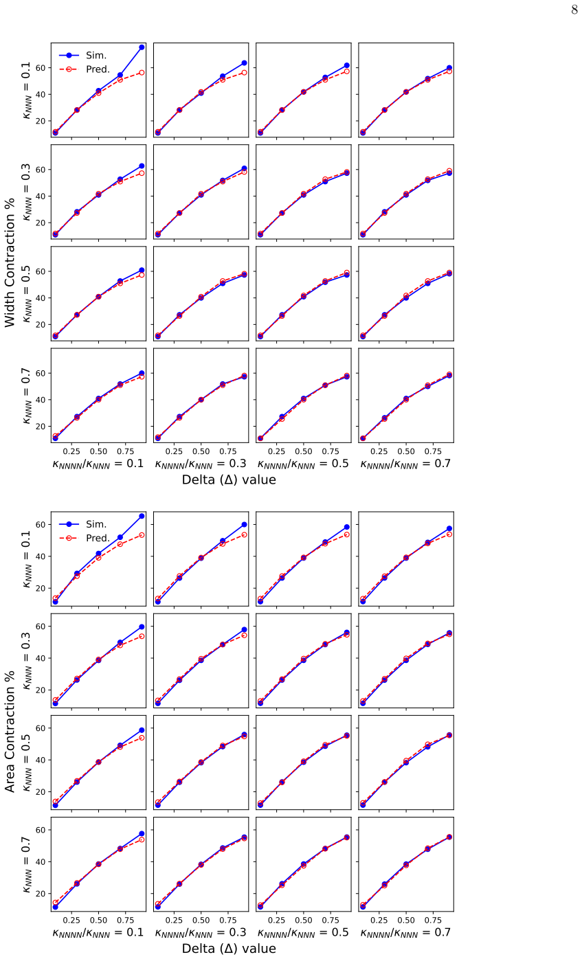
The central discovery claimed is that a single parameter-conditioned GAN can reproduce the output fields of a biophysical tissue model across most of its parameter space and thereby predict the organisation of real cultured tissues. RAPTOR maps five input channels — mould depression, tether placement, and constant-valued maps of $\Delta$, $\kappa_{\mathrm{NNN}}$ and $\kappa_{\mathrm{NNNN}}$ — to eight output channels: cell density, six products of the cell-orientation Q tensor ($S_x^2$, $S_y^2$, $S_z^2$, $S_xS_y$, $S_xS_z$, $S_yS_z$), and average bond tension. Trained on 3,124 CONDOR simulations augmented to 12,653 examples, it closely matches CONDOR on a held-out test set and in grid scans over the parameter space, with Pearson correlations above 0.96 for bulk properties; the clear exception is the high-contraction corner (large $\Delta$, small $\kappa$), where RAPTOR under-predicts contraction. The paper further claims that nonlinear least-squares fitting of the three parameters to the width and area ratios of cultured glial, fibroblast and corneal tissues yields parameter sets for which both CONDOR and RAPTOR reproduce the experimental tissue shapes, with fitted $\Delta$ values that are lower for glial tissue and higher for fibroblast and corneal tissue, consistent with their different contractility.
Load-bearing premise
The load-bearing premise is that the three cell and matrix parameters, fitted to only the width and area ratios of one experimental tissue, are physically transferable: the same values predict density, alignment, and tension in other mould geometries and across the whole tissue, rather than merely reproducing the two scalar numbers used in the fit.
Editorial extensions
If this is right
- RAPTOR predictions run in a fraction of a second, so mould designs can be screened against density, alignment, and tension criteria without running day-long CONDOR simulations, making high-throughput and automated design practical.
- The fitted parameter values for a given cell type can be reused in subsequent CONDOR or RAPTOR runs, so a small number of calibration experiments could parameterise the model for new cell lines or matrix conditions.
- The network generalises to at least one mould geometry absent from the training set (a long continuous tethering bar), indicating that predictions are not limited to memorised shapes; accuracy degrades only in the high-$\Delta$, low-$\kappa$ corner of parameter space.
- The demonstration for glial, fibroblast, and corneal tissues suggests the same workflow — train once, then fit parameters per tissue type — could be applied to other engineered tissues that can be grown in tethered moulds.
Reading between the lines
- The authors leave implicit that RAPTOR could be inverted for inverse design: rather than checking a mould shape, one could optimise the mould and tether layout directly against desired alignment and tension maps, since the forward map is cheap enough to embed in an evolutionary loop.
- Because the parameter fit targets only two scalar quantities, the fitted triple ($\Delta$, $\kappa_{\mathrm{NNN}}$, $\kappa_{\mathrm{NNNN}}$) may not be identifiable; our inference is that adding shape-based or spatially resolved residuals, such as local width profiles or alignment maps, to the fit would tighten parameter estimates and make transferability claims testable.
- The failure mode at high $\Delta$ and small $\kappa$ is localised in a corner of parameter space, which suggests a targeted remedy the authors do not pursue: oversample that corner in the training data or use a separate network specialised to high-contraction cases.
- The same parameter-conditioned image-translation design could in principle be applied to other biophysical tissue models, not just CONDOR, whenever a model can generate enough training simulations; this would let experimentalists choose the cheapest or most faithful simulator and still get real-time predictions.
Signed reviews
Editorial analysis
A structured set of objections, weighed in public.
Referee Report
Summary. The manuscript presents RAPTOR, a conditional pix2pix generative model trained on outputs of the CONDOR biophysical model, with the three CONDOR parameters Δ, κ_NNN, and κ_NNNN supplied as additional input channels. Training data consist of roughly 3,569 CONDOR simulations with random mould and tether layouts; the held-out test split of 445 simulations yields high Pearson correlations (0.96–0.99) for mean density, area, tension, and alignment quantities. The authors then exploit the speed of RAPTOR to fit CONDOR parameters to experimental glial, fibroblast, and corneal tissues by minimizing the two-residual objective in Eq. (6), and compare the resulting RAPTOR and CONDOR width and area ratios with measurements. They report excellent agreement and propose RAPTOR as a near-real-time tool for tethered mould design.
Significance. If the claims hold, RAPTOR would be a practically valuable extension of CONDOR-ML: it conditions a fast surrogate on physically relevant model parameters, and the systematic grid comparison in Sec. III B usefully characterises the surrogate's domain of validity. The held-out CONDOR test set and the explicit identification of the high-Δ/low-κ failure region are genuine strengths. However, the experimental validation does not yet establish transferable predictive power: the fitted parameters are not identifiable from the two scalar observables used, and no independent experimental outcomes are predicted. The central application claim therefore needs additional evidence before the paper can be accepted.
major comments (3)
- [III C, Eq. (6)] Section III C fits the three CONDOR parameters (Δ, κ_NNN, κ_NNNN) by minimising Eq. (6), whose residual contains only two scalar quantities: relative area and relative width. Three unknowns constrained by two scalars is an underdetermined inverse problem, so the parameters are not identifiable from this fitting procedure alone. The paper itself notes in Sec. III C 1 that part of the parameter variation across the three glial cultures 'may arise since the parameter fit is only made for width and area, rather than the overall shape of the tissue,' and the reported spread in Δ (mean 0.2227, σ 0.06784) is consistent with multiple nearly equivalent optima. This is load-bearing because the proposed use of RAPTOR for mould design assumes that parameters fitted to one tissue and mould transfer to other geometries and to all output fields (density, alignment, tension). Please demonstrate identifiability, for example through profile likelihoods, joint fitting to several moulds, or leave-one-out experimental validation, before claiming transferable parameters.
- [III C 3, Table I] The corneal comparison in Sec. III C 3 is the weakest experimental test and does not support the statement of 'excellent agreement'. Because the area was not reported in Ref. [17], the fit uses only one observable (w/w0 = 0.35) to determine three parameters. The optimised parameters give RAPTOR w/w0 = 0.459 and CONDOR w/w0 = 0.525, so neither model reproduces the measured contraction, and the fitted Δ = 0.8298 lies near the high-Δ, low-κ_NNNN/κ_NNN boundary of the region where Sec. III B documents systematic RAPTOR/CONDOR disagreement. This case therefore illustrates the identifiability problem rather than providing an independent validation of the method.
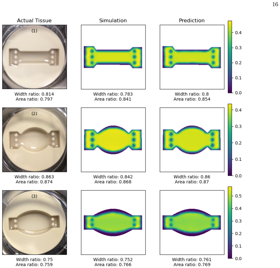
- [III C, Table I, Figs. 9–11] The experimental validation is partly circular: the quantities reported in Table I (relative tissue area and width) are exactly the two residuals minimised in Eq. (6), so the agreement for those quantities is a consequence of the fitting procedure rather than an independent prediction. No experimental tissue is held out for a different mould geometry, and no full-field outputs (density, alignment, tension) are compared with experiment. The strong RAPTOR-vs-CONDOR test on the held-out CONDOR test set (Fig. 2) verifies emulation of the simulator but not the physical transferability of fitted parameters. Please add at least one independent experimental validation, such as fitting on one mould and predicting a second mould, comparing a measured alignment or density field not used in the loss, or performing leave-one-mould-out analysis across the three glial cultures.
minor comments (6)
- [II B] The heading 'T raining Data' and the duplicated 'of' in 'A total of of 3569 unique simulations' are typos that should be corrected.
- [III A / Fig. 2] The text in Sec. III A defines N(P < 0.2) as the number of pixels with density less than 0.2, whereas the Fig. 2 caption says 'number of pixels with density exceeding 0.2'; please clarify which convention is intended and use it consistently.
- [II C, Eq. (5)] The definition of N_c as 'P ip wi,p' is garbled; it should read N_c = Σ_{i,p} w_{i,p} (or an equivalent explicit expression), and the relationship between barred and unbarred field averages should be stated cleanly.
- [Table I] Table I uses 'κ3n' and 'κ4n' in the header without definition; please define these abbreviations in the caption or use κ_NNN and κ_NNNN consistently with the text.
- [IV / Abstract] The phrases 'predictions for arbitrary mould designs' (Abstract) and 'allowing predictions for arbitrary choices of parameter values' (Sec. II B) are too strong given the failure region at high Δ and low κ documented in Sec. III B; please qualify the claim to the parameter range covered by the training data.
- [Fig. 9 / Table I] The Fig. 9 caption does not identify which experimental image corresponds to G1, G2, and G3 in Table I; please add labels so the reader can connect the visual comparisons to the tabulated values.
Circularity Check
The experimental 'validation' of RAPTOR and CONDOR against tissue width and area reduces to an in-sample fit because Eq. (6) fits the model parameters to exactly those experimental widths and areas, and Table I then reports the fitted outputs as predictions.
-
fitted input called prediction
[Section III C, Eq. (6), and Table I (Secs. III C 1–III C 3)]
"During the fit, the residual sum of squares (RSS) is minimised: RSS = (AM − AE)2 + (wM − wE)2 , (6) where AM and wM are the relative area and width calculated from results of the predicted model operated on the selected mould shape, and AE and wE are the corresponding relative area and width measured from the experimental results. CONDOR simulations are then carried out with these parameters. This provides additional validation for CONDOR and RAPTOR."
The fitting target in Eq. (6) is the pair (AE, wE), which is exactly the experimental A/A0 and w/w0 reported in Table I. The nonlinear least-squares fit selects ∆, κNNN, and κNNNN to minimize the RAPTOR residuals against those two scalars; the same optimized RAPTOR outputs are then presented as 'Predictions', and CONDOR is run at the fitted parameters and its outputs are also presented as 'Predictions'. Agreement in width and area is therefore enforced by the fitting objective rather than independently tested. The corneal case is even more circular: only width was used in the fit because area was not measured, so three parameters were constrained by one scalar, and the reported RAPTOR w/w0 = 0.459 is simply the best-fit value to the measured 0.35.
full rationale
Most of the paper is not circular: RAPTOR is a pix2pix GAN trained on CONDOR simulations, and the held-out test-set comparisons in Figs. 1–2 evaluate the surrogate against a disjoint set of simulator outputs, which is a legitimate internal consistency check. The parameter-conditioned extension over CONDOR-ML is new and independently testable. The circularity is localized to the experimental validation pipeline. Section III C fits the three CONDOR parameters to the experimental width and area via Eq. (6), and Table I then reports the resulting width and area as RAPTOR and CONDOR 'predictions'. Because the residuals being minimized are exactly the discrepancies reported as agreement, the quantitative 'validation' against glial, fibroblast, and corneal tissues reduces to an in-sample fit for those scalar observables. The paper itself acknowledges the limitation: parameter variation among glial cultures 'may arise since the parameter fit is only made for width and area, rather than the overall shape of the tissue.' No load-bearing self-citation chain was found: Refs. [11] and [12] supply the underlying biophysical model and prior ML implementation, but they are not invoked as an external uniqueness theorem or as a substitute for the present validation. The central claim of a fast surrogate has independent content, so the score is 6 rather than higher: partial circularity in the experimental validation, while the surrogate-vs-simulator component remains self-contained.
Assumptions & free parameters
free parameters (4)
- delta (cell-matrix interaction strength) =
glial 0.2227 +/- 0.0678; fibroblast 0.6578; corneal 0.8298; tenocyte/myoblast 0.95
- kappa_NNN (next-next-nearest neighbour spring constant) =
glial 0.6589 +/- 0.0321; fibroblast 0.4438; corneal 0.6465
- kappa_NNNN (next-next-next-nearest neighbour spring constant) =
glial 0.3794 +/- 0.0196; fibroblast 0.2526; corneal 0.06678
- Gaussian smoothing width sigma =
3*lp
assumptions (5)
- domain assumption CONDOR energy model (Eqs 1-2) adequately represents cell-matrix self-organisation in tethered hydrogels.
- domain assumption Simulated annealing reaches the minimum-energy configuration for every training mould.
- domain assumption Randomly generated moulds and uniform parameter ranges cover the design space of interest.
- standard math pix2pix with parameter input channels can learn the mapping from mould plus parameters to tissue property fields.
- domain assumption Experimental width and area ratios extracted from images or literature are accurate representatives of tissue contraction.
Cite this review
Pith. "Pith review of Rapid prediction of organisation in engineered corneal, glial and fibroblast tissues using machine learning and biophysical models." pith.science (2026). https://pith.science/paper/WRKMQ62O
@misc{pith2026250208062,
  author       = {Pith},
  title        = {Pith review of: Rapid prediction of organisation in engineered corneal, glial and fibroblast tissues using machine learning and biophysical models},
  year         = {2026},
  howpublished = {\url{https://pith.science/paper/WRKMQ62O}},
  note         = {Machine review of arXiv:2502.08062}
}
read the original abstract
We present a machine learning approach for predicting the organisation of corneal, glial and fibroblast cells in 3D cultures used for tissue engineering. Our machine-learning-based method uses a powerful generative adversarial network architecture called pix2pix, which we train using results from biophysical contractile network dipole orientation (CONDOR) simulations. In the following, we refer to the machine learning method as the RAPTOR (RApid Prediction of Tissue ORganisation) approach. A training data set containing a range of CONDOR simulations is created, covering a range of underlying model parameters. Validation of the trained neural network is carried out by comparing predictions with cultured glial, corneal, and fibroblast tissues, with good agreements for both CONDOR and RAPTOR approaches. An approach is developed to determine CONDOR model parameters for specific tissues using a fit to tissue properties. RAPTOR outputs a variety of tissue properties, including cell densities of cell alignments and tension. Since it is fast, it could be valuable for the design of tethered moulds for tissue growth.
Figures


Figures from the paper (8 more)




Reference graph
Works this paper leans on
-
[17]
J. P. Hague, P. W. Mieczkowski, C. O’Rourke, A. J. Loughlin, and J. B. Phillips, Microscopic biophysical model of self-organization in tissue due to feedback be- tween cell- and macroscopic-scale forces, Phys. Rev. Res. 2, 043217 (2020)
work page 2020
-
[1]
Predictive capability for structures with no analogue in the training data The I-shaped mould examined in this section contains a large continuous tethering bar, which is used to show how RAPTOR handles unexpected tethering arrange- ments. The training data set contains random arrange- ments of many small circular tethers, so the case of a long continuous...
-
[2]
An additional set of 500 simulations (approximately 1/8th of the total) was created using instances where ∆ was set to either 0.05 or 0 .95, in order to improve the representation of cases at the edges of the parameter space. CONDOR simulations were made to simulate growth of cultured tissues in the randomly created moulds for a set of randomly selected p...
-
[3]
Glial cultures In this section, comparisons are made with cultured glial tissue (glial cell populated hydrogels in tethered moulds). Glial cells play an important role in the nervous system, and direct the growth and support of neurons. Nerve repair guides are an application of highly aligned glial tissue [19, 20]. We have validated against lab grown glia...
-
[4]
Fibroblast cultures The results from RAPTOR for fibroblasts were com- pared to relative tissue widths and areas derived from images in Ref. [18]. Fibroblasts synthesise collagen and play in important role in tissue healing and repair. Fi- broblast tissue is relatively contractile, an important fea- ture related to its role in wound closure. Using image da...
-
[5]
Corneal cultures We use the values provided in Ref. [17] to compare RAPTOR predictions with experimental results for en- gineered tissue containing corneal stromal cells. Corneal tissue is an important part of the eye, and high alignment of cells and particularly matrix is important for the opti- cal properties of the cornea. Ref. [17] does not provide a ...
-
[6]
Tenocyte and myoblast cultures This section concludes with a very brief discussion of tissue cultures of tenocytes and myoblasts. Represen- tative examples of artificial tendon and muscle tissues grown in tethered moulds can be found in Refs. [21] and [22]. Both types of tissue are highly contractile. The maximum ∆ = 0.95 is returned from the parameter se...
work page 2022
-
[7]
A. Grosberg, P.-L. Kuo, C.-L. Guo, N. Geisse, M.- A. Bray, W. Adams, S. Sheehy, and K. Parker, Self- organization of muscle cell structure and function, PLoS Comput Biol 7, e1001088 (2011)
work page 2011
Show all 28 references
-
[8]
Shimizu, H
K. Shimizu, H. Fujita, and E. Nagamori, Alignment of skeletal muscle myoblasts and myotubes using linear mi- cropatterned surfaces ground with abrasives, Biotechnol Bioeng. 103, 631 (2009). 15
2009
-
[9]
R. M. Gouveia, E. Koudouna, J. Jester, F. Figueiredo, and C. J. Connon, Template curvature influences cell alignment to create improved human corneal tissue equiv- alents, Advanced Biosystems 1, 1700135 (2017)
2017
-
[10]
Werner, T
S. Werner, T. Krieg, and H. Smola, Keratinocyte- fibroblast interactions in wound healing, J Invest Der- matol. 127, 998 (2007)
2007
-
[11]
Bajaj, R
P. Bajaj, R. M. Schweller, A. Khademhosseini, J. L. West, and R. Bashir, 3D biofabrication strategies for tis- sue engineering and regenerative medicine, Annu. Rev. Biomed. Eng. 16, 247 (2014)
2014
-
[12]
Ben-Arye and S
T. Ben-Arye and S. Levenberg, Tissue engineering for clean meat production, Front. Sustain. Food Syst. 3, 00046 (2019)
2019
-
[13]
Weinhart, A
M. Weinhart, A. Hocke, S. Hippenstiel, J. Kurreck, and S. Hedtrich, 3D organ models - revolution in pharmaco- logical research?, Pharmacol. Res. 139, 446 (2019)
2019
-
[14]
Jensen, C
G. Jensen, C. Morrill, and Y. Huang, 3D tissue engineer- ing, an emerging technique for pharmaceutical research, Acta Pharm. Sin. B. 8, 756 (2018)
2018
-
[15]
K. Lu, Y. Qian, J. Gong, Z. Zhu, J. Yin, L. Ma, M. Yu, and H. Wang, Biofabrication of aligned structures that guide cell orientationand applications in tissue engineer- ing, Bio-Design and Manufacturing 4, 258 (2021)
2021
-
[16]
Jensen and Y
C. Jensen and Y. Teng, Is it time to start transitioning from 2D to 3D cell culture?, Frontiers in Molecular Bio- sciences 7 (2020)
2020
-
[18]
Andrews, H.Dickinson, and J
A. Andrews, H.Dickinson, and J. Hague, Rapid predic- tion of lab-grown tissue properties using deep learning, Physical Biology 20, 066005 (2023)
2023
-
[19]
Andrews, and H.Dickinson, High- throughput design of cultured tissue moulds using a bio- physical model: optimising cell alignment, Physical Biol- ogy 20, 066006 (2023)
J.P.Hague, A. Andrews, and H.Dickinson, High- throughput design of cultured tissue moulds using a bio- physical model: optimising cell alignment, Physical Biol- ogy 20, 066006 (2023)
2023
-
[20]
Isola, J.-Y
P. Isola, J.-Y. Zhu, T. Zhou, and A. A. Efros, Image-to- image translation with conditional adversarial networks, in 2017 IEEE Conference on Computer Vision and Pat- tern Recognition, CVPR 2017, Honolulu, HI, USA, July 21-26, 2017 (IEEE Computer Society, 2017) pp. 5967– 5976
2017
-
[21]
O’Rourke, R
C. O’Rourke, R. A. Drake, G. W. Cameron, J. A. Lough- lin, and J. B. Phillips, Optimising contraction and align- ment of cellular collagen hydrogels to achieve reliable and consistent engineered anisotropic tissue., J. Biomater. Appl. 30, 599 (2015)
2015
-
[22]
E. East, D. B. De Oliviera, J. P. Golding, and J. B. Phillips, Alignment of astrocytes increases neuronal growth in three-dimensional collagen gels and is main- tained following plastic compression to form a spinal cord repair conduit, Tissue Eng. Part A 16, 3173 (2010)
2010
-
[23]
Mukhey, J
D. Mukhey, J. Phillips, J. Daniels, and A. Kureshi, Con- trolling human corneal stromal stem cell contraction to mediate rapid cell and matrix organization of real archi- tecture for 3-dimensional tissue equivalents, Acta Bio- mater. 67, 229 (2018)
2018
-
[24]
Kostyuk and R
O. Kostyuk and R. A. Brown, Novel spectroscopic tech- nique for in situ monitoring of collagen fibril alignment in gels, Biophysical Journal 87, 648 (2004)
2004
-
[25]
Georgiou, S
M. Georgiou, S. C. Bunting, H. A. Davies, A. J. Loughlin, J. P. Golding, and J. B. Phillips, Engineered neural tissue for peripheral nerve repair, Biomater. 34, 7335 (2013)
2013
-
[26]
Phillips, ‘EngNT’ – engineering live neural tissue for nerve replacement, Emerg
J. Phillips, ‘EngNT’ – engineering live neural tissue for nerve replacement, Emerg. Top. Life. Sci. 5, 699 (2021)
2021
-
[27]
Garvin, J
J. Garvin, J. Qi, M. Maloney, and A. J. Banes, Novel sys- tem for engineering bioartificial tendons and application of mechanical load, Tissue Eng. Part A 9, 967 (2003)
2003
-
[28]
A. J. Capel, R. P. Rimington, J. W. Fleming, D. J. Player, L. A. Baker, M. C. Turner, J. M. Jones, N. R. W. Martin, R. A. Ferguson, V. C. Mudera, and M. P. Lewis, Scalable 3D printed molds for human tissue en- gineered skeletal muscle, Front. Bioeng. Biotechnol. 7, 00020 (2019...
2019
Reviewed August 8, 2026 · model on record in the stance chip above.
Discussion (0). Continue with ORCID to comment.