REVIEW 5 major objections 4 minor 13 references
A 2D Hydrothermodynamic Analytical Model for Rapid Tumor Ablation using High-Intensity Focused Ultrasound
T0 review · 5 major / 4 minor · reviewed 2026-08-16 · deepseek-v4-flash
Pith's one-line read A stationary tumor matrix suppresses acoustic streaming, converting absorbed ultrasound momentum into localized heat with a sharp lesion boundary at 0.75 w0.
desk verdict The paper's central numbers are imposed rather than derived, and the post-pulse containment proof is internally inconsistent with the paper's own heating profile. read the letter →
The pith
A machine-rendered reading of the paper's core claim, the machinery that carries it, and where it could break.
The reading
What carries the argument
The carrying mechanism is the second-order expansion of the compressible viscous Navier-Stokes equations with the zero-streaming constraint $v_2=0$, imposed because the tumor matrix is treated as fixed and anchored. This converts the acoustic force density $F_2=2\alpha I(r)/c_0\,\hat{r}$ into a static pressure gradient and removes the convective term $\rho_0 C_p v_2\cdot\nabla T$ from the energy balance. The derived optimum $\alpha=1/(2x_0)$, the spherical focusing factor $(a/r)^2$, the parabolic focal temperature profile $T(r,\tau)=T_0+\Delta T_{\max}(1-r^2/w_0^2)$, the Arrhenius injury integral, and the 2D free-space Green's function convolution then combine to produce the lesion radius and the containment statement.
What would settle it
Measure the time-averaged second-order velocity in a dense anchored tumor or tissue-mimicking phantom during a 1 s HIFU pulse. A nonzero $v_2$ with $\rho_0 C_p v_2\cdot\nabla T$ comparable to $2\alpha I$ would break the pressure-gradient heat conversion. Alternatively, record the radial temperature at $t=1$ s and shortly after: the model's cooling law (Eq. 39) predicts a monotone drop at $r_b=0.75\,w_0$, so any measured temperature rise outside $r_b$ after pulse cutoff would falsify the containment claim.
Extended reading notes
Core claim
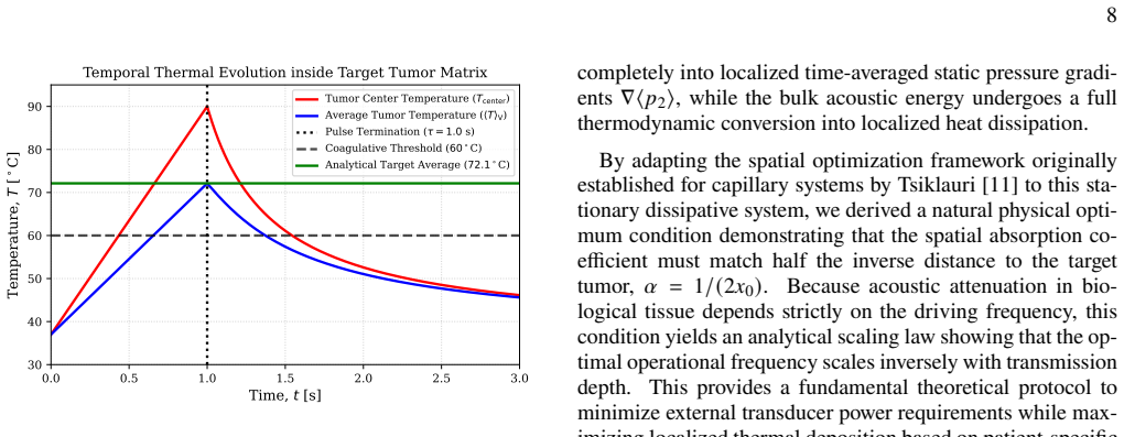
The central claim is that the kinematic constraint $v_2=0$ inside a stationary tumor makes the time-averaged acoustic momentum flux $F_2 = 2\alpha I(r)/c_0\,\hat{r}$ transfer entirely into a static second-order pressure gradient $\nabla\langle p_2\rangle$, so the absorbed energy appears as heat rather than as acoustic wind. With that constraint, the Pennes bioheat equation reduces to a local, non-diffusive source on the pulse timescale; a spherical focusing geometry with geometric convergence $\propto 1/r^2$ overrides exponential attenuation within a critical radius $r_{\mathrm{crit}}=2x_0$; and the non-isothermal Arrhenius integral gives $r_b=0.75\,w_0$. The stated thermal outcome is a peak focal temperature of $90^\circ$C, a volumetric average of $72.1^\circ$C inside the lesion, and post-pulse Green's-function cooling that stays monotonically below $60^\circ$C at the boundary.
Load-bearing premise
The whole argument rests on assuming, rather than deriving, that the tumor matrix is rigid enough to make the second-order streaming velocity exactly zero; if even a small $v_2$ exists, convective heat transport reappears and the claimed sharp containment no longer follows.
Editorial extensions
If this is right
- Focal lesions in anchored tumors should have a sharp necrotic boundary at $r_b=0.75\,w_0$ rather than a gradual damage gradient.
- Optimal operating frequency for a given patient scales inversely with target depth through $\alpha = 1/(2x_0)$.
- Plane-wave HIFU is inherently limited because upstream tissue receives about $e$ times more heat than the tumor core, so spherical focusing is not optional.
- After pulse termination the boundary temperature falls monotonically below $60^\circ$C, which is why residual conduction injury is negligible on this timescale.
Reading between the lines
- If streaming suppression is real, tumors with stiffer, more collagenous extracellular matrices should exhibit sharper thermal lesions than soft or fluid-rich tumors; comparing lesion sharpness across tumor stiffness would test this directly.
- The $\alpha=1/(2x_0)$ scaling suggests a simple patient-specific tuning rule: choose the HIFU frequency from target depth alone, then check whether the measured lesion radius matches $0.75\,w_0$ across a range of depths.
- The cooling phase replaces the parabolic heating profile by a Gaussian initial condition; whether real focal profiles are closer to Eq. (33) or Eq. (36) will determine how literally the $0.75\,w_0$ boundary and the monotone cooling statement should be read.
Signed reviews
Editorial analysis
A structured set of objections, weighed in public.
Referee Report
Summary. The manuscript presents an analytical 2D hydrothermodynamic model of HIFU ablation. It argues that in a dense anchored tumor, second-order acoustic streaming is exactly zero, so absorbed acoustic momentum becomes static pressure gradients and heat; that with a spherical focusing geometry and alpha=1/(2x0), a 1 s top-hat pulse produces a parabolic temperature profile; that the Arrhenius injury integral gives a lesion radius r_b=0.75w0; and that post-pulse Green's-function cooling proves thermal containment, with a volume-averaged tumor temperature of 72.1 C and a focal peak of 90 C. The paper is transparent about many of its assumptions and provides closed-form expressions throughout.
Significance. If the zero-streaming premise and the thermal predictions were established, the model would offer a simple analytical design rule for HIFU frequency-versus-depth and a sharp lesion criterion, which could be useful for device optimization. The paper is clearly written, and the closed-form expressions are easy to check. However, the central quantitative claims are not derived from the model equations in the way claimed: the lesion radius, the average temperature, and the containment proof follow from imposed input values or from an initial condition inconsistent with the heating profile. The potential significance is therefore not realized by the present manuscript.
major comments (5)
- [II, Eqs. (16)-(18)] The zero-streaming constraint v2=0 is asserted from the statement that 'structural biological boundaries are fixed and anchored'; it is not derived from the tissue mechanics or from any equation. This is load-bearing because Eq. (19) is simplified by dropping the convective term rho0 Cp v2 dot grad T, and the entire localization argument relies on that drop. The manuscript provides no estimate of a residual second-order velocity or the timescale on which v2 relaxes; without that, the claim of complete suppression is an assumption rather than a result.
- [III, Eq. (34)] The lesion radius r_b=0.75w0 is not obtained by solving the Arrhenius injury integral. The text sets Omega(r_b)=1, but then substitutes T(r_b,tau)=60 C and T(0,tau)=90 C into the parabolic profile Eq. (33) and solves algebraically. The Arrhenius expression (29)-(32) plays no role in determining r_b beyond supplying the fixed 60 C threshold, and the 90 C peak is an assumed input chosen to match clinical practice. Thus Eq. (34) is a rearrangement of assumed boundary values, not a predicted lesion boundary.
- [III, Eqs. (33), (36), (41)-(42)] The post-pulse problem is initialized with the Gaussian theta(r,0)=DeltaT_max exp(-r^2/w0^2), but the end-of-pulse profile from Eq. (33) is the parabola theta(r,tau)=DeltaT_max(1-r^2/w0^2). These agree only at r=0; at r_b=0.75w0 the Gaussian gives 37+53 exp(-0.5625)=67.2 C, not the 60 C that defines r_b. Consequently Eqs. (41)-(42) describe cooling of a different initial state, and the monotone decay at r_b does not imply containment for the actual parabolic profile. Furthermore, the derivative test only at r=r_b does not bound Omega_cooling(r) for r>r_b, where a diffusing Gaussian can initially heat; the paper never evaluates the cooling injury integral outside r_b.
- [III, Eqs. (43)-(46)] The reported average 72.1 C is computed from the same assumed DeltaT_max=53 C and gamma=0.75, so it is not an independent output. It also uses a 3D spherical volume average in a model that is otherwise presented as 2D and uses a 2D Green's function (Eq. (37)). Had the average been taken over a 2D disk, the prefactor would be different and the value would change (roughly 75.1 C for the same parabolic profile). The paper should either treat the problem consistently in 2D or provide a 3D derivation of the averaged quantity.
- [III, Eq. (40)] The cooling injury integral Omega_cooling(r) is not evaluated. The text asserts that it 'drops to near zero within milliseconds' based on the derivative sign at r_b. This is not a calculation; the Arrhenius integrand is exponentially sensitive, and the paper supplies no bound on the integral over t' for r>=r_b. Without such a bound, the central claim of complete structural containment is unproven.
minor comments (4)
- [II, Eqs. (15), (25)-(26)] The normalization of the intensity I0 is not defined in relation to the transducer aperture radius a and focal radius R_f; please specify exactly how I0 in Eq. (15) and Eq. (26) is obtained from the source parameters in Eq. (25).
- [III, Fig. 1 caption] The angular brackets in the average temperature label are rendered as 'T V' rather than as an explicit averaged quantity; please fix the notation for clarity.
- [Data Availability Statement] The statement says that 'data and numerical codes' are available, but no code or repository is described anywhere in the paper; please either provide the code or revise the statement to match the content.
- [III, Eq. (32)] The 'multi-scale asymptotic expansion' leading to Eq. (32) is stated without an error estimate; since the claim is that the Arrhenius integral is solved analytically, please quantify the neglected terms or cite a standard asymptotic result.
Circularity Check
The lesion-boundary 'prediction' r_b=0.75w0 is an algebraic consequence of assumed 60°C/90°C endpoints, and the post-pulse containment proof uses a Gaussian cooling profile inconsistent with the computed end-of-pulse profile.
-
fitted input called prediction
[Section III, Eqs. (32)-(34)]
"To locate the sharp structural damage boundary radius r_b matching the experimental cytotoxic boundaries achieved by the FMBA apparatus, we set Ω(r_b)=1.0. At this structural perimeter, the peak temperature reaches the transition threshold (T(r_b,τ)=T_necrosis≈60°C). ... r_b = w_0 sqrt(1 - (T_necrosis-T_0)/ΔT_max) = w_0 sqrt(1 - (60-37)/(90-37)) = w_0 sqrt(1 - 23/53) ≈ 0.75 w_0."
The Arrhenius integral in Eq. (32) is never solved for the damage boundary. Instead, the paper inserts T(r_b,τ)=60°C and ΔT_max=53°C (from an assumed 90°C peak) and algebraically rearranges the parabolic profile to obtain r_b=0.75w0. The 'predicted' boundary therefore contains no information beyond the assumed cytotoxic threshold and peak temperature, which are themselves taken from the clinical 60-90°C window the paper claims to explain. The result is a fitted input renamed as a derivation.
-
self citation load bearing
[Section II, Eq. (23); also Eq. (28) and Conclusion]
"Adapting the optimization framework established by Tsiklauri [11] to our stationary dissipative framework, we maximize the core energy deposition rate at the targeted focal spot. Evaluating the non-oscillatory damping and convergence profile at the depth boundary yields the optimal amplitude attenuation criterion: α = 1/(2x_0). ... By applying the Tsiklauri optimization frequency condition α=1/(2x_0) from Eq. (23), the cross-over threshold becomes r_crit=2x_0."
The optimal frequency-depth law is not derived in this paper; it is imported from the author's own prior work [11]. This condition is load-bearing because it sets the operational frequency, produces r_crit=2x0, and underlies the claim that a localization rate exceeding 90% is 'mathematically guaranteed.' The self-citation is the only stated justification for the central optimization criterion. Although the condition could in principle be derived from Eq. (13), the paper does not do so, so the self-citation chain carries the argument.
1 more flagged steps
-
other
[Section III, Eqs. (33), (36), (41)-(42)]
"The initial condition for this cooling phase is the spatial temperature profile at the end of the pulse window, T(r,τ), established in Eq. (33). ... The initial temperature field at t′=0 is confined to the focal zone and can be modeled as a localized 2D Gaussian distribution that matches the central peak ΔT_max=53°C and vanishes at the beam margins: θ(r,0)=ΔT_max exp(−r^2/w_0^2)."
Eq. (33) gives an end-of-pulse parabolic profile θ(r,τ)=ΔT_max(1−r^2/w0^2), while Eq. (36) replaces it with a Gaussian. At the claimed boundary r_b=0.75w0, the Gaussian initial temperature is 37+53 exp(−0.5625)≈67°C, not 60°C. The 'immediate monotonic decay below 60°C' is therefore not a continuation of the heating solution; it is an artifact of the newly assumed Gaussian. The derivative test at r_b<w0 only shows that a centered Gaussian cools at that point, and it says nothing about r>w0 where the Gaussian initially heats. The containment conclusion is built into an inconsistent ansatz rather than derived from the model's own thermal state.
full rationale
The hydrodynamic expansion and the plane-wave-versus-spherical focusing comparison are standard and largely self-contained, and the suppression v2=0, while assumed rather than derived, is a stated kinematic constraint rather than a circular reduction. The circularity is concentrated in the thermal damage claims. The lesion radius r_b=0.75w0 is obtained by inserting T_necrosis=60°C and ΔT_max=53°C into the parabolic profile and rearranging; the Arrhenius integral plays no role in locating r_b. The average 72.1°C is then the same assumed profile integrated over that boundary, so it adds no independent content. The optimal frequency condition α=1/(2x0) is taken from the author's earlier self-citation and is load-bearing for the frequency-depth scaling and the localization-rate guarantee. Finally, the post-pulse containment 'proof' substitutes a Gaussian initial condition that contradicts the computed parabolic end-of-pulse profile, so the claimed immediate monotone drop below 60°C at the boundary is an artifact of the chosen ansatz rather than a consequence of the model. Collectively, the central quantitative predictions reduce to assumed clinical temperature endpoints and a self-cited optimization criterion, warranting a circularity score of 7.
Assumptions & free parameters
free parameters (5)
- Delta T_max (focal peak temperature rise) =
53 C (implied by T_peak=90 C)
- I0 (source acoustic intensity) =
not specified
- w0 (beam waist radius) =
not specified
- chi/w0^2 (thermal diffusivity over waist squared) =
not specified
- integration boundary rb=0.75w0 =
0.75w0
assumptions (6)
- domain assumption v2=0 in tumor matrix
- domain assumption Non-diffusive pulse window (K grad^2 T approx 0, w_b approx 0)
- ad hoc to paper Parabolic axial intensity profile I_axial(r) approx I_max (1 - r^2/w0^2)
- ad hoc to paper Gaussian initial condition for cooling (Eq. 36)
- standard math Linear equation of state and harmonic driving
- standard math Arrhenius high-barrier asymptotics (Delta Ea / (Rg T) >> 1)
Cite this review
Pith. "Pith review of A 2D Hydrothermodynamic Analytical Model for Rapid Tumor Ablation using High-Intensity Focused Ultrasound." pith.science (2026). https://pith.science/paper/WZLCGB5T
@misc{pith2026260812200,
  author       = {Pith},
  title        = {Pith review of: A 2D Hydrothermodynamic Analytical Model for Rapid Tumor Ablation using High-Intensity Focused Ultrasound},
  year         = {2026},
  howpublished = {\url{https://pith.science/paper/WZLCGB5T}},
  note         = {Machine review of arXiv:2608.12200}
}
abstract
We establish a self-consistent 2D hydrothermodynamic analytical model for the localized thermal ablation of dense human tumors using high-intensity focused ultrasound. By expanding compressible Navier-Stokes equations up to second order, we demonstrate that within a structurally stationary cellular tumor matrix, acoustic streaming (acoustic wind) velocity is suppressed. This constraint forces the absorbed wave momentum flux to transfer entirely into localized, time-averaged, static, second order, pressure gradients, converting the bulk acoustic energy directly into localized heat. Using a short, 1 s, duration, high-amplitude top-hat pulse, we solve the simplified Pennes bioheat transfer equation within non-diffusive timescales. Adapting the hydrodynamic optimization framework established by Tsiklauri~(2026), we derive a natural physical criterion where the acoustic absorption coefficient matches half the inverse target depth, $\alpha = 1/(2x_0)$, proving that the optimal operational frequency scales inversely with transmission distance. We show that while incident plane waves overheat upstream tissues due to exponential decay, a spherically focusing wave geometry effectively bypasses healthy tissue boundaries via geometric convergence ($\propto 1/r^2$). Analytically solving the non-isothermal Arrhenius injury integral yields a sharp lesion boundary radius at $r_b = 0.75\,w_0$. Volumetric averaging bounded strictly within this necrosis perimeter demonstrates that the average tumor temperature reaches $72.1^\circ\text{C}$ while central point values peak at $90^\circ{\rm C}$. Finally, convolving the post-pulse thermal profile with a 2D free-space Green's function verifies immediate, monotonic temperature decay below $60^\circ{\rm C}$ at the boundary, demonstrating complete structural containment and explaining the $>90\%$ localization rates observed in clinical applications.
Figures
Reference graph
Works this paper leans on
-
[1]
Z. Izadifar, Z. Izadifar, D. Chapman, and P. Babyn, Journal of Clinical Medicine9, 10.3390/jcm9020460 (2020)
-
[2]
Federal Medical and Biological Agency (FMBA) of Rus- sia, Meduza ultrasound-based therapeutic diagnostic unit (hifu): High-tech non-invasive tumor ablation system (2022), verifiable registration trace available at TAdviser Tech- nology Database: https://tadviser.com/index.php/Product :Rostec_and_FMBA_of_Russia:_MedUza_Ultrasound- Based_Therapeutic_Diagnos...
work page 2022
- [3]
- [4]
-
[5]
J.-F.Aubry,M.Tanter,andM.Fink,StrokeVascularNeurology 6, 312 (2021)
work page 2021
-
[6]
A. D. Maxwell, C. A. Cain, and S. Visual, Ultrasound in Medicine & Biology49, 1145 (2023)
work page 2023
-
[7]
Overgaard, International Journal of Hyperthermia11, 1 (1995)
J. Overgaard, International Journal of Hyperthermia11, 1 (1995)
work page 1995
-
[8]
J. E. Kennedy, Nature Reviews Cancer5, 321 (2005)
work page 2005
Show all 13 references
-
[9]
J. W. Jenne, A. Preusser, and M. Günther, Cancer Imaging22, 14 (2022)
2022
-
[10]
W.J.Elias,N.Lipsman,A.Feldman,andV.Krishna,Radiology: Imaging Cancer2, e200015 (2020)
2020
- [11]
-
[12]
T. D. Mast, The Journal of the Acoustical Society of America 108, 3021 (2000)
2000
-
[13]
C.R.Hill,J.C.Bamber,andG.R.terHaar,Physical Principles of Medical Ultrasonics,2nded.(JohnWiley&Sons,Chichester, UK, 2004)
2004
Reviewed August 16, 2026 · model on record in the stance chip above.
Discussion (0). Continue with ORCID to comment.