REVIEW 4 major objections 8 minor 55 references
Integrating nano- and micrometer-scale energy deposition models for mechanistic prediction of radiation-induced DNA damage and cell survival
T0 review · 4 major / 8 minor · reviewed 2026-08-06 · deepseek-v4-flash
Pith's one-line read MT-GSM2 builds survival domains directly from DBSCAN clusters of simulated DNA double-strand breaks, predicting proton RBE10 across 4–20 keV/µm from parameters fit at a single LET.
desk verdict The cross-LET proton RBE10 prediction is a genuine non-circular test and it passes; the paper's main soft spot is the never-tested DBSCAN clustering rule, not the core integration. read the letter →
The pith
A machine-rendered reading of the paper's core claim, the machinery that carries it, and where it could break.
The reading
What carries the argument
The central object is the chromosome-aware DBSCAN cluster of DSBs, which replaces GSM2's artificial domains. Under the clustering rules, weighted DSBs within 1 µm of each other and on the same chromosome form a cluster, and the number of sublethal lesions in each cluster enters the GSM2 survival product $$S_n(z_n|D_{\text{abs}}) = \prod_j \prod_x \frac{r x}{(a+r)x + b x(x-1)}.$$ This turns spatial damage coordinates into biophysical domains with no fitted domain parameters, allowing the LET dependence of survival to emerge from the simulated DSB topology rather than from per-LET calibration.
What would settle it
Take a proton LET in the covered range that was not used for fitting, say 16 keV/µm, and compare MT-GSM2 predictions—with parameters fixed at LET = 11.1 keV/µm—against new H460 clonogenic survival data; a deviation larger than experimental uncertainty would falsify the fixed-parameter transferability claim.
Extended reading notes
Core claim
The paper's discovery is that treating each DBSCAN cluster of DSBs—restricted to DSBs on the same chromosome and within 1 µm of each other—as an independent GSM2 domain makes the fitted repair and interaction rates transferable across LET and across radiation type. DSBs weighted by their complexity index are grouped into clusters; each cluster then contributes a factor to cell survival through GSM2's reaction scheme, and averaging over a cell population gives the survival curve. With parameters fit only to H460 survival at LET = 11.1 keV/µm, the model reproduces RBE10 values over the full tested proton LET range, with predicted RBE10 exceeding the clinical reference of 1.1 and approaching experimental values at high LET. The authors present this as one of the first consistent multiscale models linking nanodosimetric and microdosimetric representations of radiation to cell survival.
Load-bearing premise
The load-bearing premise is that only DSBs within 1 µm of each other and on the same chromosome can interact lethally, and that distinct DBSCAN clusters never interact; if this clustering rule is wrong, predicted survival changes even with identical fitted rates.
Editorial extensions
If this is right
- At LET around 20 keV/µm, predicted RBE10 exceeds 3, well above the fixed clinical value of 1.1; the paper argues that constant-RBE proton therapy underestimates biological effects near the distal edge of the spread-out Bragg peak.
- Parameters fit only at LET = 11.1 keV/µm for H460 reproduce survival across the whole tested LET range, so the cluster-based domain structure, not per-LET recalibration, carries the LET dependence.
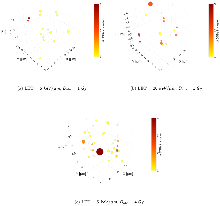
- The ratio of clusters to total DSBs and the mean cluster size both grow with LET, and per-nucleus survival tracks clustering, supporting the claim that nanodosimetric damage topology drives the RBE rise.
- Because the same framework reproduces 220 kV X-ray survival for HUVEC cells, a single parameter set can span photon and proton qualities, supporting use in mixed-field treatment planning.
- Expanding the simulator's energy database to clinical proton energies, helium ions, and carbon ions is presented as the direct path to broader clinical applicability.
Reading between the lines
- The model's reliance on the 1 µm and same-chromosome rules implies a testable prediction: controlled changes in chromosome territory organization should shift survival for the same LET and dose, even when total DSB yield is unchanged.
- If the cluster-independence assumption is right, pairwise DSB interactions are the dominant lethal channel at high LET; one could estimate the interaction rate b directly from time-resolved co-localization of repair foci.
- Extending the framework to carbon ions would provide a sharper test, because MT-GSM2 would predict an RBE–LET curve for heavier ions with no new free parameters beyond the DBSCAN rules.
- The fitted second-order rate b becoming comparable to the repair rate r suggests that approaches neglecting pairwise damage interactions may need artificial domain sizes to compensate; data-derived clusters could replace those domains more generally.
Signed reviews
Editorial analysis
A structured set of objections, weighed in public.
Referee Report
Summary. The manuscript presents MT-GSM2, a multiscale model that feeds DSB coordinates and chromosome identities from the MINAS-TIRITH Geant4-DNA simulation into the GSM2 survival model. DBSCAN groups DSBs on the same chromosome within 1 um into clusters, which replace GSM2's artificial domains; Eq. (2) computes survival as a product over clusters. GSM2 parameters a, b, and r are fitted to HUVEC 220 kV X-ray survival data and to H460 proton survival at 11.1 keV/um, and the model is then used to predict H460 RBE10 across approximately 4-20 keV/um. The central quantitative claim is that this cross-LET prediction matches the Patel/Bronk experiments, supporting the clustering-based domain concept.
Significance. The design has real strengths: the H460 prediction is a genuine cross-LET test because a, b, and r are fixed at a single LET; the chromosome-aware DBSCAN domain definition is a conceptually important alternative to fitted MKM domains; the Geant4 phase-space replication and the residual comparison with TLK are useful benchmarking elements. If the clustering rule is robust, the framework would be a valuable mechanistic bridge between nanodosimetric damage patterns and cell survival. However, the paper does not currently demonstrate that robustness: the domain-defining parameters are not varied, no uncertainties are attached to predicted survival or RBE, and the HUVEC agreement is a fit rather than a prediction. These gaps must be closed before the "excellent agreement" claim can be accepted.
major comments (4)
- [Section 2.3 and Eq. (2)] The DBSCAN clustering rule is load-bearing for the cross-LET prediction. Because a, b, and r are fit at 11.1 keV/um, the fit can absorb the cluster statistics at that single LET, so the predicted LET dependence is carried almost entirely by the number and size of the DBSCAN clusters. The 1 um threshold and the same-chromosome restriction are not fitted, but they are also not varied; Section 2.3 states the threshold is chosen a priori, and Section 2.5 calls the domain definition "parameter-free," which is inaccurate. Please provide a sensitivity analysis over epsilon (e.g., 0.5-2 um) and over the chromosome restriction, showing RBE10 and SF curves, and state how the conclusions change. Without this, the agreement in Fig. 3.4 could be an artifact of the clustering rule.
- [Sections 3.3 and 3.6] No statistical uncertainties are reported for the predicted survival fractions, RBE10 values, or fitted parameters a, b, and r. Figures 3.3 and 3.4 show point predictions only, and Eq. (4) is minimized without confidence intervals. Since Ncells = 2000 and the phase-space sampling are stochastic inputs, the "excellent agreement" in Fig. 3.4 is not quantitatively assessable. Please add error bars or confidence intervals, e.g., by bootstrapping over simulated cells or by repeating the parameter fit on resampled data.
- [Section 3.2 and Table 1] The HUVEC X-ray comparison is not an independent validation. The endothelial a, b, and r are fitted to the same 220 kV X-ray survival data shown in Fig. 3.3a, so the small residuals in Fig. 3.3b are a goodness-of-fit result, not a predictive test. The text should label this as calibration, and the claim in Section 4.2 that MT-GSM2 "predicts the biological effects of photons... from first principles" should be softened or supported by a withheld-dose or independent-photon test.
- [Section 2.4 and Eq. (2)] The mapping from the DSB complexity index i to the number of sublethal lesions x_j is not written explicitly. The sentence "DSB-type damages with i and i+1 account for the same number of sublethal lesions" implies x_j = sum over DSBs of ceil((i+1)/2), but the paper never gives the formula. Because survival in Eq. (2) depends exponentially on x_j, this mapping is as important as the clustering rule. Please state the mapping in equation form and test its sensitivity to a plausible alternative (e.g., x_j = sum (i+1)).
minor comments (8)
- [Abstract] The abstract and several section headers contain rendering artifacts such as "keV=—m" and "\gsm"; please fix the typography.
- [Section 2.2] "cathegorized" should be "categorized"; use consistent spelling of "sublethal" throughout.
- [Section 3.2] In the caption of Figure 3.3, "MINAS-TIRTIH" should be "MINAS-TIRITH".
- [Equation (4)] The quantity is labeled NMSE, but the expression is a dose-weighted squared error normalized by S(D); please clarify the normalization or rename the quantity.
- [Section 4.1] The claim of a "strong correlation between the number of DSB clusters and RBE10" is not directly shown by Figure 3.6, which plots cluster-to-total ratio versus LET; either add a direct RBE10-versus-clustering panel or rephrase the statement.
- [Section 3.5 and Figure 3.7] The correlation between high clustering and reduced survival is built into Eq. (2), since survival is computed from the clusters; presenting it as independent support for the model's mechanism is misleading.
- [Section 2.6] Please specify which proton experiments were excluded because their phase space exceeded the MINAS-TIRITH database limits, and whether Figure 3.4 includes all remaining experimental points.
- [Table 1] The row for HUVEC cells is labeled "Endothelial"; use the same cell-line name as in the text for consistency.
Circularity Check
The proton cross-LET prediction is independent, but the HUVEC X-ray comparison is an in-sample fit and the clustering-survival correlation is built into Eq. (2).
-
fitted input called prediction
[Section 3.2 and Section 3.6, Table 1]
"The model’s predicted SF curve (MT-GSM2, red) is compared to experimental data (blue) from Paget et al. [33] in Figure 3.3a. The results demonstrate strong agreement across all tested doses. [...] Table 1 reports the fitted GSM2 parameters for the studied cell lines."
The endothelial cell line row in Table 1 gives a, b, r for the HUVEC 220 kV X-ray data. Section 2.5 states that a, b, r are calibrated by minimizing the NMSE between predicted and experimental cell survival. Therefore the red curve in Figure 3.3a is the fitted model evaluated on the very survival data used for fitting; calling it a 'prediction' and using its agreement as validation is circular. The agreement is a consequence of the optimization, not an independent test.
-
self definitional
[Eq. (2) and Section 3.5, Figure 3.7]
"Sn(zn|Dabs) = product over clusters of xj x=1 rx / ((a+r)x + bx(x-1)) [...] The colormap encodes a clustering index defined as the ratio of number of clusters to total DSBs within each nucleus. Lower values of this index correspond to more severe clustering (i.e., more DSBs per cluster). This analysis reveals a correlation between high clustering and reduced cell survival, particularly pronounced at higher LET values."
Equation (2) defines the survival probability as a product over clusters where each cluster with x sublethal lesions contributes a factor containing x and x(x-1). Thus, for fixed parameters, a nucleus with more DSBs per cluster necessarily has a lower computed survival. The correlation displayed in Figure 3.7 is therefore not an empirical discovery but a mathematical consequence of the model's defining equation. Presenting it as evidence that 'nanodosimetric features shape the biological response' is circular: the response was constructed to depend on cluster size in exactly this way.
full rationale
The proton RBE10 predictions for H460 cells are the strongest independent part of the paper: the GSM2 parameters a, b, r were fitted only to the 11.1 keV/um survival curve and then used to predict survival at other LET values without refitting. That cross-LET transfer is a genuine predictive test, though its validity depends on the a priori DBSCAN clustering rules. However, two load-bearing presentations are circular. First, the HUVEC X-ray 'prediction' in Figure 3.3a is a fit to the same survival data that supplied the endothelial parameters in Table 1, so the reported agreement is not independent validation. Second, the claimed correlation between clustering and reduced survival in Figure 3.7 is built directly into Eq. (2), where survival is a product over clusters with per-cluster lesion count in the argument; this is a definitional identity, not an emergent finding. These are partial circularities that affect the presentation of validation evidence, while the central cross-LET proton prediction retains independent content. The self-citations to GSM2 and MINAS-TIRITH are normal methodological references and do not, by themselves, make the derivation circular; the circularity is in the fitted-input-as-prediction and self-definitional correlation steps.
Assumptions & free parameters
free parameters (9)
- a (H460) =
6.45e-3 h^-1
- b (H460) =
2.90 h^-1
- r (H460) =
1.74 h^-1
- a (HUVEC) =
7.21e-2 h^-1
- b (HUVEC) =
2.83e-1 h^-1
- r (HUVEC) =
4.84 h^-1
- DBSCAN distance threshold (epsilon) =
1 um
- DBSCAN minimum cluster size =
1 DSB
- Complexity-to-sublethal-lesion mapping =
floor(i/2)+1
assumptions (5)
- domain assumption GSM2 reaction kinetics and survival formula Eq. (2) are valid.
- ad hoc to paper DBSCAN clusters behave as independent GSM2 domains.
- ad hoc to paper Only DSBs on the same chromosome and within 1 um can interact lethally.
- domain assumption MINAS-TIRITH precomputed databases and Geant4 physics lists accurately model DSB induction.
- domain assumption Sampling specific energy z_n from the microdosimetric multi-event distribution captures cellular stochasticity.
Cite this review
Pith. "Pith review of Integrating nano- and micrometer-scale energy deposition models for mechanistic prediction of radiation-induced DNA damage and cell survival." pith.science (2026). https://pith.science/paper/YUPX43CV
@misc{pith2026250700929,
  author       = {Pith},
  title        = {Pith review of: Integrating nano- and micrometer-scale energy deposition models for mechanistic prediction of radiation-induced DNA damage and cell survival},
  year         = {2026},
  howpublished = {\url{https://pith.science/paper/YUPX43CV}},
  note         = {Machine review of arXiv:2507.00929}
}
read the original abstract
We present an integrated modeling framework that combines the Generalized Stochastic Microdosimetric Model (GSM2), used to predict cell survival fractions, with MINAS-TIRITH, a fast and efficient Geant4 DNA-based tool for simulating radiation-induced DNA damage in cell populations. This approach enables the generation of spatially and structurally resolved double-strand break (DSB) distributions, capturing key features such as damage complexity and chromosome specificity. A novel application of the DBSCAN clustering algorithm is introduced to group DSBs at the micrometer scale. This allows the identification of physical aggregates of DNA damage and their association with subnuclear domains, providing a direct link to the cell survival probability as predicted by \gsm. The model was validated using experimental data from HUVEC cells irradiated with 220 kV X-rays and H460 cells exposed to protons over a wide linear energy transfer (LET) range, from approximately 4 keV/{\mu}m to over 20 keV/{\mu}m. Results show excellent agreement between simulations and experimental survival probabilities, making this one of the first consistent multi-scale models to bridge nanodosimetric and microdosimetric representations of radiation with biological outcomes such as cell survival. By incorporating the inherent stochastic nature of radiation-matter interactions, this framework effectively connects the physical properties of the radiation field to the biological response at the cellular level. Its accuracy across various radiation types and energies supports its potential for use in biologically optimized radiotherapy.
Figures


Figures from the paper (4 more)


Reference graph
Works this paper leans on
- [1]
-
[2]
M. Durante, J. Flanz, Charged particle beams to cure cancer: strengths and challenges, in: Seminars in Oncology, Vol. 46, Elsevier, 2019, pp. 219–225
work page 2019
-
[3]
N. Ipe, G. Fehrenbacher, I. Gudowska, H. Paganetti, J. Schippers, S. Roesler, et al., Ptcog publica- tions sub-committee task group on shielding design and radiation safety of charged particle therapy facilities, PTCOG Report 1 (2010). 14
work page 2010
-
[4]
R. B. Hawkins, A statistical theory of cell killing by radiation of varying linear energy transfer, Radiation research 140 (3) (1994) 366–374
work page 1994
-
[5]
Y. Kase, T. Kanai, Y. Matsumoto, Y. Furusawa, H. Okamoto, T. Asaba, M. Sakama, H. Shinoda, Microdosimetric measurements and estimation of human cell survival for heavy-ion beams, Radiation research 166 (4) (2006) 629–638
work page 2006
-
[6]
O. N. Vassiliev, Formulation of the multi-hit model with a non-poisson distribution of hits, Interna- tional Journal of Radiation Oncology* Biology* Physics 83 (4) (2012) 1311–1316
work page 2012
-
[7]
T. Friedrich, M. Durante, M. Scholz, The local effect model—principles and applications, The Health Risks of Extraterrestrial Environments (2013)
work page 2013
-
[8]
L. Manganaro, G. Russo, R. Cirio, F. Dalmasso, S. Giordanengo, V. Monaco, S. Muraro, R. Sacchi, A. Vignati, A. Attili, A monte carlo approach to the microdosimetric kinetic model to account for dose rate time structure effects in ion beam therapy with application in treatment planning simulations, Medical physics 44 (4) (2017) 1577–1589
work page 2017
Show all 55 references
-
[9]
O. N. Vassiliev, Microdosimetry. elements of stochastic transport theory, in: Monte Carlo Methods for Radiation Transport, Springer, 2017, pp. 195–223
2017
-
[10]
Inaniwa, N
T. Inaniwa, N. Kanematsu, Adaptation of stochastic microdosimetric kinetic model for charged- particle therapy treatment planning, Physics in Medicine & Biology 63 (9) (2018) 095011
2018
-
[11]
Cordoni, M
F. Cordoni, M. Missiaggia, A. Attili, S. Welford, E. Scifoni, C. La Tessa, Generalized stochastic microdosimetric model: The main formulation, Physical Review E 103 (1) (2021) 012412
2021
-
[12]
F. G. Cordoni, M. Missiaggia, C. La Tessa, E. Scifoni, Multiple levels of stochasticity accounted for in different radiation biophysical models: from physics to biology, International Journal of Radiation Biology (2022) 1–16
2022
-
[13]
S. J. McMahon, A. L. McNamara, J. Schuemann, H. Paganetti, K. M. Prise, A general mechanistic model enables predictions of the biological effectiveness of different qualities of radiation, Scientific reports 7 (1) (2017) 1–14
2017
-
[14]
A. L. McNamara, J. Schuemann, H. Paganetti, A phenomenological relative biological effectiveness (rbe) model for proton therapy based on all published in vitro cell survival data, Physics in Medicine & Biology 60 (21) (2015) 8399
2015
-
[15]
F. G. Cordoni, M. Missiaggia, E. Scifoni, C. La Tessa, An artificial intelligence-based model for cell killing prediction: development, validation and explainability analysis of the anakin model, Physics in Medicine & Biology 68 (8) (2023) 085017
2023
-
[16]
L. Tian, C. Hahn, A. Lühr, An ion-independent phenomenological relative biological effectiveness (rbe) model for proton therapy, Radiotherapy and Oncology 174 (2022) 69–76
2022
-
[17]
D. B. Flint, S. J. Bright, C. McFadden, T. Konishi, D. K. Martinus, M. Manandhar, M. B. Kacem, L. Bronk, G. O. Sawakuchi, An empirical model of carbon-ion relative biological effectiveness based on the linear correlation between radiosensitivity to photons and carbon ions, Phy...
2024
-
[18]
A. Hu, W. Zhou, X. Luo, R. Qiu, J. Li, Correlation between dna double-strand break distribution in 3d genome and ionizing radiation-induced cell death, Radiation Research (2025)
2025
-
[19]
Sakata, R
D. Sakata, R. Hirayama, W.-G. Shin, M. Belli, M. A. Tabocchini, R. D. Stewart, O. Belov, M. A. Bernal, M.-C. Bordage, J. Brown, et al., Prediction of dna rejoining kinetics and cell survival after proton irradiation for v79 cells using geant4-dna (vol 105, 102508, 2023), PHYSI...
2024
-
[20]
dsbandrepair
T. N. Hoang, Y. Thibaut, K. Chatzipapas, D. Sakata, S. Incerti, C. Villagrasa, Y. Perrot, et al., “dsbandrepair”–an updated geant4-dna simulation tool for evaluating the radiation-induced dna dam- age and its repair, Physica Medica 124 (2024) 103422
2024
-
[21]
F. G. Cordoni, M. Missiaggia, E. Scifoni, C. La Tessa, Cell survival computation via the generalized stochastic microdosimetric model (gsm2); part i: The theoretical framework, Radiation research 197 (3) (2022) 218–232. 15
2022
-
[22]
F. G. Cordoni, On the emergence of the deviation from a poisson law in stochastic mathematical models for radiation-induced dna damage: A system size expansion, Entropy 25 (9) (2023) 1322
2023
-
[23]
F. G. Cordoni, A spatial measure-valued model for radiation-induced dna damage kinetics and repair under protracted irradiation condition, Journal of Mathematical Biology 88 (2) (2024) 21
2024
-
[24]
Missiaggia, F
M. Missiaggia, F. Cordoni, E. Scifoni, C. L. Tessa, Cell survival computation via the generalized stochastic microdosimetric model (gsm2); part ii: Numerical results, Radiation Research (2024)
2024
-
[25]
Bordieri, M
G. Bordieri, M. Missiaggia, G. Cartechini, M. Battestini, L. Bronk, F. Guan, D. Grosshans, P. Rai, E. Scifoni, C. La Tessa, et al., Validation of the generalized stochastic microdosimetric model (gsm2) over a broad range of let and particle beam type: a unique model for accura...
2024
-
[26]
Thibaut, G
Y. Thibaut, G. Gonon, J. S. Martinez, M. Petit, A. Vaurijoux, G. Gruel, C. Villagrasa, S. Incerti, Y.Perrot, Minastirith: anewtoolforsimulatingradiation-induceddnadamageatthecellpopulation level, Physics in Medicine & Biology 68 (3) (2023) 034002
2023
-
[27]
Thibaut, G
Y. Thibaut, G. Gonon, J. Martinez, M. Petit, R. Babut, A. Vaurijoux, G. Gruel, C. Villagrasa, S. Incerti, Y. Perrot, Experimental validation in a neutron exposure frame of the minas tirith for cell damage simulation, Physics in Medicine & Biology 68 (22) (2023) 225008
2023
-
[28]
Friedrich, K
T. Friedrich, K. Ilicic, C. Greubel, S. Girst, J. Reindl, M. Sammer, B. Schwarz, C. Siebenwirth, D. W. Walsh, T. E. Schmid, et al., Dna damage interactions on both nanometer and micrometer scale determine overall cellular damage, Scientific Reports 8 (1) (2018) 16063
2018
-
[29]
R. Grün, T. Friedrich, E. Traneus, M. Scholz, Is the dose-averaged let a reliable predictor for the relative biological effectiveness?, Medical physics 46 (2) (2019) 1064–1074
2019
-
[30]
Paganetti, C
H. Paganetti, C. B. Simone II, W. R. Bosch, D. Haas-Kogan, D. G. Kirsch, H. Li, X. Liang, W. Liu, A. Mahajan, M. D. Story, et al., Nrg oncology white paper on the relative biological effectiveness in proton therapy, International Journal of Radiation Oncology* Biology* Physics (2024)
2024
-
[31]
V. E. Bellinzona, F. Cordoni, M. Missiaggia, F. Tommasino, E. Scifoni, C. La Tessa, A. Attili, Linking microdosimetric measurements to biological effectiveness in ion beam therapy: A review of theoretical aspects of mkm and other models, Frontiers in Physics (2021) 623
2021
-
[32]
Kundrát, W
P. Kundrát, W. Friedland, J. Becker, M. Eidemüller, A. Ottolenghi, G. Baiocco, Analytical formu- las representing track-structure simulations on dna damage induced by protons and light ions at radiotherapy-relevant energies, Scientific Reports 10 (1) (2020) 15775
2020
-
[33]
Paget, M
V. Paget, M. Ben Kacem, M. Dos Santos, M. A. Benadjaoud, F. Soysouvanh, V. Buard, T. Georges, A.Vaurijoux, G.Gruel, A.François, etal., Multiparametricradiobiologicalassaysshowthatvariation of x-ray energy strongly impacts relative biological effectiveness: comparison between 2...
2019
-
[34]
Patel, L
D. Patel, L. Bronk, F. Guan, C. R. Peeler, S. Brons, I. Dokic, A. Abdollahi, C. Rittmüller, O. Jäkel, D. Grosshans, et al., Optimization of monte carlo particle transport parameters and validation of a novel high throughput experimental setup to measure the biological effects ...
2017
-
[35]
Bronk, F
L. Bronk, F. Guan, D. Patel, D. Ma, B. Kroger, X. Wang, K. Tran, J. Yiu, C. Stephan, J. Debus, et al., Mapping the relative biological effectiveness of proton, helium and carbon ions with high- throughput techniques, Cancers 12 (12) (2020) 3658
2020
-
[36]
Agostinelli, J
S. Agostinelli, J. Allison, K. a. Amako, J. Apostolakis, H. Araujo, P. Arce, M. Asai, D. Axen, S. Banerjee, G. Barrand, et al., Geant4—a simulation toolkit, Nuclear instruments and methods in physics research section A: Accelerators, Spectrometers, Detectors and Associated Equ...
2003
-
[37]
Allison, K
J. Allison, K. Amako, J. Apostolakis, H. Araujo, P. A. Dubois, M. Asai, G. Barrand, R. Capra, S. Chauvie, R. Chytracek, et al., Geant4 developments and applications, IEEE Transactions on nuclear science 53 (1) (2006) 270–278. 16
2006
-
[38]
Allison, K
J. Allison, K. Amako, J. Apostolakis, P. Arce, M. Asai, T. Aso, E. Bagli, A. Bagulya, S. Banerjee, G. Barrand, et al., Recent developments in geant4, Nuclear instruments and methods in physics research section A: Accelerators, Spectrometers, Detectors and Associated Equipment ...
2016
-
[39]
Incerti, G
S. Incerti, G. Baldacchino, M. Bernal, R. Capra, C. Champion, Z. Francis, P. Guèye, A. Man- tero, B. Mascialino, P. Moretto, et al., The geant4-dna project, International Journal of Modeling, Simulation, and Scientific Computing 1 (02) (2010) 157–178
2010
-
[40]
Incerti, A
S. Incerti, A. Ivanchenko, M. Karamitros, A. Mantero, P. Moretto, H. Tran, B. Mascialino, C. Cham- pion, V. Ivanchenko, M. Bernal, et al., Comparison of geant4 very low energy cross section models with experimental data in water, Medical physics 37 (9) (2010) 4692–4708
2010
-
[41]
Lampe, S
S.Incerti, I.Kyriakou, M.Bernal, M.Bordage, Z.Francis, S.Guatelli, V.Ivanchenko, M.Karamitros, N. Lampe, S. B. Lee, et al., Geant4-dna example applications for track structure simulations in liquid water: a report from the geant4-dna project, Medical physics 45 (8) (2018) e722–e739
2018
-
[42]
H. N. Tran, J. Archer, G. Baldacchino, J. M. Brown, F. Chappuis, G. A. P. Cirrone, L. Desorgher, N. Dominguez, S. Fattori, S. Guatelli, et al., Review of chemical models and applications in geant4- dna: Report from the esa biorad iii project, Medical Physics 51 (9) (2024) 5873–5889
2024
-
[43]
Ester, H.-P
M. Ester, H.-P. Kriegel, J. Sander, X. Xu, et al., A density-based algorithm for discovering clusters in large spatial databases with noise, in: kdd, Vol. 96, 1996, pp. 226–231
1996
-
[44]
Ester, H.-P
M. Ester, H.-P. Kriegel, J. Sander, X. Xu, Density-connected sets and their application for trend detection in spatial databases., in: KDD, Vol. 97, 1997, pp. 10–15
1997
-
[45]
Francis, C
Z. Francis, C. Villagrasa, I. Clairand, Simulation of dna damage clustering after proton irradiation using an adapted dbscan algorithm, Computer methods and programs in biomedicine 101 (3) (2011) 265–270
2011
-
[46]
R. H. Byrd, P. Lu, J. Nocedal, C. Zhu, A limited memory algorithm for bound constrained opti- mization, SIAM Journal on scientific computing 16 (5) (1995) 1190–1208
1995
-
[47]
R. B. Hawkins, A microdosimetric-kinetic model for the effect of non-poisson distribution of lethal lesions on the variation of rbe with let, Radiation research 160 (1) (2003) 61–69
2003
-
[48]
F. Guan, L. Bronk, U. Titt, S. H. Lin, D. Mirkovic, M. D. Kerr, X. R. Zhu, J. Dinh, M. Sobieski, C. Stephan, et al., Spatial mapping of the biologic effectiveness of scanned particle beams: towards biologically optimized particle therapy, Scientific reports 5 (1) (2015) 9850
2015
-
[49]
R. D. Stewart, Two-lesion kinetic model of double-strand break rejoining and cell killing, Radiation research 156 (4) (2001) 365–378
2001
-
[50]
Friedrich, U
T. Friedrich, U. Scholz, T. Elsässer, M. Durante, M. Scholz, Calculation of the biological effects of ion beams based on the microscopic spatial damage distribution pattern, International journal of radiation biology 88 (1-2) (2012) 103–107
2012
-
[51]
Pfuhl, T
T. Pfuhl, T. Friedrich, M. Scholz, Comprehensive comparison of local effect model iv predictions with the particle irradiation data ensemble, Medical Physics 49 (1) (2022) 714–726
2022
-
[52]
P. J. Deschavanne, B. Fertil, A review of human cell radiosensitivity in vitro, International Journal of Radiation Oncology* Biology* Physics 34 (1) (1996) 251–266
1996
-
[53]
J. F. Fowler, Is repair of dna strand break damage from ionizing radiation second-order rather than first-order? a simpler explanation of apparently multiexponential repair, Radiation research 152 (2) (1999) 124–136
1999
-
[54]
Carabe-Fernandez, R
A. Carabe-Fernandez, R. Dale, H. Paganetti, Repair kinetic considerations in particle beam radio- therapy, The British journal of radiology 84 (1002) (2011) 546–555
2011
-
[55]
T. S. Dexheimer, Dna repair pathways and mechanisms, in: DNA repair of cancer stem cells, Springer, 2012, pp. 19–32. 17
2012
Reviewed August 6, 2026 · model on record in the stance chip above.
Discussion (0). Continue with ORCID to comment.