REVIEW 3 major objections 6 minor 16 references
Adversarial Convolutional Networks with Weak Domain-Transfer for Multi-Sequence Cardiac MR Images Segmentation
T0 review · 3 major / 6 minor · reviewed 2026-08-14 · deepseek-v4-flash
Pith's one-line read The paper claims that pseudo masks from bSSFP and T2 MRI, transferred to LGE through a weak domain-transfer loss, improve heart-chamber segmentation where LGE labels are scarce.
desk verdict The transfer-loss idea is reasonable and clearly presented, but the headline result on the challenge test set is confounded by training on LGE images from 30 of the 40 test patients. read the letter →
The pith
A machine-rendered reading of the paper's core claim, the machinery that carries it, and where it could break.
The reading
What carries the argument
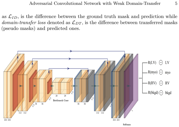
The load-bearing mechanism is the weak domain-transfer loss $L_{DT}$, a combination of cross-entropy and Dice loss computed between the network's prediction on an LGE slice and a pseudo mask obtained by mapping the corresponding bSSFP or T2 annotation through $j = \lfloor i n / m \rfloor$. The pseudo masks are used as training targets only for LGE slices without manual labels, and the loss is combined with the individual-domain loss as $L_G = \lambda L_{ID} + (1-\lambda) L_{DT}$ with $\lambda=0.9$, which keeps the transferred supervision weak. The adversarial component drives the generator to produce masks that the discriminator cannot distinguish from ground-truth masks conditioned on the image, which sharpens the boundaries.
What would settle it
Replace the index-based slice correspondence in the U+A+D+T pipeline with random pairings between bSSFP/T2 masks and LGE slices; if the reported Dice and surface-distance gains persist, the domain-transfer loss is not contributing the way the paper claims.
Extended reading notes
Core claim
The paper claims that pseudo masks derived from bSSFP and T2 images, transferred to LGE via the linear slice correspondence $j = \lfloor i n / m \rfloor$, provide enough training signal to improve LGE segmentation when combined with ground-truth supervision at a low weight. In the proposed end-to-end framework, the generator and discriminator are optimized jointly; the total generator loss weighs the individual-domain loss at $\lambda = 0.9$ and the domain-transfer loss at $0.1$, making the transfer deliberately weak. The reported experiments on the challenge test set (patients 6 to 45) show that adding this loss to the adversarial model improves average Dice from 76.6% to 82.4% for the left ventricle and from 42.0% to 61.0% for the myocardium, with consistent gains in Jaccard and surface distance and mixed Hausdorff results.
Load-bearing premise
The whole pipeline rests on the premise that pseudo masks transferred from bSSFP or T2 to LGE via the simple slice-index formula $j = \lfloor i n / m \rfloor$ align well enough to act as training targets, despite ignoring slice spacing, field of view, orientation, and motion differences between modalities.
Editorial extensions
If this is right
- LGE segmentation can be trained with only a handful of annotated LGE cases by borrowing annotations from bSSFP and T2 sequences of the same patient.
- The slice-correspondence formula provides a label source that costs nothing beyond the existing annotations and integrates directly into the loss, with no pre-trained model or separate registration step required.
- The transfer weight $\lambda = 0.9$ suggests that a deliberately weak transfer loss is more effective than treating pseudo masks as ground truth.
- Adversarial training and cross-modality transfer are complementary: each alone helps on different structures, and together they improve both left ventricle and myocardium segmentation.
Reading between the lines
- If the index mapping were replaced by a proper registration step that recovers true slice correspondence, the weak-transfer loss might yield larger gains or allow a higher transfer weight.
- The same pseudo-mask transfer could apply to other cross-modality segmentation problems where one modality is expensive to annotate, as long as the two modalities share structural layout.
- The gain concentrated in the myocardium (42% to 61%) compared with the right ventricle (69.5% to 71.0%) suggests the weak transfer mainly helps structures with low contrast in LGE; a testable extension is to weight the transfer loss per structure.
Signed reviews
Editorial analysis
A structured set of objections, weighed in public.
Referee Report
Summary. The paper proposes an end-to-end adversarial segmentation framework for multi-sequence cardiac magnetic resonance (CMR) images, targeting Late Gadolinium Enhancement (LGE) segmentation when LGE annotations are scarce. The generator is a dilated residual U-shape network trained with cross-entropy and Dice losses; a CNN discriminator is trained adversarially to distinguish predicted from ground-truth masks; and a 'weak domain-transfer loss' transfers masks from bSSFP and T2 modalities to LGE slices via slice-index normalization j = floor(i*n/m). The authors evaluate on the MICCAI 2019 challenge data: a two-patient ablation in Table 1, and challenge test results for patients 6-45 in Tables 2 and 3, reporting Dice, Jaccard, surface distance, and Hausdorff distance. The abstract concludes that the proposed adversarial pipeline outperforms baseline deep-learning methods.
Significance. The idea of using weakly aligned cross-modality masks as a weak supervision signal is interesting and practically relevant, and the release of code and use of a public challenge dataset are strengths. If the reported gains were causally attributable to the transfer loss, this would be a useful contribution to multi-modal cardiac segmentation. However, the primary experimental comparison is confounded by training-set composition, and the only controlled ablation is too small to support the strength of the claims. The paper needs substantial additional analysis or tempering of its conclusions before it meets the standard for publication.
major comments (3)
- [§3.1, Tables 2 and 3] The comparison between U+A+D and U+A+D+T on test patients 6-45 is not a controlled test of the transfer loss. Because patients 6-35 have manual T2/bSSFP annotations, U+A+D+T uses those annotations to create pseudo masks for LGE slices from exactly these test patients, thereby including LGE images of patients 6-35 in its training set. U+A+D, lacking the transfer loss, has no supervision signal on those LGE slices and is effectively trained on a smaller dataset that does not include the LGE images of patients 6-35. The large myocardium Dice gain (42.0% to 61.0%) may simply reflect additional target-domain training data rather than the transfer mechanism. The authors should report results separately for truly unseen patients 36-45, who have no annotations in any modality, or add a control baseline that uses unlabeled LGE images from patients 6-35 without the transfer loss, and adjust the abstract's claims accordingly.
- [§3.1, Table 1] The only non-confounded comparison is on two held-out patients (patients 4 and 5), with no error bars, no repeated runs, and no significance test. The observed differences are not statistically established: for example, the right ventricle Dice is lower for U+A+D+T than for U+A+D (71.7% vs 74.7%), and the myocardium improvement of 5.7 percentage points could easily be within the variability expected for n=2. This table alone cannot support the abstract's claim that the proposed pipeline outperforms baseline deep-learning methods.
- [§2, Eq. (1)] The pseudo-mask alignment assumption in Eq. (1) is directly testable using patients 1-5, for whom all three modalities are manually annotated. The authors could quantitatively evaluate the overlap between bSSFP/T2 masks mapped by j = floor(i*n/m) and the actual LGE masks for these patients. Without such a validation, the adequacy of the weak domain-transfer loss remains unquantified, especially given the footnote acknowledging that 'Ideally, registration could be performed to find the correspondence.' This is particularly important because the main experimental evidence (Table 2) depends on the pseudo masks being accurate enough to serve as training targets.
minor comments (6)
- [§3, Implementation] The text states 'α is set as 0.9' and then refers to weights for cross-entropy and Dice loss, but α is not defined in the equations (Eqs. (2)-(3) use β1 and β2). This appears to be a typo; please clarify the notation consistently.
- [Algorithm 1] In Algorithm 1, 'update G by LIN' is written; the subscript 'IN' is not defined anywhere, and it should likely be 'LID' (individual-domain loss). Please correct this typo.
- [§3.1] The phrase 'we performe a very preliminary experiment' contains a typo ('performe' should be 'perform').
- [Figure 4] The row label 'O' for original images is ambiguous; consider using 'Input' or 'LGE' to match the modality being segmented.
- [Tables 1-3] Hausdorff distance is reported without units and without specifying whether the average or maximum Hausdorff distance is used; please define the metric and specify units (millimeters).
- [Abstract and §4] The abstract's statement that the proposed pipeline 'outperforms baseline deep-learning methods' overstates the evidence, since the only comparisons are the authors' own U+D and U+A+D variants. Please qualify the claim to refer to the specific baselines evaluated, or include comparisons with external methods.
Circularity Check
No circularity: the reported improvement is an empirical comparison, not a derivation forced by the loss definitions or by self-citation.
full rationale
The paper's central claim is an empirical result: adding the weak domain-transfer loss LDT to the adversarial segmentation pipeline improves Dice and surface-distance metrics on the MICCAI 2019 test set. The loss is defined as a weighted combination of cross-entropy and Dice losses between predictions and pseudo masks (Eqs. 2-4), and the total adversarial objective is a sum of generator and discriminator losses (Eq. 6). Nothing in these equations forces the reported improvement; the transfer loss could in principle degrade performance, and indeed the adversarial baseline without transfer performs worse on the left ventricle in Table 1. The comparison is conducted on an external challenge test set, not on the training objective. No parameter is fitted to the test metrics, no uniqueness theorem is invoked, and no load-bearing claim is justified solely by a self-citation. The dilated residual U-shape architecture is attributed to prior work by one of the co-authors, but that citation supplies the backbone structure, not the conclusion that the proposed transfer loss works. A separate evaluation concern exists: patients 6-35 have manual bSSFP/T2 annotations, and those annotations are used to construct pseudo masks for LGE slices of the same patients, while the baseline U+A+D receives no supervision signal on those LGE images. This means the reported average over patients 6-45 may reflect transductive exposure to the test cohort rather than a purely generalizable cross-modality transfer effect. That is a data-contamination and experimental-validity issue, not a circularity in the derivation-chain sense required by the analysis: the improvement is still measured empirically on external metrics and is not equivalent by construction to the inputs of the loss function. Accordingly, no circular step is identified and the circularity score is 0.
Assumptions & free parameters
free parameters (4)
- lambda (λ) =
0.9
- beta1 (β1) =
0.9
- beta2 (β2) =
0.9
- alpha (α) =
0.9
assumptions (3)
- ad hoc to paper The annotation masks of corresponding slices from bSSFP, T2, and LGE overlap sufficiently that masks from bSSFP/T2 can serve as pseudo masks for LGE.
- ad hoc to paper The slice index normalization j = floor(i*n/m) establishes correct correspondence between axial slices of different modalities.
- domain assumption Z-score normalization calibrates intensity ranges across modalities so the network can use multi-modality inputs.
Cite this review
Pith. "Pith review of Adversarial Convolutional Networks with Weak Domain-Transfer for Multi-Sequence Cardiac MR Images Segmentation." pith.science (2026). https://pith.science/paper/Z63ODEO2
@misc{pith2026190809298,
  author       = {Pith},
  title        = {Pith review of: Adversarial Convolutional Networks with Weak Domain-Transfer for Multi-Sequence Cardiac MR Images Segmentation},
  year         = {2026},
  howpublished = {\url{https://pith.science/paper/Z63ODEO2}},
  note         = {Machine review of arXiv:1908.09298}
}
read the original abstract
Analysis and modeling of the ventricles and myocardium are important in the diagnostic and treatment of heart diseases. Manual delineation of those tissues in cardiac MR (CMR) scans is laborious and time-consuming. The ambiguity of the boundaries makes the segmentation task rather challenging. Furthermore, the annotations on some modalities such as Late Gadolinium Enhancement (LGE) MRI, are often not available. We propose an end-to-end segmentation framework based on convolutional neural network (CNN) and adversarial learning. A dilated residual U-shape network is used as a segmentor to generate the prediction mask; meanwhile, a CNN is utilized as a discriminator model to judge the segmentation quality. To leverage the available annotations across modalities per patient, a new loss function named weak domain-transfer loss is introduced to the pipeline. The proposed model is evaluated on the public dataset released by the challenge organizer in MICCAI 2019, which consists of 45 sets of multi-sequence CMR images. We demonstrate that the proposed adversarial pipeline outperforms baseline deep-learning methods.
Figures


Figures from the paper (1 more)

Reference graph
Works this paper leans on
-
[1]
U-net: Convolutional networks for biomedical image segmentation
Ronneberger, O., Fischer, P., Brox, T. U-net: Convolutional networks for biomedical image segmentation. InMICCAI, 234-241, Springer, Cham, 2015
work page 2015
-
[2]
Goodfellow, I., Pouget-Abadie, J., Mirza, M., Xu, B., Warde-Farley, D., Ozair, S., ... and Bengio, Y.. Generative adversarial nets. InNIPS., 2672-2680, 2014
work page 2014
-
[3]
Multivariate mixture model for myocardial segmentation combining multi-source images
Xiahai Zhuang. Multivariate mixture model for myocardial segmentation combining multi-source images. InTPAMI., 2018
work page 2018
-
[4]
Multivariate mixture model for cardiac segmentation from multi-sequence MRI
Zhuang, X. Multivariate mixture model for cardiac segmentation from multi-sequence MRI. In MICCAI, pp.581-588, 2016
work page 2016
-
[5]
Multi-scale patch and multi-modality atlases for whole heart segmentation of MRI
Zhuang, X., and Shen, J.. Multi-scale patch and multi-modality atlases for whole heart segmentation of MRI. InMedical image analysis, 31, 77–87, 2016 , Elsevier
work page 2016
-
[6]
Isola, P., Zhu, J. Y., Zhou, T., and Efros, A. A. Image-to-image translation with conditional adversarial networks. InCVPR, 1125–1134, 2017
work page 2017
-
[7]
Semantic segmentation using adversarial networks
Luc, P., Couprie, C., Chintala, S., and Verbeek, J. Semantic segmentation using adversarial networks. InarXiv preprint arXiv:1611.08408 , 2016
arXiv 2016
-
[8]
Evaluation of Algorithms for Multi-Modality Whole Heart Segmentation: An Open-Access Grand Challenge
Zhuang, X., Li, L., Payer, C., Stern, D., Urschler, M., Heinrich, M. P.and others. Evaluation of Algorithms for Multi-Modality Whole Heart Segmentation: An Open- Access Grand Challenge. InarXiv preprint arXiv:1902.07880 , 2019
work page Pith review arXiv 1902
Show all 16 references
-
[9]
A., Bai, W., Dacher, J
Petitjean, C., Zuluaga, M. A., Bai, W., Dacher, J. N., Grosgeorge, D., Caudron, J and others. Right ventricle segmentation from cardiac MRI: a collation study. In Medical image analysis, 19, 1, 187–202, 2015, Elsevier
2015
-
[10]
A combined deep- learning and deformable-model approach to fully automatic segmentation of the left ventricle in cardiac MRI
Avendi, MR and Kheradvar, Arash and Jafarkhani, Hamid. A combined deep- learning and deformable-model approach to fully automatic segmentation of the left ventricle in cardiac MRI. InMedical image analysis, 30, 108–119, 2016, Elsevier
2016
-
[11]
Deep learning
LeCun, Y., Bengio, Y., and Hinton, G. Deep learning. Innature, 521, 7553, 436, 2015, Nature Publishing Group
2015
-
[12]
Deep residual learning for image recognition
He, K., Zhang, X., Ren, S., and Sun, J. Deep residual learning for image recognition. In CVPR, 770–778, 2016
2016
-
[13]
Fully convolutional networks for semantic segmentation
Long, J., Shelhamer, E., and Darrell, T.. Fully convolutional networks for semantic segmentation. In CVPR, 3431–3440, 2015
2015
-
[14]
Automatic Brain Structures Segmentation Using Deep Residual Dilated U-Net
Li, H., Zhygallo, A., Menze, B. Automatic Brain Structures Segmentation Using Deep Residual Dilated U-Net. InBiomedical optics express, 385-393, 2018
2018
-
[15]
Distilling the knowledge in a neural network
Hinton, G., Vinyals, O., Dean, J. Distilling the knowledge in a neural network. In arXiv preprint arXiv:1503.02531 , 2015
2015 arXiv
-
[16]
Moon, J. C. What is late gadolinium enhancement in hypertrophic cardiomyopathy? In Revista Española de Cardiología , 2007
2007
Reviewed August 14, 2026 · model on record in the stance chip above.
Discussion (0). Continue with ORCID to comment.