Brain Vascular Age Prediction Using Cerebral Blood Flow Velocity and Machine Learning Algorithms
Pith reviewed 2026-05-19 20:19 UTC · model grok-4.3
The pith
Features from transcranial Doppler measurements of cerebral blood flow velocity enable machine learning models to predict brain vascular age and identify accelerated aging in diseased subjects.
A machine-rendered reading of the paper's core claim, the machinery that carries it, and where it could break.
Core claim
Regression models trained exclusively on healthy subjects using MOCAIP-generated features from TCD and heart rate variability predict subjects' chronological age, and when tested on diseased subjects, reveal accelerated cerebrovascular aging, with the differences between healthy and diseased performances suggesting TCD features' relevance for evaluating such acceleration.
What carries the argument
MOCAIP algorithm for morphological analysis and clustering of signals from TCD recordings, producing features combined with heart rate variability for input to regression models predicting age.
If this is right
- Healthy subjects' cerebrovascular age is predicted to be 3.69 years above their chronological age on average.
- Subjects with acute stroke, post-stroke, Alzheimer's disease, and mild cognitive impairment exhibit different levels of age acceleration.
- TCD features may be relevant for evaluating accelerated cerebrovascular aging.
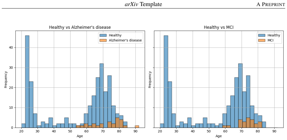
- Imbalanced datasets affect the performance of machine-learning-based brain age prediction models.
Where Pith is reading between the lines
- This method could potentially be used to monitor cerebrovascular aging in clinical settings for early intervention.
- Further studies might explore combining TCD with other imaging modalities for more accurate predictions.
- Calibrating the model to reduce the 3.69-year over-prediction in healthy subjects could improve its baseline accuracy.
Load-bearing premise
The assumption that regression models trained only on healthy subjects yield an unbiased baseline for normal cerebrovascular aging, even as they over-predict healthy subjects' ages by 3.69 years on average.
What would settle it
Observing no significant difference in predicted minus chronological age between diseased and healthy subjects when using the same trained models would falsify the claim of detecting accelerated aging via TCD features.
Figures


read the original abstract
Defining vascular age in terms of physiological function has become one focal point of the extensive studies to categorize and track chronological age. Transcranial Doppler (TCD) is a method by which cerebral blood flow velocity is measured along the major arteries feeding the human brain. This study aims to use features extracted from TCD to estimate chronological age and assess accelerated aging in subjects with various brain diseases. We predict subjects with various brain diseases to present with accelerated cerebrovascular aging when tested on various regression models trained by healthy subjects. 168 healthy subjects and 277 diseased subjects with bilateral TCD recordings of the middle cerebral artery were analyzed using the Morphological Analysis and Clustering of Intracranial Pressure (MOCAIP) algorithm. MOCAIP-generated features and heart rate variability features were used as input features for regression models to predict the brain vascular age. 66 subjects with acute stroke, 27 subjects with post stroke, 26 subjects with Alzheimer's disease, 23 subjects with mild cognitive impairment, and 135 established subjects were tested against the machine learning model to assess for accelerated cerebrovascular age. The trained model, on average, predicted healthy subjects' cerebrovascular age to be 3.69 years above their chronological age. Subjects with different disease conditions exhibited varying levels of age acceleration. The differences in healthy and diseased subjects' performances suggest that features generated using TCD may be relevant when evaluating accelerated cerebrovascular aging. Moreover, imbalanced datasets have been observed to affect the performance of machine-learning-based brain age prediction models.
Editorial analysis
A structured set of objections, weighed in public.
Referee Report
Summary. The manuscript claims that features extracted from transcranial Doppler (TCD) recordings of the middle cerebral artery via the MOCAIP algorithm, combined with heart rate variability features, can be used to train regression models on 168 healthy subjects to predict cerebrovascular (vascular) age. These models are then applied to 277 subjects with brain diseases (66 acute stroke, 27 post-stroke, 26 Alzheimer's, 23 mild cognitive impairment, and 135 'established' subjects) to demonstrate accelerated cerebrovascular aging, with the model over-predicting healthy subjects' age by an average of 3.69 years and showing varying acceleration levels across disease groups. The authors conclude that TCD/MOCAIP features are relevant for assessing accelerated cerebrovascular aging, while noting that imbalanced datasets affect model performance.
Significance. If the reported bias can be corrected and proper validation metrics provided, the work could contribute a non-invasive, TCD-based approach to quantifying cerebrovascular aging and its acceleration in neurological conditions, potentially complementing existing brain-age prediction methods that rely on MRI or other modalities. The use of MOCAIP for feature extraction from cerebral blood flow velocity is a specific strength that could enable broader clinical translation if reproducibility is demonstrated.
major comments (3)
- [Abstract] Abstract: The central claim that diseased subjects exhibit accelerated cerebrovascular aging relative to a healthy baseline is undermined by the reported 3.69-year over-prediction of age in the healthy training population itself. This systematic positive bias indicates that the learned mapping from TCD/MOCAIP features to age does not center on chronological age, so excess predictions in disease groups may partly reflect the same offset rather than disease-specific acceleration. Without explicit bias correction, calibration plots, or a demonstration that the offset is negligible after proper validation, the cross-group differences do not securely support the relevance of TCD features for accelerated aging assessment.
- [Abstract] Abstract and Methods (implied): No model performance metrics (MAE, RMSE, R²), cross-validation procedure, or statistical tests for group differences are reported, despite concrete numerical claims such as the 3.69-year offset and 'varying levels of age acceleration.' This absence prevents evaluation of whether the regression models are reliable or whether the observed differences exceed what would be expected from noise or imbalance.
- [Abstract] Abstract: The manuscript acknowledges that 'imbalanced datasets have been observed to affect the performance' but provides no details on how class imbalance was addressed during training (e.g., weighting, resampling, or stratified cross-validation) or whether results were reported separately for balanced subsets. Given that the healthy training set (n=168) is smaller than the combined diseased test set (n=277), this omission is load-bearing for interpreting the acceleration claims.
minor comments (2)
- [Abstract] The term 'established subjects' in the abstract is undefined and should be clarified (e.g., is this a control or specific disease subgroup?).
- [Abstract] The abstract states 'We predict subjects with various brain diseases to present with accelerated cerebrovascular aging' but does not specify the exact statistical comparison or null hypothesis used to support this prediction.
Simulated Author's Rebuttal
We thank the referee for the constructive and detailed comments, which highlight important aspects for improving the clarity and rigor of our analysis on TCD-based cerebrovascular age prediction. We address each major comment point by point below and will incorporate revisions to strengthen the manuscript where feasible.
read point-by-point responses
-
Referee: [Abstract] Abstract: The central claim that diseased subjects exhibit accelerated cerebrovascular aging relative to a healthy baseline is undermined by the reported 3.69-year over-prediction of age in the healthy training population itself. This systematic positive bias indicates that the learned mapping from TCD/MOCAIP features to age does not center on chronological age, so excess predictions in disease groups may partly reflect the same offset rather than disease-specific acceleration. Without explicit bias correction, calibration plots, or a demonstration that the offset is negligible after proper validation, the cross-group differences do not securely support the relevance of TCD features for accelerated aging assessment.
Authors: We acknowledge the systematic positive bias observed in the healthy training cohort (average over-prediction of 3.69 years), which is explicitly reported in the manuscript. This offset may arise from the regression fitting process or inherent characteristics of the TCD/MOCAIP features. Our core claim focuses on relative differences and varying acceleration levels across disease groups compared to this healthy baseline. To address the concern, we will add calibration plots in the revised manuscript and implement a simple bias correction by subtracting the mean healthy offset from all predictions, thereby centering the healthy excess age at zero for clearer interpretation of disease-specific acceleration. We will also explicitly clarify in the text that reported accelerations represent excess beyond the observed healthy bias. This revision should better isolate disease-related effects while preserving the reported numerical findings. revision: yes
-
Referee: [Abstract] Abstract and Methods (implied): No model performance metrics (MAE, RMSE, R²), cross-validation procedure, or statistical tests for group differences are reported, despite concrete numerical claims such as the 3.69-year offset and 'varying levels of age acceleration.' This absence prevents evaluation of whether the regression models are reliable or whether the observed differences exceed what would be expected from noise or imbalance.
Authors: We agree that the absence of standard performance metrics and validation details limits the ability to fully assess model reliability. In the revised manuscript, we will expand the Methods section to describe the cross-validation procedure (e.g., 5-fold or leave-one-out CV on the healthy cohort) and report quantitative metrics including MAE, RMSE, and R² for the regression models. We will also add statistical analyses, such as ANOVA or pairwise t-tests with multiple-comparison corrections, to evaluate the significance of differences in predicted age acceleration between the healthy group and each disease subgroup. These additions will directly support the numerical claims and allow readers to judge whether group differences are statistically meaningful beyond potential noise. revision: yes
-
Referee: [Abstract] Abstract: The manuscript acknowledges that 'imbalanced datasets have been observed to affect the performance' but provides no details on how class imbalance was addressed during training (e.g., weighting, resampling, or stratified cross-validation) or whether results were reported separately for balanced subsets. Given that the healthy training set (n=168) is smaller than the combined diseased test set (n=277), this omission is load-bearing for interpreting the acceleration claims.
Authors: We recognize that dataset imbalance, particularly the disparity between the healthy training set (n=168) and the larger diseased cohort (n=277), requires explicit handling and discussion. In the revision, we will detail any techniques applied during model training, such as class weighting in the regression models or stratified sampling where relevant. If these were not previously used, we will re-evaluate and report performance on balanced subsets of the data or using imbalance-robust metrics. We will also expand the discussion to quantify the potential impact of imbalance on the observed acceleration differences, ensuring the claims are interpreted with appropriate caveats. revision: yes
Circularity Check
No significant circularity: vascular age predictions use independent diseased-subject data
full rationale
The paper extracts MOCAIP and HRV features from TCD recordings, fits regression models exclusively on the 168 healthy subjects to map features to chronological age, then applies the fitted models to the separate 277 diseased subjects to obtain predicted vascular ages and compute group-wise differences in (predicted minus chronological) age. This chain does not reduce to self-definition or fitted-input renaming because the diseased cohort supplies independent feature vectors whose outputs are not constrained by the healthy training distribution; the reported 3.69-year average over-prediction on healthy subjects is an observed validation statistic rather than a definitional identity, and the cross-group comparison therefore retains external empirical content.
Axiom & Free-Parameter Ledger
free parameters (2)
- Regression model hyperparameters
- Feature selection from MOCAIP output
axioms (2)
- domain assumption TCD velocity waveforms from the middle cerebral artery contain information about cerebrovascular aging processes
- domain assumption Regression models trained on healthy subjects can serve as an unbiased reference for normal vascular aging
Reference graph
Works this paper leans on
-
[1]
Julian Libiseller-Egger, Jody E Phelan, Zachi I Attia, Ernest Diez Benavente, Susana Campino, Paul A Friedman, Francisco Lopez-Jimenez, David A Leon, and Taane G Clark. Deep learning-derived cardiovascular age shares a genetic basis with other cardiac phenotypes.Scientific reports, 12(1):22625, 2022. ISSN 2045-2322
work page 2022
-
[2]
Hangsik Shin, Gyujeong Noh, and Byung-Moon Choi. Photoplethysmogram based vascular aging assessment using the deep convolutional neural network.Scientific Reports, 12(1):11377, 2022. ISSN 2045-2322
work page 2022
-
[3]
Hongfang Han, Sheng Ge, and Haixian Wang. Prediction of brain age based on the community structure of functional networks.Biomedical Signal Processing and Control, 79:104151, 2023. ISSN 1746-8094
work page 2023
-
[4]
Tamoghna Chattopadhyay, Pavithra Senthilkumar, Rahul H Ankarath, Christopher Patterson, Emma J Gleave, Sophia I Thomopoulos, Heng Huang, Li Shen, Lei You, and Degui Zhi. Deep learning to predict future cognitive decline: a multimodal approach using brain mri and clinical data.Frontiers in Neuroimaging, 5:1726037, 2026
work page 2026
-
[5]
Katia Maria Poloni, Ricardo José Ferrari, and Alzheimer’s Disease Neuroimaging Initiative. A deep ensemble hip- pocampal cnn model for brain age estimation applied to alzheimer’s diagnosis.Expert Systems with Applications, 195:116622, 2022. ISSN 0957-4174
work page 2022
-
[6]
Predicting age from cortical structure across the lifespan
Christopher R Madan and Elizabeth A Kensinger. Predicting age from cortical structure across the lifespan. European Journal of Neuroscience, 47(5):399–416, 2018. ISSN 0953-816X
work page 2018
-
[7]
Bing Wang and Tuan D Pham. Mri-based age prediction using hidden markov models.Journal of neuroscience methods, 199(1):140–145, 2011. ISSN 0165-0270
work page 2011
-
[8]
Federation of brain age estimation in structural neuroimaging data
Sunitha Basodi, Rajikha Raja, Bhaskar Ray, Harshvardhan Gazula, Jingyu Liu, Eric Verner, and Vince D Calhoun. Federation of brain age estimation in structural neuroimaging data. In2021 43rd Annual International Conference of the IEEE Engineering in Medicine & Biology Society (EMBC), pages 3854–3857. IEEE, 2021. ISBN 172811179X
work page 2021
-
[9]
Peyman Hosseinzadeh Kassani, Alexej Gossmann, and Yu-Ping Wang. Multimodal sparse classifier for adolescent brain age prediction.IEEE journal of biomedical and health informatics, 24(2):336–344, 2019. ISSN 2168-2194
work page 2019
-
[10]
A review on brain age prediction models.Brain Research, 1823:148668,
LK Soumya Kumari and R Sundarrajan. A review on brain age prediction models.Brain Research, 1823:148668,
-
[11]
Yanxue Li, Hongjian Gao, Lan Lin, Yutong Wu, and Xinyu Zhu. Uk biobank-centric advances in brain age prediction: a comprehensive review.Reviews in the Neurosciences, 37(1):19–42, 2026. ISSN 0334-1763
work page 2026
-
[12]
Vishnu M Bashyam, Guray Erus, Jimit Doshi, Mohamad Habes, Ilya M Nasrallah, Monica Truelove-Hill, Dhivya Srinivasan, Liz Mamourian, Raymond Pomponio, and Yong Fan. Mri signatures of brain age and disease over the lifespan based on a deep brain network and 14 468 individuals worldwide.Brain, 143(7):2312–2324, 2020. ISSN 0006-8950
work page 2020
-
[13]
Matt L Miller, Paolo Ghisletta, Bradley S Jacobs, Cheryl L Dahle, and Naftali Raz. Changes in cerebral arterial pulsatility and hippocampal volume: a transcranial doppler ultrasonography study.Neurobiology of Aging, 108: 110–121, 2021. ISSN 0197-4580. 14 arXivTemplateA PREPRINT
work page 2021
-
[14]
Guangkun Nie, Qinghao Zhao, Gongzheng Tang, Yaxin Li, and Shenda Hong. Artificial intelligence-derived photoplethysmography age as a digital biomarker for cardiovascular health.Communications Medicine, 5(1):481,
-
[15]
Denis A Engemann, Apolline Mellot, Richard Höchenberger, Hubert Banville, David Sabbagh, Lukas Gemein, Tonio Ball, and Alexandre Gramfort. A reusable benchmark of brain-age prediction from m/eeg resting-state signals.Neuroimage, 262:119521, 2022. ISSN 1053-8119
work page 2022
-
[16]
Shiang Hu, Xue Xiang, Xiaolong Huang, Yan Lu, Xulai Zhang, Dezhong Yao, and Pedro A Valdes-Sosa. Lifespan brain age prediction based on multiple eeg oscillatory features and sparse group lasso.Frontiers in Aging Neuroscience, 17:1559067, 2025. ISSN 1663-4365
work page 2025
-
[17]
Matthew P Pase, Natalie A Grima, Con Stough, Andrew Scholey, and Andrew Pipingas. Association of pulsatile and mean cerebral blood flow velocity with age and neuropsychological performance.Physiology & behavior, 130:23–27, 2014. ISSN 0031-9384
work page 2014
-
[18]
Xiao Hu, Peng Xu, Fabien Scalzo, Paul Vespa, and Marvin Bergsneider. Morphological clustering and analysis of continuous intracranial pressure.IEEE Transactions on Biomedical Engineering, 56(3):696–705, 2008
work page 2008
-
[19]
Aichi Chien, Fernando Vinuela, Viktor Szeder, Geoffrey Colby, Reza Jahan, Anthony Wang, Satoshi Tateshima, Gary Duckwiler, and Noriko Salamon. Cerebrovascular longitudinal atlas: Changes in cerebral arteries in unruptured intracranial aneurysm patients followed with mra.NeuroImage: Clinical, 46:103766, 2025. ISSN 2213-1582
work page 2025
-
[20]
Samuel G Thorpe, Corey M Thibeault, Nicolas Canac, Kian Jalaleddini, Amber Dorn, Seth J Wilk, Thomas Devlin, Fabien Scalzo, and Robert B Hamilton. Toward automated classification of pathological transcranial doppler waveform morphology via spectral clustering.PloS one, 15(2):e0228642, 2020. ISSN 1932-6203
work page 2020
-
[21]
Hongyin Ma, Zhen-Ni Guo, Hang Jin, Xiuli Yan, Jia Liu, Shan Lv, Peng Zhang, Xin Sun, and Yi Yang. Preliminary study of dynamic cerebral autoregulation in acute ischemic stroke: association with clinical factors.Frontiers in Neurology, 9:1006, 2018. ISSN 1664-2295
work page 2018
-
[22]
Jongyeol Kim. Pictorial essay: transcranial doppler findings of the intracranial and extracranial diseases.Journal of Neurosonology and Neuroimaging, 11(1):2–21, 2019. ISSN 2635-425X
work page 2019
-
[23]
Shadnaz Asgari, Nestor Gonzalez, Andrew W Subudhi, Robert Hamilton, Paul Vespa, Marvin Bergsneider, Robert C Roach, and Xiao Hu. Continuous detection of cerebral vasodilatation and vasoconstriction using intracranial pulse morphological template matching.Plos one, 7(11):e50795, 2012. ISSN 1932-6203
work page 2012
-
[24]
Machine learning for health services researchers.Value in Health, 22(7):808–815, 2019
Patrick Doupe, James Faghmous, and Sanjay Basu. Machine learning for health services researchers.Value in Health, 22(7):808–815, 2019. ISSN 1098-3015
work page 2019
-
[25]
Nianzong Hou, Mingzhe Li, Lu He, Bing Xie, Lin Wang, Rumin Zhang, Yong Yu, Xiaodong Sun, Zhengsheng Pan, and Kai Wang. Predicting 30-days mortality for mimic-iii patients with sepsis-3: a machine learning approach using xgboost.Journal of translational medicine, 18(1):462, 2020. ISSN 1479-5876
work page 2020
-
[26]
Peng Zhao, Illhoi Yoo, and Syed H Naqvi. Early prediction of unplanned 30-day hospital readmission: model development and retrospective data analysis.JMIR medical informatics, 9(3):e16306, 2021
work page 2021
-
[27]
Negar Darabi, Niyousha Hosseinichimeh, Anthony Noto, Ramin Zand, and Vida Abedi. Machine learning-enabled 30-day readmission model for stroke patients.Frontiers in neurology, 12:638267, 2021. ISSN 1664-2295
work page 2021
-
[28]
Multimodal machine learning for 10-year dementia risk prediction: the framingham heart study
Huitong Ding, Amiya Mandapati, Alexander P Hamel, Cody Karjadi, Ting FA Ang, Weiming Xia, Rhoda Au, and Honghuang Lin. Multimodal machine learning for 10-year dementia risk prediction: the framingham heart study. Journal of Alzheimer’s Disease, 96(1):277–286, 2023. ISSN 1387-2877
work page 2023
-
[29]
Nima Safaei, Babak Safaei, Seyedhouman Seyedekrami, Mojtaba Talafidaryani, Arezoo Masoud, Shaodong Wang, Qing Li, and Mahdi Moqri. E-catboost: An efficient machine learning framework for predicting icu mortality using the eicu collaborative research database.Plos one, 17(5):e0262895, 2022. ISSN 1932-6203
work page 2022
-
[30]
Shih-Yi Lin, Kin-Man Law, Yi-Chun Yeh, Kuo-Chen Wu, Jhih-Han Lai, Chih-Hsueh Lin, Wu-Huei Hsu, Cheng- Chieh Lin, and Chia-Hung Kao. Applying machine learning to carotid sonographic features for recurrent stroke in patients with acute stroke.Frontiers in Cardiovascular Medicine, 9:804410, 2022. ISSN 2297-055X
work page 2022
-
[31]
Javier Mar, Ania Gorostiza, Oliver Ibarrondo, Carlos Cernuda, Arantzazu Arrospide, Álvaro Iruin, Igor Larrañaga, Mikel Tainta, Enaitz Ezpeleta, and Ane Alberdi. Validation of random forest machine learning models to predict dementia-related neuropsychiatric symptoms in real-world data.Journal of Alzheimer’s Disease, 77(2):855–864,
-
[32]
John P Corradi, Stephen Thompson, Jeffrey F Mather, Christine M Waszynski, and Robert S Dicks. Prediction of incident delirium using a random forest classifier.Journal of medical systems, 42(12):261, 2018. ISSN 0148-5598. 15 arXivTemplateA PREPRINT
work page 2018
-
[33]
Juan C Rojas, Kyle A Carey, Dana P Edelson, Laura R Venable, Michael D Howell, and Matthew M Churpek. Predicting intensive care unit readmission with machine learning using electronic health record data.Annals of the American Thoracic Society, 15(7):846–853, 2018. ISSN 2329-6933
work page 2018
-
[34]
Accurate predictions on small data with a tabular foundation model.Nature, 637 (8045):319–326, 2025
Noah Hollmann, Samuel Müller, Lennart Purucker, Arjun Krishnakumar, Max Körfer, Shi Bin Hoo, Robin Tibor Schirrmeister, and Frank Hutter. Accurate predictions on small data with a tabular foundation model.Nature, 637 (8045):319–326, 2025. ISSN 0028-0836
work page 2025
-
[35]
Yunhua Li, Jianfeng Yang, Pan Xiao, Haibo Liu, Yingjun Zhou, Xiuqi Yang, Gangwen Chen, and Zhichao Zuo. Mri delta-radiomics and morphological feature-driven tabpfn model for preoperative prediction of lymphovascular invasion in invasive breast cancer.Technology in Cancer Research & Treatment, 24:15330338251362050, 2025. ISSN 1533-0346
work page 2025
-
[36]
Jiaqi Luo, Yuan Yuan, and Shixin Xu. Time: Tabpfn-integrated multimodal engine for robust tabular-image learning.arXiv preprint arXiv:2506.00813, 2025
-
[37]
Vinh Quang Tran and Haewon Byeon. Predicting dementia in parkinson’s disease on a small tabular dataset using hybrid lightgbm–tabpfn and shap.Digital Health, 10:20552076241272585, 2024. ISSN 2055-2076
work page 2024
-
[38]
Scikit-learn: Machine learning in python
Fabian Pedregosa, Gaël Varoquaux, Alexandre Gramfort, Vincent Michel, Bertrand Thirion, Olivier Grisel, Mathieu Blondel, Peter Prettenhofer, Ron Weiss, and Vincent Dubourg. Scikit-learn: Machine learning in python. the Journal of machine Learning research, 12:2825–2830, 2011. ISSN 1532-4435. 16
work page 2011
discussion (0)
Sign in with ORCID, Apple, or X to comment. Anyone can read and Pith papers without signing in.