A Systematic Benchmark of Intraoperative Ultrasound-to-MR Synthesis for Brain Tumour Surgery
Pith reviewed 2026-06-28 19:01 UTC · model grok-4.3
The pith
No single model wins all metrics in ultrasound-to-MRI synthesis, but perceptual quality tracks downstream tumor segmentation utility while SSIM does not.
A machine-rendered reading of the paper's core claim, the machinery that carries it, and where it could break.
Core claim
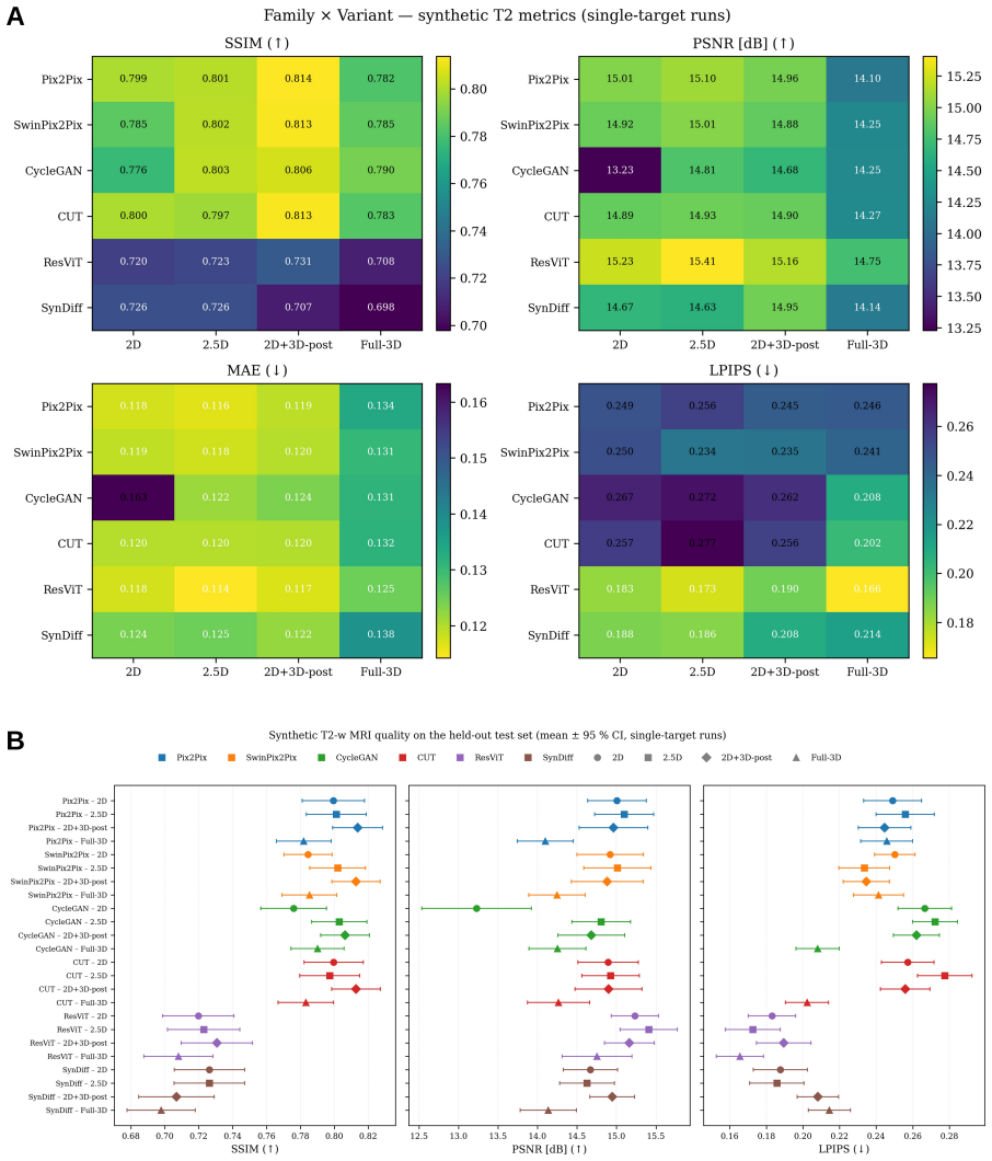
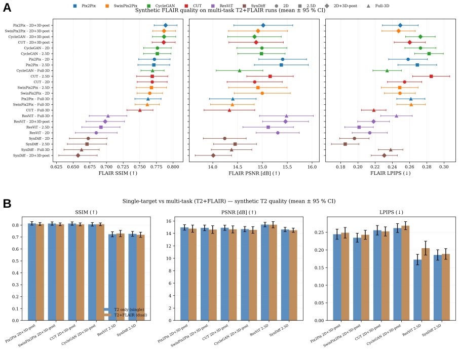
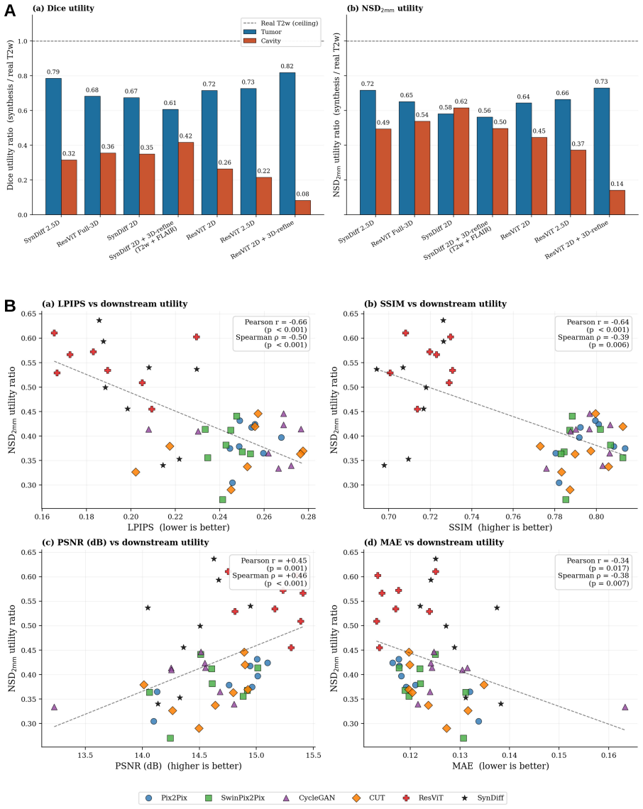
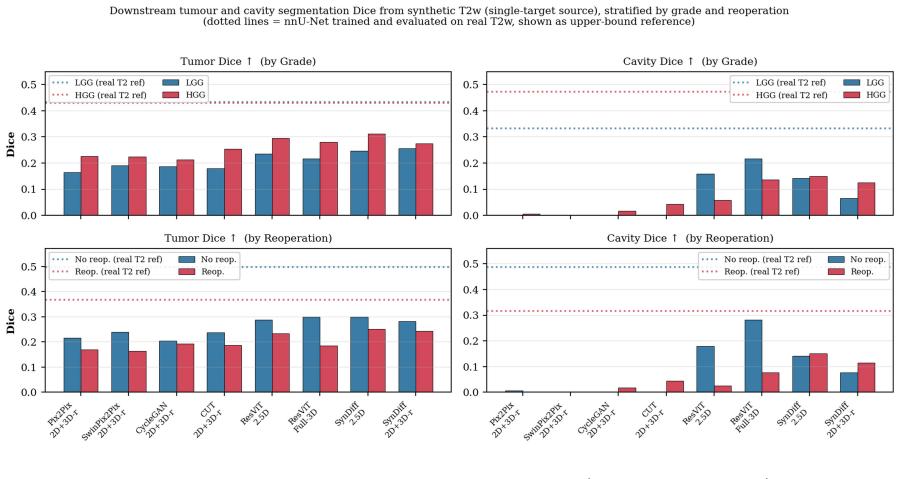
Across six generators trained under four inference regimes and two targets on 76 patients, no architecture dominated every evaluation axis; perceptual quality tracked downstream utility most closely with LPIPS showing r=-0.66 against segmentation Dice while SSIM showed r=-0.64 in the opposite direction, and SynDiff-2.5D reached the highest U_Dice of 0.55 on tumor and resection cavity segmentation.
What carries the argument
Systematic multi-regime benchmark of generators (Pix2Pix, SwinPix2Pix, CycleGAN, CUT, ResViT, SynDiff) paired with nnU-Net v2 segmentation as the downstream utility measure on paired ioUS/MRI data.
If this is right
- Perceptual and downstream-task metrics should be reported alongside or instead of global SSIM for synthesis evaluation.
- Architecture selection for synthesis should be conditioned on surgical phase, patient history, and specific clinical objective.
- The 2.5D regime with SynDiff preserves segmentation utility better than the other tested combinations.
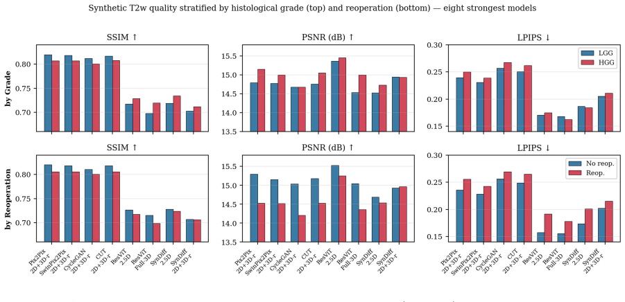
- Subgroup performance by histological grade and reoperation status provides guidance for targeted deployment.
Where Pith is reading between the lines
- Synthesis models could be trained with explicit perceptual losses to improve downstream clinical utility rather than optimizing pixel-wise or structural metrics alone.
- The negative SSIM-utility link suggests that high-SSIM outputs may be overly smoothed and lose the fine details needed for accurate tumor boundary segmentation.
- The same multi-axis protocol could be applied to other intraoperative-to-preoperative translation tasks to check whether perceptual metrics reliably predict task performance beyond this dataset.
Load-bearing premise
The 60/16 patient-level split on the ReMIND dataset represents real-world variability in histological grade and reoperation cases, and nnU-Net v2 segmentation performance serves as a valid proxy for clinical utility.
What would settle it
A follow-up experiment on a larger held-out cohort where models with the highest SSIM scores produce the highest downstream Dice scores would falsify the reported negative correlation between SSIM and utility.
Figures
read the original abstract
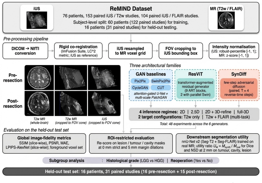
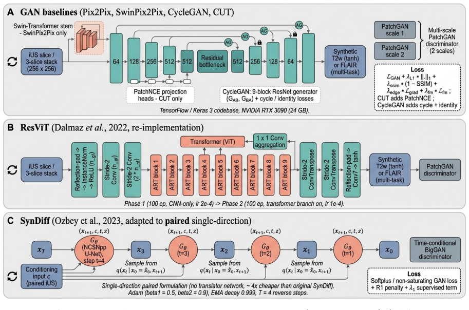
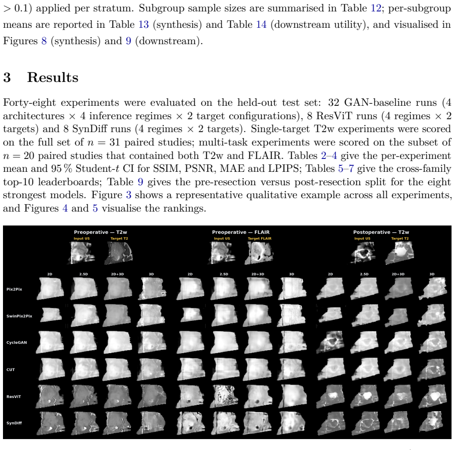
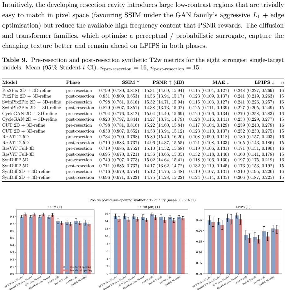
Intraoperative ultrasound (ioUS) is a versatile, cost-effective modality in brain tumour surgery, but its interpretation is difficult: acquisition planes are non-standard, artefacts are modality-specific, and its appearance differs markedly from the preoperative MRI on which surgical-planning tools, segmentation models and the surgeon's experience rely. Synthesising MRI-like images from ioUS could let this MRI-based infrastructure be reused intraoperatively without an extra scan. Most prior work evaluates a single architecture in isolation; to our knowledge, no benchmark has spanned architectural paradigms, inference regimes and downstream-task endpoints under a common protocol. We address this gap on the public ReMIND data set (76 patients; 153 paired ioUS/T2w and 104 paired ioUS/FLAIR studies; 60/16 patient-level train/held-out split). Six generators (four GAN baselines: Pix2Pix, SwinPix2Pix, CycleGAN, CUT; the transformer-augmented ResViT; and the few-step diffusion model SynDiff) were each trained under four inference regimes (2D, 2.5D, 2D + 3D-refinement, full-3D) and two targets (T2w only; T2w + FLAIR multi-task), yielding 48 experiments. Image-fidelity metrics (SSIM, PSNR, MAE, LPIPS) were complemented by an nnU-Net v2 downstream segmentation evaluation (tumour and resection cavity) and by subgroup analyses by histological grade and reoperation. No architecture dominated every axis, and, critically, perceptual quality tracked downstream utility most closely (LPIPS, r=-0.66, p<0.001), whereas higher SSIM was associated with worse utility (r=-0.64, p<0.001); SynDiff-2.5D best preserved downstream segmentation (U_Dice=0.55). Perceptual and downstream-task metrics should therefore be reported alongside or in preference to global SSIM, and architecture choice conditioned on surgical phase, patient history and clinical objective.
Editorial analysis
A structured set of objections, weighed in public.
Referee Report
Summary. The paper presents a systematic benchmark of six generators (Pix2Pix, SwinPix2Pix, CycleGAN, CUT, ResViT, SynDiff) for intraoperative ultrasound-to-T2w/FLAIR MRI synthesis on the public ReMIND dataset (76 patients, 60/16 patient-level split). It evaluates 48 configurations across 2D/2.5D/3D regimes and single/multi-task targets using image metrics (SSIM, PSNR, MAE, LPIPS) plus nnU-Net v2 downstream segmentation (tumour/resection cavity Dice) and reports that no architecture dominates all axes, LPIPS correlates most strongly with utility (r=-0.66), SSIM correlates negatively (r=-0.64), and SynDiff-2.5D achieves the highest U_Dice=0.55.
Significance. If the downstream correlations hold, the work provides actionable guidance that perceptual metrics should be prioritised over global SSIM for synthesis tasks whose value is measured by reuse of MRI-based tools in surgery. The scale (48 experiments, multiple paradigms, public data, subgroup analyses) and explicit comparison of fidelity versus task metrics are strengths that could influence evaluation protocols in medical image translation.
major comments (1)
- [Abstract] Abstract: the headline correlations (LPIPS r=-0.66, SSIM r=-0.64 with U_Dice) and the recommendation to prefer perceptual metrics rest on nnU-Net v2 segmentation Dice being a faithful proxy for intraoperative clinical utility; no evidence or discussion is supplied that this auto-segmentation task captures surgeon-relevant factors such as artefact interpretation, non-standard plane navigation or real-time resection guidance.
minor comments (1)
- [Abstract] Abstract and Methods: the 60/16 patient-level split should include explicit discussion of whether it captures histological-grade and reoperation variability; the current description leaves open whether the held-out set is representative for the claimed generalisability.
Simulated Author's Rebuttal
We thank the referee for the constructive comment on the abstract. We respond point-by-point below.
read point-by-point responses
-
Referee: [Abstract] Abstract: the headline correlations (LPIPS r=-0.66, SSIM r=-0.64 with U_Dice) and the recommendation to prefer perceptual metrics rest on nnU-Net v2 segmentation Dice being a faithful proxy for intraoperative clinical utility; no evidence or discussion is supplied that this auto-segmentation task captures surgeon-relevant factors such as artefact interpretation, non-standard plane navigation or real-time resection guidance.
Authors: We agree that nnU-Net v2 Dice is used as a proxy for utility and that the manuscript does not supply direct evidence linking it to every surgeon-relevant factor. The endpoint was selected because tumour and resection-cavity segmentation quantifies preservation of the anatomical information required to reuse MRI-based planning tools—the central motivation for ioUS-to-MRI synthesis. The reported correlations (LPIPS r=-0.66, SSIM r=-0.64) therefore demonstrate that perceptual metrics better predict performance on this specific task. We will revise the abstract and add an explicit limitations paragraph in the discussion stating that the proxy does not capture artefact interpretation, non-standard navigation or real-time guidance, and that surgeon-in-the-loop validation remains necessary. This is a partial revision; the experimental design and quantitative findings are unchanged. revision: partial
Circularity Check
No circularity: empirical benchmark on held-out data
full rationale
The paper conducts a systematic benchmark by training six generators under multiple regimes on a 60-patient training split of the public ReMIND dataset and evaluating image metrics plus nnU-Net v2 downstream segmentation on a 16-patient held-out set. All reported findings (correlations between LPIPS/SSIM and U_Dice, architecture rankings) are direct statistical summaries of these independent test-set measurements. No equations, fitted parameters renamed as predictions, self-citations, or uniqueness theorems appear in the derivation chain; the work contains no first-principles derivations that could reduce to their inputs by construction.
Axiom & Free-Parameter Ledger
free parameters (1)
- Model-specific training hyperparameters for the six generators
axioms (1)
- domain assumption The ReMIND dataset with its 60/16 patient split provides a representative and unbiased test of generalization for brain tumor surgery cases.
Forward citations
Cited by 1 Pith paper
-
What neurosurgeons need to see: synthetic intra-operative MRI from ultrasound for brain-shift compensation in brain tumour surgery
End-to-end pipeline uses ResViT-2.5D to synthesize post-resection MRI from ioUS then anchors deformable registration, yielding 5.86 mm TRE on 14 ReMIND subjects while producing an integrated whole-brain volume reflect...
Reference graph
Works this paper leans on
-
[1]
Multitask weakly supervised generative network for MR-US registration
Azampour, M.F., Mach, K., Fatemizadeh, E., Demiray, B., Westenfelder, K., Steiger, K., Eiber, M., Wendler, T., Kainz, B., Navab, N., 2024. Multitask weakly supervised generative network for MR-US registration. IEEE Transactions on Medical Imaging 43, 3780–3793. https://doi.org/10.1109/TMI.2024.3400899
-
[2]
DiffUS: differentiable ultrasound rendering from volumetric imaging
Bertramo, N., Duguey, G., Gopalakrishnan, V., 2025. DiffUS: differentiable ultrasound rendering from volumetric imaging. arXiv:2508.06768
-
[3]
ResViT: residual vision transformers for multimodal medical image synthesis
Dalmaz, O., Yurt, M., Çukur, T., 2022. ResViT: residual vision transformers for multimodal medical image synthesis. IEEE Transactions on Medical Imaging 41, 2598–2614.https: //doi.org/10.1109/TMI.2022.3167808
-
[4]
Unified brain MR-ultrasound synthesis using multi-modal hierarchical representations
Dorent, R., Haouchine, N., Kögl, F., Joutard, S., Juvekar, P., Torio, E., Golby, A., Ourselin, S., Frisken, S., Vercauteren, T., Kapur, T., Wells, W.M., 2023. Unified brain MR-ultrasound synthesis using multi-modal hierarchical representations. In: Medical Image Computing and Computer-Assisted Intervention – MICCAI 2023, LNCS 14229. Springer, pp. 448–458....
-
[5]
Patient-specific real-time segmentation in trackerless brain ultrasound
Dorent, R., Torio, E., Haouchine, N., Galvin, C., Frisken, S., Golby, A., Kapur, T., Wells, W., 2024. Patient-specific real-time segmentation in trackerless brain ultrasound. In: Medical Image Computing and Computer-Assisted Intervention – MICCAI 2024, LNCS 15006. Springer, pp. 477–487.https://doi.org/10.1007/978-3-031-72089-5_45
-
[6]
The Brain Resection Multimodal Image Registration (ReMIND2Reg) 2025 challenge
Dorent, R., Rigolo, L., Galvin, C.P., Chen, J., Heinrich, M.P., Carass, A., Colliot, O., Wassermann, D., Golby, A., Kapur, T., Wells, W., 2025. The Brain Resection Multimodal Image Registration (ReMIND2Reg) 2025 challenge. arXiv:2508.09649
-
[7]
Unified cross-modal medical image synthesis with hierarchical mixture of product-of-experts
Dorent, R., Haouchine, N., Golby, A., Frisken, S., Kapur, T., Wells, W., 2026. Unified cross-modal medical image synthesis with hierarchical mixture of product-of-experts. IEEE Transactions on Pattern Analysis and Machine Intelligence 48, 1641–1656.https://doi. org/10.1109/TPAMI.2025.3616632
-
[8]
Automatic ultrasound-MRI registration for neurosurgery using the 2D and 3D LC2 metric
Fuerst, B., Wein, W., Müller, M., Navab, N., 2014. Automatic ultrasound-MRI registration for neurosurgery using the 2D and 3D LC2 metric. Medical Image Analysis 18, 1312–1319. https://doi.org/10.1016/j.media.2014.04.008
-
[9]
Learn2Reg 2024: new benchmark datasets driving progress on new challenges
Hansen, L., Heyer, W., Großbröhmer, C., et al., 2025. Learn2Reg 2024: new benchmark datasets driving progress on new challenges. Journal of Machine Learning for Biomedical Imaging (MELBA) 2025:034. arXiv:2509.01217
-
[10]
MIND: modality independent neighbourhood descriptor for multi-modal de- formable registration
Heinrich, M.P., Jenkinson, M., Bhushan, M., Matin, T., Gleeson, F.V., Brady, M., Schnabel, J.A., 2012. MIND: modality independent neighbourhood descriptor for multi-modal de- formable registration. Medical Image Analysis 16, 1423–1435.https://doi.org/10.1016/ j.media.2012.05.008
2012
-
[11]
To- 36 wards realtime multimodal fusion for image-guided interventions using self-similarities
Heinrich, M.P., Jenkinson, M., Papież, B.W., Brady, S.M., Schnabel, J.A., 2013. To- 36 wards realtime multimodal fusion for image-guided interventions using self-similarities. In: MICCAI 2013, LNCS 8149. Springer, pp. 187–194. https://doi.org/10.1007/ 978-3-642-40811-3_24
2013
-
[12]
Maximizing safe resection of low- and high- grade glioma
Hervey-Jumper, S.L., Berger, M.S., 2016. Maximizing safe resection of low- and high- grade glioma. Journal of Neuro-Oncology 130, 269–282. https://doi.org/10.1007/ s11060-016-2110-4
2016
-
[13]
Synth- Morph: learning contrast-invariant registration without acquired images
Hoffmann, M., Billot, B., Greve, D.N., Iglesias, J.E., Fischl, B., Dalca, A.V., 2022. Synth- Morph: learning contrast-invariant registration without acquired images. IEEE Transactions on Medical Imaging 41, 543–558.https://doi.org/10.1109/TMI.2021.3116879
-
[14]
Nat Methods18(2), 203–211 (Feb 2021)
Isensee, F., Jaeger, P.F., Kohl, S.A.A., Petersen, J., Maier-Hein, K.H., 2021. nnU-Net: a self-configuring method for deep learning-based biomedical image segmentation. Nature Methods 18, 203–211.https://doi.org/10.1038/s41592-020-01008-z
-
[15]
Isola, P., Zhu, J.Y., Zhou, T., Efros, A.A., 2017. Image-to-image translation with conditional adversarial networks. In: IEEE Conference on Computer Vision and Pattern Recognition (CVPR). pp. 1125–1134.https://doi.org/10.1109/CVPR.2017.632
-
[16]
Cross-modal conditional latent diffusion model for brain MRI to ultrasound image translation
Jiang, S., Wang, L., Li, Y., Yang, Z., Zhou, Z., Li, B., 2025. Cross-modal conditional latent diffusion model for brain MRI to ultrasound image translation. Physics in Medicine & Biology 70, 155005.https://doi.org/10.1088/1361-6560/adf0bc
-
[17]
Anatomy-aware self-supervised fetal MRI synthesis from unpaired ultrasound images
Jiao, J., Namburete, A.I.L., Papageorghiou, A.T., Noble, J.A., 2019. Anatomy-aware self-supervised fetal MRI synthesis from unpaired ultrasound images. In: Machine Learning in Medical Imaging (MLMI 2019), LNCS 11861. Springer, pp. 178–186.https://doi.org/ 10.1007/978-3-030-32692-0_21
-
[18]
Self-supervised ultrasound to MRI fetal brain image synthesis
Jiao, J., Namburete, A.I.L., Papageorghiou, A.T., Noble, J.A., 2020. Self-supervised ultrasound to MRI fetal brain image synthesis. IEEE Transactions on Medical Imaging 39, 4413–4424.https://doi.org/10.1109/TMI.2020.3018560
-
[19]
ReMIND: the brain resection multimodal imaging database
Juvekar, P., Dorent, R., Kögl, F., Torio, E., Barr, C., Rigolo, L., Galvin, C., Jowkar, N., Kazi, A., Haouchine, N., Cheema, H., Navab, N., Pieper, S., Wells, W.M., Bi, W.L., Golby, A., Frisken, S., Kapur, T., 2024. ReMIND: the brain resection multimodal imaging database. Scientific Data 11, 494.https://doi.org/10.1038/s41597-024-03295-z
-
[20]
Multi-task learning using uncertainty to weigh losses for scene geometry and semantics
Kendall, A., Gal, Y., Cipolla, R., 2018. Multi-task learning using uncertainty to weigh losses for scene geometry and semantics. In: IEEE Conference on Computer Vision and Pattern Recognition (CVPR). pp. 7482–7491.https://doi.org/10.1109/CVPR.2018.00781
-
[21]
Registration of 3D fetal neurosonography and MRI
Kuklisova-Murgasova, M., Cifor, A., Napolitano, R., Papageorghiou, A., Quaghebeur, G., Rutherford, M.A., Hajnal, J.V., Noble, J.A., Schnabel, J.A., 2013. Registration of 3D fetal neurosonography and MRI. Medical Image Analysis 17, 1137–1150. https: //doi.org/10.1016/j.media.2013.07.004
-
[22]
Lasala, A., Fiorentino, M.C., Bandini, A., Moccia, S., Giannarou, S., 2026. Two-step latent 37 diffusion modelling for morphology-guided synthesis of glioma intraoperative ultrasound images. Biomedical Signal Processing and Control 120, 110037. https://doi.org/10. 1016/j.bspc.2026.110037
-
[23]
Li, Y., Jiang, S., Yang, Z., Wang, L., Wang, S., Zhou, Z., 2026. Brain tumor segmentation via cross-modality semi-supervised transfer learning with 3D MRI diffusion model synthetic ultrasound. Information Fusion 127, 103757.https://doi.org/10.1016/j.inffus.2025. 103757
-
[24]
Machado, I., Toews, M., George, E., Unadkat, P., Essayed, W., Luo, J., Teodoro, P., Carvalho, H., Martins, J., Golland, P., Pieper, S., Frisken, S., Golby, A., Wells, W., Ou, Y.,
-
[25]
NeuroImage 202, 116094.https://doi.org/10.1016/j.neuroimage.2019.116094
Deformable MRI-ultrasound registration using correlation-based attribute matching for brain shift correction: accuracy and generality in multi-site data. NeuroImage 202, 116094.https://doi.org/10.1016/j.neuroimage.2019.116094
-
[26]
Metrics Reloaded: recommendations for image analysis validation
Maier-Hein, L., Reinke, A., Godau, P., et al., 2024. Metrics Reloaded: recommendations for image analysis validation. Nature Methods 21, 195–212.https://doi.org/10.1038/ s41592-023-02151-z
2024
-
[27]
Least squares generative adversarial networks
Mao, X., Li, Q., Xie, H., Lau, R.Y.K., Wang, Z., Smolley, S.P., 2017. Least squares generative adversarial networks. In: IEEE International Conference on Computer Vision (ICCV). pp. 2794–2802.https://doi.org/10.1109/ICCV.2017.304
-
[28]
Online database of clinical MR and ultrasound images of brain tumors
Mercier, L., Del Maestro, R.F., Petrecca, K., Araujo, D., Haegelen, C., Collins, D.L., 2012. Online database of clinical MR and ultrasound images of brain tumors. Medical Physics 39, 3253–3261.https://doi.org/10.1118/1.4709600
-
[29]
Fast free-form deformation using graphics processing units
Modat, M., Ridgway, G.R., Taylor, Z.A., Lehmann, M., Barnes, J., Hawkes, D.J., Fox, N.C., Ourselin, S., 2010. Fast free-form deformation using graphics processing units. Computer Methods and Programs in Biomedicine 98, 278–284.https://doi.org/10.1016/j.cmpb. 2009.09.002
-
[30]
A 3D cross-modal keypoint descriptor for MR-US matching and registration
Morozov, D., Dorent, R., Haouchine, N., 2025. A 3D cross-modal keypoint descriptor for MR-US matching and registration. arXiv:2507.18551
-
[31]
Clinically applicable segmentation of head and neck anatomy for radiotherapy: deep learning algorithm development and validation study
Nikolov, S., Blackwell, S., Zverovitch, A., et al., 2021. Clinically applicable segmentation of head and neck anatomy for radiotherapy: deep learning algorithm development and validation study. Journal of Medical Internet Research 23, e26151.https://doi.org/10. 2196/26151
2021
-
[32]
Özbey, M., Dalmaz, O., Dar, S.U.H., Bedel, H.A., Öztürk, Ş., Güngör, A., Çukur, T.,
-
[33]
IEEE Transactions on Medical Imaging 42, 3524–3539.https://doi.org/10.1109/TMI.2023
Unsupervised medical image translation with adversarial diffusion models. IEEE Transactions on Medical Imaging 42, 3524–3539.https://doi.org/10.1109/TMI.2023. 3290149
-
[34]
Contrastive learning for unpaired 38 image-to-image translation
Park, T., Efros, A.A., Zhang, R., Zhu, J.Y., 2020. Contrastive learning for unpaired 38 image-to-image translation. In: European Conference on Computer Vision (ECCV), LNCS 12354. Springer, pp. 319–345.https://doi.org/10.1007/978-3-030-58545-7_19
-
[35]
Rahmani, M., Moghaddasi, H., Pour-Rashidi, A., Ahmadian, A., Najafzadeh, E., Farnia, P.,
-
[36]
D2BGAN: dual discriminator Bayesian generative adversarial network for deformable MR-ultrasound registration applied to brain shift compensation. Diagnostics 14, 1319. https://doi.org/10.3390/diagnostics14131319
-
[37]
Rai, A., Singh, V., Shetty, P., Moiyadi, A.V., 2025. Brainshift correction using navigated intraoperative ultrasound informs intraoperative decision-making during glioma surgery. Acta Neurochirurgica 167, 124.https://doi.org/10.1007/s00701-025-06457-z
-
[38]
Learning to match 2D keypoints across preoperative MR and intraoperative ultrasound
Rasheed, H., Dorent, R., Fehrentz, M., Kapur, T., Wells, W.M., Golby, A., Frisken, S., Navab, N., Haouchine, N., 2024. Learning to match 2D keypoints across preoperative MR and intraoperative ultrasound. In: Simplifying Medical Ultrasound (ASMUS 2024, MICCAI Workshop), LNCS 15186. Springer, pp. 78–87. https://doi.org/10.1007/ 978-3-031-73647-6_8
2024
-
[39]
Influence of high-performance image-to-image translation networks on clinical visual assessment and outcome prediction: utilizing ultrasound to MRI translation in prostate cancer
Salmanpour, M.R., Mousavi, A., Xu, Y., Weeks, W.B., Hacihaliloglu, I., 2026. Influence of high-performance image-to-image translation networks on clinical visual assessment and outcome prediction: utilizing ultrasound to MRI translation in prostate cancer. International Journal of Computer Assisted Radiology and Surgery 21, 125–135.https://doi.org/10. 100...
2026
-
[40]
Glioma extent of resection and its impact on patient outcome
Sanai, N., Berger, M.S., 2008. Glioma extent of resection and its impact on patient outcome. Neurosurgery 62, 753–764.https://doi.org/10.1227/01.neu.0000318159.21731.cf
-
[41]
Shetty, P., Yeole, U., Singh, V., Moiyadi, A., 2021. Navigated ultrasound-based image guidance during resection of gliomas: practical utility in intraoperative decision-making and outcomes. Neurosurgical Focus 50, E14.https://doi.org/10.3171/2020.10.FOCUS20550
-
[42]
ConvexAdam: self- configuring dual-optimization-based 3D multitask medical image registration
Siebert, H., Großbröhmer, C., Hansen, L., Heinrich, M.P., 2025. ConvexAdam: self- configuring dual-optimization-based 3D multitask medical image registration. IEEE Trans- actions on Medical Imaging 44, 738–748.https://doi.org/10.1109/TMI.2024.3462248
-
[43]
Translation of fetal brain ultrasound images into pseudo-MRI images using artificial intelligence
Silverstein, N., Beloosesky, R., Leibowitz, E., Azhari, H., 2025. Translation of fetal brain ultrasound images into pseudo-MRI images using artificial intelligence. arXiv:2504.02408
-
[44]
BrainVoxGen: deep learning framework for synthesis of ultrasound to MRI
Singh, S., Bewoor, M., Ranapurwala, A., Rai, S., Patil, S., 2023. BrainVoxGen: deep learning framework for synthesis of ultrasound to MRI. arXiv:2310.08608
-
[45]
Unsgård, G., Selbekk, T., Brostrup Müller, T., Ommedal, S., Torp, S.H., Myhr, G., Bang, J., Nagelhus Hernes, T.A., 2005. Ability of navigated 3D ultrasound to delineate gliomas and metastases: comparison of image interpretations with histopathology. Acta Neurochirurgica 147, 1259–1269.https://doi.org/10.1007/s00701-005-0624-1
-
[46]
Enhancing pix2pix with Swin 39 Transformer for cross-modal brain CT-MR synthesis
Verdicchio, M., Isgrò, F., Salvatore, M., Aiello, M., 2025. Enhancing pix2pix with Swin 39 Transformer for cross-modal brain CT-MR synthesis. Research Square preprint rs.3.rs- 7565545/v1.https://doi.org/10.21203/rs.3.rs-7565545/v1
-
[47]
IEEE Transactions on Image Processing 13(4), 600–612 (Apr 2004)
Wang, Z., Bovik, A.C., Sheikh, H.R., Simoncelli, E.P., 2004. Image quality assessment: from error visibility to structural similarity. IEEE Transactions on Image Processing 13, 600–612.https://doi.org/10.1109/TIP.2003.819861
-
[48]
High-resolution image synthesis and semantic manipulation with conditional GANs
Wang, T.C., Liu, M.Y., Zhu, J.Y., Tao, A., Kautz, J., Catanzaro, B., 2018. High-resolution image synthesis and semantic manipulation with conditional GANs. In: IEEE Conference on Computer Vision and Pattern Recognition (CVPR). pp. 8798–8807.https://doi.org/ 10.1109/CVPR.2018.00917
-
[49]
Wang, J., Chen, X., Zhang, Y., Liu, M., Zhang, H., 2024. Unsupervised multimodal 3D med- ical image registration with multilevel correlation balanced optimization. arXiv:2409.05040. Learn2Reg 2024 challenge submission
-
[50]
Coarse-to-fine joint registration of MR and ultrasound images via imaging style transfer
Wang, J., Zhang, Y., Liu, M., Chen, X., Wang, Y., Zhang, H., 2025. Coarse-to-fine joint registration of MR and ultrasound images via imaging style transfer. arXiv:2508.05240. ReMIND2Reg 2024 challenge submission
-
[51]
Unsupervised MR-US multimodal image registration with multilevel correlation pyramidal optimization
Wang, J., Liu, Z., Liu, M., Chen, X., Yu, X., Wang, Y., Zhang, H., 2026. Unsupervised MR-US multimodal image registration with multilevel correlation pyramidal optimization. arXiv:2602.06288
-
[52]
Globalregistration of ultrasound to MRI using the LC2 metric for enabling neurosurgical guidance
Wein, W., Ladikos, A., Fuerst, B., Shah, A., Sharma, K., Navab, N., 2013. Globalregistration of ultrasound to MRI using the LC2 metric for enabling neurosurgical guidance. In: MICCAI 2013, LNCS 7908. Springer, pp. 34–41.https://doi.org/10.1007/978-3-642-40811-3_ 5
-
[53]
Xiao, Y., Fortin, M., Unsgård, G., Rivaz, H., Reinertsen, I., 2017. REtroSpective Evaluation of Cerebral Tumors (RESECT): a clinical database of pre-operative MRI and intra-operative ultrasound in low-grade glioma surgeries. Medical Physics 44, 3875–3882.https://doi. org/10.1118/1.4986620
-
[54]
Xiao, Y., Rivaz, H., Chabanas, M., Fortin, M., Machado, I., Ou, Y., Heinrich, M.P., Schnabel, J.A., Zhong, X., Maier, A., Wein, W., Shams, R., Kadoury, S., Drobny, D., Modat, M., Reinertsen, I., 2020. Evaluation of MRI to ultrasound registration methods for brain shift correction: the CuRIOUS2018 challenge. IEEE Transactions on Medical Imaging 39, 777–786...
-
[55]
Tackling the generative learning trilemma with denoising diffusion GANs
Xiao, Z., Kreis, K., Vahdat, A., 2022. Tackling the generative learning trilemma with denoising diffusion GANs. In: International Conference on Learning Representations (ICLR). arXiv:2112.07804
-
[56]
Towards automated correction of brain shift using deep deformable MRI-ioUS 40 registration
Zeineldin, R.A., Karar, M.E., Coburger, J., Wirtz, C.R., Mathis-Ullrich, F., Burgert, O., 2020. Towards automated correction of brain shift using deep deformable MRI-ioUS 40 registration. Current Directions in Biomedical Engineering 6, 20200039.https://doi.org/ 10.1515/cdbme-2020-0039
-
[57]
Deep Residual Learning for Image Recognition
Zhang, R., Isola, P., Efros, A.A., Shechtman, E., Wang, O., 2018. The unreasonable effectiveness of deep features as a perceptual metric. In: IEEE Conference on Computer Vision and Pattern Recognition (CVPR). pp. 586–595.https://doi.org/10.1109/CVPR. 2018.00068
-
[58]
Unpaired image-to-image translation using cycle-consistent adversarial networks
Zhu, J.Y., Park, T., Isola, P., Efros, A.A., 2017. Unpaired image-to-image translation using cycle-consistent adversarial networks. In: IEEE International Conference on Computer Vision (ICCV). pp. 2223–2232.https://doi.org/10.1109/ICCV.2017.244. 41
discussion (0)
Sign in with ORCID, Apple, or X to comment. Anyone can read and Pith papers without signing in.