REVIEW 4 major objections 6 minor 45 references
Deep Learning Algorithms to Isolate and Quantify the Structures of the Anterior Segment in Optical Coherence Tomography Images
T0 review · 4 major / 6 minor · reviewed 2026-08-14 · deepseek-v4-flash
Pith's one-line read A deep convolutional neural network localizes the scleral spur on anterior-segment OCT images at expert level and segments anterior-chamber structures with a Dice coefficient of 95.7%.
desk verdict A useful ASOCT segmentation and scleral-spur localization pipeline whose headline accuracy is real but measured against the same lab's annotations, so it needs revision rather than rejection. read the letter →
The pith
A machine-rendered reading of the paper's core claim, the machinery that carries it, and where it could break.
The reading
What carries the argument
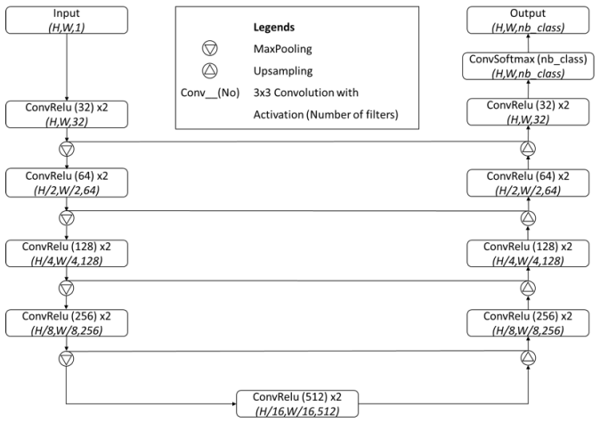
The load-bearing mechanism is a hybrid network called FRRUnet, which combines the U-Net skip-connection encoder-decoder design (for local texture plus contextual arrangement) with the full-resolution residual pathway of the full-resolution residual network (for precise boundary localization). For landmark detection the paper turns localization into a three-class segmentation problem that predicts a focus region (a small square centered on the scleral spur), an attention region, and background; the spur is taken as the focus-region center, which avoids coordinate regression's need for many training examples. For tissue segmentation, predictions from the three base networks are blended by a tiny ensemble network with frozen base weights. A two-step quality check then filters scans whose spur confidence (intersection over union at least 0.80) or contour structure indicates poor image quality, so that only reliable parameter measurements are reported.
What would settle it
One test: gather a new ASOCT dataset in which multiple fellowship-trained ophthalmologists independently mark the scleral spur and segment the tissues on the same scans. If the machine's spur locations and Dice scores against the pooled expert consensus fall outside the human-human agreement range, or if the machine systematically tracks one annotator's bias rather than the consensus, the claim of expert-level accuracy fails. A second, harder test would compare spur locations implied by the OCT images against the same eyes' gonioscopic or histologic angle anatomy, the clinical reference for true closure.
Extended reading notes
Core claim
The paper's central claim is that a segmentation-based deep-learning pipeline can replace the human grader for both scleral-spur localization and anterior-segment tissue segmentation in ASOCT images, despite limited training data. Rather than regressing coordinates directly, the network is trained to label a small focus region around the spur together with an attention region and background; this segmentation formulation is what lets a modest dataset reach expert-level accuracy. On unseen images the machine's spur placement agreed with a fellowship-trained glaucoma expert at intraclass correlation coefficients of 0.984 (X) and 0.993 (Y), and in parameter measurements the machine's ACW, TISA, and AOD values agreed with experts to a similar degree as experts agreed with each other, while the machine was more repeatable across repeat runs than human graders. The same pipeline segments the anterior-chamber structures at a mean Dice of 95.7%, then computes eight clinical parameters and passes or rejects each scan based on an automated confidence and contour-count quality check.
Load-bearing premise
The load-bearing assumption is that the human-annotated scleral spur and the single-annotator tissue labels used as training targets are reliable enough proxies for the true anatomy, since the paper concedes there is no absolute ground truth for labeling; if those labels carry systematic bias or error, the network simply reproduces that bias and "as accurate as an ophthalmologist" does not mean anatomically correct.
Editorial extensions
If this is right
- Because the spur and segmentations are produced automatically for every scan, all 128 radial sections of a swept-source ASOCT volume can be quantified, yielding a 360-degree map of angle metrics such as AOD and TISA instead of a few hand-picked sections.
- The machine's spur localization agreed with human experts about as well as experts agreed with each other, while being more consistent across repeat runs, so automated measurements should improve reproducibility of angle-closure assessment.
- The built-in quality check rejects scans with low spur confidence or implausible contour counts before parameters are reported, which is necessary for deploying automated quantification in a screening workflow.
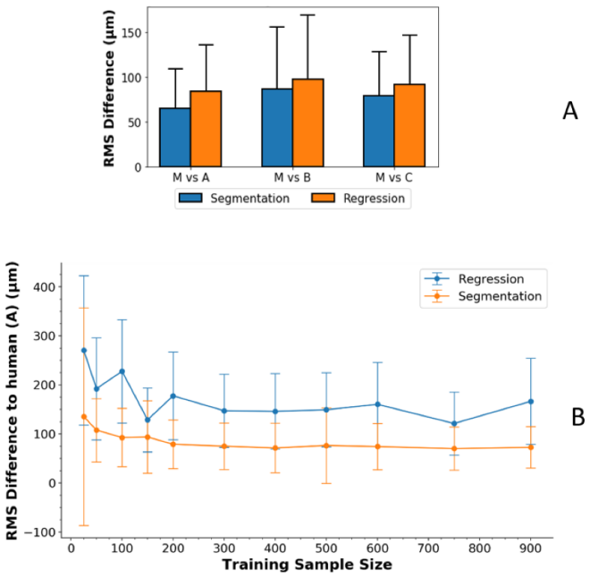
- The segmentation-based formulation reaches expert-level spur detection with roughly 100 images, so the approach can be adapted to other anterior-segment landmarks without a large annotation campaign.
Reading between the lines
- An extension the paper leaves implicit: averaging labels from several ophthalmologists during training should shrink the inherited human bias the authors discuss, and could make the machine's spur location closer to a consensus ground truth than any single expert.
- The same "detect a landmark by segmenting a small region around it" trick may transfer to other small anatomical landmarks in OCT, ultrasound, or X-ray where coordinate regression needs excessive training data.
- A testable consequence: retraining and testing the pipeline on scans from other devices and populations, or on eyes with more severe angle closure, would show whether the expert-level agreement and 95.7% Dice generalize beyond the single hospital cohort used here.
Signed reviews
Editorial analysis
A structured set of objections, weighed in public.
Referee Report
Summary. This manuscript presents a deep-learning pipeline for anterior-segment optical coherence tomography (ASOCT) images. A hybrid U-Net/FRRnet architecture (FRRUnet) is used to detect the scleral spur via a segmentation approach, an ensemble of U-Net, FRRnet, and FRRUnet segments the iris, corneo-sclera shell, and anterior chamber, and the outputs are used to compute eight clinically relevant ASOCT parameters with an automated quality check. The authors report that, with limited training data, the DCNN localizes the scleral spur on unseen ASOCT images as accurately as an experienced ophthalmologist and achieves a mean Dice coefficient of 95.7% for tissue segmentation. Validation includes inter- and intra-observer comparisons using ICC and Bland-Altman plots, training-size experiments, and parameter-level ICCs.
Significance. The clinical motivation is strong: objective, automated 360-degree quantification of ASOCT images could support angle-closure glaucoma screening and monitoring. The paper's strengths are the patient-level split between training and test sets, the explicit comparison of machine-human agreement with human-human agreement, the training-size ablation, and the use of Bland-Altman and ICC for reproducibility. However, the headline claims are about accuracy, whereas the study design can only establish agreement with human annotators, and the segmentation Dice is computed against a single annotator. The contribution is therefore better framed as a reproducible automated measurement pipeline whose accuracy relative to anatomical truth remains to be established. The reported specificity formula is also incorrect, and the intra-observer comparison for the machine is not apples-to-apples with the human intra-observer comparison.
major comments (4)
- [§3.2, Specificity/Sensitivity definitions] The specificity and sensitivity are defined by identical formulas: Specificity = |D∩M|/|M| and Sensitivity = |D∩M|/|M|. The correct specificity is the true-negative rate, e.g., |D^c ∩ M^c|/|M^c|. Since the Discussion explicitly claims 'high sensitivity and specificity', the reported specificity values in Figure 11 cannot be interpreted. Please correct the formulas and verify the reported numbers.
- [§4 and §2.2] The limitations paragraph concedes 'the lack of an absolute ground truth in labelling' and, for ASOCT segmentation, 'we only had one trainer and observer.' Yet the abstract states that the DCNN detects the scleral spur 'as accurately as an experienced ophthalmologist' and reports a 95.7% Dice coefficient. These claims are not supported: for SSL, the machine was trained on labels from observers A and B and then compared with those same observers plus observer C, so the comparison measures agreement with the training annotators, not accuracy against anatomical truth. For segmentation, the Dice score is agreement with a single annotator, not a measure of anatomical accuracy. In addition, §2.2 says manual segmentations were prepared by two trainers while §4 says there was only one trainer/observer for segmentation; this inconsistency must be resolved. Please reframe the claims as agreement/reproducibility, or provide a consensus or independent reference.
- [§3.1, Intra-observer test] The machine intra-observer test is performed by training a second model from scratch and comparing it with the first model. Since the network is deterministic after training, repeated application of the same model to the same image would yield zero intra-observer variability; comparing two separately trained models measures training stochasticity, not observer repeatability. The statement that the machine 'generally had lower variability than that of human grader' is therefore not established by this comparison. Please report repeated-inference consistency separately, or explicitly frame the second-model comparison as a training-stability analysis and avoid direct claims of lower intra-observer variability relative to humans.
- [§3.4 and §2.6] The SSL confidence threshold in the quality check is set to 0.8 after 'visually comparison of good and failed cases' on the data under study, and the test images are stated to be 'made sure to be of usable quality clinically.' This does not validate the claim that the quality check 'asserts the reliability' of the extracted parameters on poor-quality scans. Please validate the quality check on an independent set containing poor-quality images, or temper the claim accordingly.
minor comments (6)
- [§2.1] 'Nayoga, Japan' should be 'Nagoya, Japan'.
- [Table 3] The column headed 'A vs B vs C' is ambiguous; specify whether the ICC is computed across the three human graders jointly or as pairwise averages.
- [§2.2 and §4] The number of annotators for the manual segmentations is stated inconsistently; please clarify whether one or two trainers prepared the segmentation labels.
- [§3.2] In the Dice formula, consider using TP/FP/FN notation so that readers can verify the definition and compare it with the standard Dice formulation.
- [Reference [45]] The author name in reference [45] is corrupted ('zgn iek'); it should be corrected, e.g., Özgün Çiçek.
- [§3.1] Please clarify whether the two halves of a divided image can appear in both the training and testing sets; the current text only states that images were divided in half.
Circularity Check
One self-referential quality-check metric; the central SSL and segmentation claims rest on independent human benchmarks and are not circular.
-
self definitional
[Section 2.6, Quality check]
"First, upon the detection of the SSL a square region surrounding the center of the predicted region was obtained as the reference. A confidence index was computed as the intersection over union (IoU; between 0-1) between the predicted and reference regions. Scans that yielded a confidence index greater than or equal to 0.80 were considered good, while lower values were designated as poor quality."
The 'reference' for the IoU is not an independent SSL location; it is a square drawn around the center of the network's own predicted region. Therefore the confidence index measures how much the predicted focus region overlaps a box centered on itself, i.e., a compactness/self-consistency score. A prediction that is consistently shifted from the true spur by a fixed offset would still have high IoU with its own centered box, so the quality check cannot assert the reliability of SSL-based parameters unless the prediction is already correct. This validates a prediction against itself by construction, rather than against any external ground truth.
full rationale
The central performance claims—scleral spur localization 'as accurately as an experienced ophthalmologist' and segmentation with a Dice coefficient of 95.7%—are evaluated against human annotations on unseen test images, not against the training labels. The network is not fitted to the test labels, and the inclusion of observer C, a fellowship-trained glaucoma expert who was not a trainer, provides independent human comparison for the SSL claim. The paper also reports human-human ICC values, so the machine-human agreement is benchmarked against human reproducibility. The acknowledged overlap of observers A and B as both trainers and evaluators is a limitation of label quality and independence, but it does not make the evaluation mathematically equivalent to the training objective; the test images were not used in training. I therefore do not treat supervised learning from human labels as circular per se. The one concrete reduction-by-construction I identified is the quality-check confidence index: its reference region is derived from the prediction's own center, so the IoU measures self-consistency rather than accuracy. That step is secondary to the headline results but is genuinely self-referential. No load-bearing self-citation chain, uniqueness import, or ansatz smuggling appears in the paper. The overall circularity score is 2: the central claims retain independent content, and only a minor auxiliary metric reduces to its own input.
Assumptions & free parameters
free parameters (2)
- SSL confidence threshold =
0.80
- Segmentation contour count thresholds =
iris <= 5, anterior chamber <= 6, corneo-sclera <= 10
assumptions (3)
- domain assumption Human scleral spur annotations on ASOCT images, made using three anatomical definitions, are a reliable proxy for the true scleral spur location.
- domain assumption Test-set ASOCT images are clinically usable and representative of the target population.
- domain assumption Manual ASOCT segmentation by one trained observer is a valid ground truth for computing Dice scores.
Cite this review
Pith. "Pith review of Deep Learning Algorithms to Isolate and Quantify the Structures of the Anterior Segment in Optical Coherence Tomography Images." pith.science (2026). https://pith.science/paper/3BXRNATT
@misc{pith2026190900331,
  author       = {Pith},
  title        = {Pith review of: Deep Learning Algorithms to Isolate and Quantify the Structures of the Anterior Segment in Optical Coherence Tomography Images},
  year         = {2026},
  howpublished = {\url{https://pith.science/paper/3BXRNATT}},
  note         = {Machine review of arXiv:1909.00331}
}
read the original abstract
Accurate isolation and quantification of intraocular dimensions in the anterior segment (AS) of the eye using optical coherence tomography (OCT) images is important in the diagnosis and treatment of many eye diseases, especially angle closure glaucoma. In this study, we developed a deep convolutional neural network (DCNN) for the localization of the scleral spur, and the segmentation of anterior segment structures (iris, corneo-sclera shell, anterior chamber). With limited training data, the DCNN was able to detect the scleral spur on unseen ASOCT images as accurately as an experienced ophthalmologist; and simultaneously isolated the anterior segment structures with a Dice coefficient of 95.7%. We then automatically extracted eight clinically relevant ASOCT parameters and proposed an automated quality check process that asserts the reliability of these parameters. When combined with an OCT machine capable of imaging multiple radial sections, the algorithms can provide a more complete objective assessment. This is an essential step toward providing a robust automated framework for reliable quantification of ASOCT scans, for applications in the diagnosis and management of angle closure glaucoma.
Figures


Figures from the paper (12 more)




Reference graph
Works this paper leans on
-
[1]
RJ Casson, HS Newland, J Muecke, S McGovern, LM Abraham, WK Shein, D Selva, and T Aung. Gonioscopy findings and prevalence of occludable angles in a burmese population: the meiktila eye study. British journal of ophthalmology , 91(7):856–859, 2007
work page 2007
-
[2]
Anterior segment imaging for angle closure
Sunee Chansangpetch, Prin Rojanapongpun, and Shan C Lin. Anterior segment imaging for angle closure. American journal of ophthalmology , 188:xvi–xxix, 2018
work page 2018
-
[3]
Jin-Wei Cheng, Ying Zong, You-Yan Zeng, and Rui-Li Wei. The prevalence of primary angle closure glaucoma in adult asians: a systematic review and meta-analysis. PloS one , 9(7):e103222, 2014
work page 2014
-
[4]
Cumba, Sunita Radhakrishnan, Nicholas P
Ricardo J. Cumba, Sunita Radhakrishnan, Nicholas P. Bell, Kundandeep S. Nagi, Alice Z. Chuang, Shan C. Lin, Kimberly A. Mankiewicz, and Robert M. Feldman. Reproducibility of scleral spur identi- fication and angle measurements using fourier domain anterior segment optical coherence tomography. Journal of Ophthalmology , 2012:1–14, 2012
work page 2012
-
[5]
Sripad Krishna Devalla, Khai Sing Chin, Jean-Martial Mari, Tin A Tun, Nicholas G Strouthidis, Tin Aung, Alexandre H Thiry, and Michal JA Girard. A deep learning approach to digitally stain optical coherence tomography images of the optic nerve head. Investigative ophthalmology & visual science , 59(1):63–74, 2018
work page 2018
-
[6]
Sripad Krishna Devalla, Prajwal K Renukanand, Bharathwaj K Sreedhar, Giridhar Subramanian, Liang Zhang, Shamira Perera, Jean-Martial Mari, Khai Sing Chin, Tin A Tun, and Nicholas G Strouthidis. Drunet: a dilated-residual u-net deep learning network to segment optic nerve head tissues in optical coherence tomography images. Biomedical optics express , 9(7)...
work page 2018
-
[7]
The impor- tance of skip connections in biomedical image segmentation , pages 179–187
Michal Drozdzal, Eugene Vorontsov, Gabriel Chartrand, Samuel Kadoury, and Chris Pal. The impor- tance of skip connections in biomedical image segmentation , pages 179–187. Springer, 2016
work page 2016
-
[8]
Yadollah Eslami, Golshan Latifi, Sasan Moghimi, Reza Ghaffari, Ghasem Fakhraie, Reza Zarei, Mah- mood Jabbarvand, Massood Mohammadi, and Shan Lin. Effect of adjunctive viscogonioplasty on drainage angle status in cataract surgery: a randomized clinical trial. Clinical & experimental ophthal- mology, 41(4):368–378, 2013
work page 2013
Show all 45 references
-
[9]
Epidemiology of angle-closure glaucoma
David S Friedman. Epidemiology of angle-closure glaucoma. J Curr Glaucoma Pract , 1(1):1–3, 2007
2007
-
[10]
Neural networks and the bias/variance dilemma
Stuart Geman, Elie Bienenstock, and Ren Doursat. Neural networks and the bias/variance dilemma. Neural Computation, 4(1):1–58, 1992
1992
-
[11]
Framing u-net via deep convolutional framelets: Application to sparse- view ct
Yoseob Han and Jong Chul Ye. Framing u-net via deep convolutional framelets: Application to sparse- view ct. IEEE transactions on medical imaging , 37(6):1418–1429, 2018
2018
-
[12]
Neural network ensembles
Lars Kai Hansen and Peter Salamon. Neural network ensembles. IEEE Transactions on Pattern Analysis & Machine Intelligence , 12(10):993–1001, 1990
1990
-
[13]
Brain tumor segmentation with deep neural networks
Mohammad Havaei, Axel Davy, David Warde-Farley, Antoine Biard, Aaron Courville, Yoshua Bengio, Chris Pal, Pierre-Marc Jodoin, and Hugo Larochelle. Brain tumor segmentation with deep neural networks. Medical image analysis , 35:18–31, 2017
2017
-
[14]
Deep residual learning for image recognition
Kaiming He, Xiangyu Zhang, Shaoqing Ren, and Jian Sun. Deep residual learning for image recognition. In Proceedings of the IEEE conference on computer vision and pattern recognition , pages 770–778, 2015. 19
2015
-
[15]
Predicting the outcome of laser peripheral iridotomy for primary angle closure suspect eyes using anterior segment optical coherence tomography
Victor Koh, Mohammad Reza Keshtkaran, David Hernstadt, Maria Cecilia D Aquino, Paul T Chew, and Chelvin Sng. Predicting the outcome of laser peripheral iridotomy for primary angle closure suspect eyes using anterior segment optical coherence tomography. Acta ophthalmologica, 9...
2019
-
[16]
H-denseunet: Hybrid densely connected unet for liver and liver tumor segmentation from ct volumes
Xiaomeng Li, Hao Chen, Xiaojuan Qi, Qi Dou, Chi-Wing Fu, and Pheng Ann Heng. H-denseunet: Hybrid densely connected unet for liver and liver tumor segmentation from ct volumes. arXiv preprint arXiv:1709.07330, 2017
2017 arXiv
-
[17]
Class imbalance problem , pages 171–171
Charles X Ling and Victor S Sheng. Class imbalance problem , pages 171–171. Springer, 2011
2011
-
[18]
Semantic segmentation of aerial images with an ensemble of cnns.ISPRS Annals of the Photogrammetry, Remote Sensing and Spatial Information Sciences , 3:473, 2016
Dimitrios Marmanis, Jan D Wegner, Silvano Galliani, Konrad Schindler, Mihai Datcu, and Uwe Stilla. Semantic segmentation of aerial images with an ensemble of cnns.ISPRS Annals of the Photogrammetry, Remote Sensing and Spatial Information Sciences , 3:473, 2016
2016
-
[19]
Anterior segment imaging in glaucoma: an updated review
Jessica S Maslin, Yaniv Barkana, and Syril K Dorairaj. Anterior segment imaging in glaucoma: an updated review. Indian journal of ophthalmology , 63(8):630, 2015
2015
-
[20]
Qualitative evalua- tion of anterior segment in angle closure disease using anterior segment optical coherence tomography
Sasan Moghimi, Rebecca Chen, Nikoo Hamzeh, Nassim Khatibi, and Shan C Lin. Qualitative evalua- tion of anterior segment in angle closure disease using anterior segment optical coherence tomography. Journal of current ophthalmology , 28(4):170–175, 2016
2016
-
[21]
Nolan, Jovina L
Winifred P. Nolan, Jovina L. See, Paul T. K. Chew, David S. Friedman, Scott D. Smith, Sunita Rad- hakrishnan, Ce Zheng, Paul J. Foster, and Tin Aung. Detection of primary angle closure using anterior segment optical coherence tomography in asian eyes. Ophthalmology, 114(1):33–39, 2007
2007
-
[22]
Lens vault, thickness, and position in chinese subjects with angle closure
Monisha E Nongpiur, Mingguang He, Nishani Amerasinghe, David S Friedman, Wan-Ting Tay, Mani Baskaran, Scott D Smith, Tien Yin Wong, and Tin Aung. Lens vault, thickness, and position in chinese subjects with angle closure. Ophthalmology, 118(3):474–479, 2011
2011
-
[23]
Novel association of smaller anterior chamber width with angle closure in singaporeans
Monisha E Nongpiur, Lisandro M Sakata, David S Friedman, Mingguang He, Yiong-Huak Chan, Ragha- van Lavanya, Tien Yin Wong, and Tin Aung. Novel association of smaller anterior chamber width with angle closure in singaporeans. Ophthalmology, 117(10):1967–1973, 2010
1967
-
[24]
Okamoto, K
R. Okamoto, K. & Higashita. Anterior eye three-dimensional image processing apparatus and method of anterior eye three-dimensional image processing., 2017
2017
-
[25]
Natural history of glaucoma
Ying Pan and Rohit Varma. Natural history of glaucoma. Indian journal of ophthalmology , 59(Suppl1):S19, 2011
2011
-
[26]
Fullresolution residual net- works for semantic segmentation in street scenes
Tobias Pohlen, Alexander Hermans, Markus Mathias, and Bastian Leibe. Fullresolution residual net- works for semantic segmentation in street scenes. arXiv preprint, 2017
2017
-
[27]
Role of anterior segment optical coherence tomog- raphy in angleclosure disease: a review
Natalia Porporato, Mani Baskaran, and Tin Aung. Role of anterior segment optical coherence tomog- raphy in angleclosure disease: a review. Clinical & experimental ophthalmology , 46(2):147–157, 2018
2018
-
[28]
Angle imaging: advances and challenges
Desmond TL Quek, Monisha E Nongpiur, Shamira A Perera, and Tin Aung. Angle imaging: advances and challenges. Indian journal of ophthalmology , 59(Suppl1):S69, 2011
2011
-
[29]
H. A. Quigley and A. T. Broman. The number of people with glaucoma worldwide in 2010 and 2020. The British journal of ophthalmology , 90(3):262–267, 2006
2010
-
[30]
U-net: Convolutional networks for biomedical image segmentation
Olaf Ronneberger, Philipp Fischer, and Thomas Brox. U-net: Convolutional networks for biomedical image segmentation. In International Conference on Medical image computing and computer-assisted intervention, pages 234–241. Springer, 2015. 20
2015
-
[31]
Sakata, Raghavan Lavanya, David S
Lisandro M. Sakata, Raghavan Lavanya, David S. Friedman, Han T. Aung, Hong Gao, Rajesh S. Kumar, Paul J. Foster, and Tin Aung. Comparison of gonioscopy and anterior segment ocular coherence tomog- raphy in detecting angle closure in different quadrants of the anterior chamber a...
2008
-
[32]
Assessment of the scleral spur in anterior segment optical coherence tomography images
Lisandro M Sakata, Raghavan Lavanya, David S Friedman, Han T Aung, Steve K Seah, Paul J Foster, and Tin Aung. Assessment of the scleral spur in anterior segment optical coherence tomography images. Archives of Ophthalmology , 126(2):181–185, 2008
2008
-
[33]
F. E. Seager, J. Wang, K. S. Arora, and H. A. Quigley. The effect of scleral spur identification methods on structural measurements by anterior segment optical coherence tomography. J Glaucoma, 23(1):e29–38, 2014
2014
-
[34]
Management of angle closure glaucoma
Jovina LS See, Maria Cecilia D Aquino, Joel Aduan, and Paul TK Chew. Management of angle closure glaucoma. Indian journal of ophthalmology , 59(Suppl1):S82, 2011
2011
-
[35]
Over- feat: Integrated recognition, localization and detection using convolutional networks
Pierre Sermanet, David Eigen, Xiang Zhang, Michal Mathieu, Rob Fergus, and Yann LeCun. Over- feat: Integrated recognition, localization and detection using convolutional networks. arXiv preprint arXiv:1312.6229, 2013
2013 arXiv
-
[36]
Understanding machine learning: From theory to algorithms
Shai Shalev-Shwartz and Shai Ben-David. Understanding machine learning: From theory to algorithms . Cambridge university press, 2014
2014
-
[37]
Associations of iris structural mea- surements in a chinese population: the singapore chinese eye study
Chelvin C Sng, John C Allen, Monisha E Nongpiur, Li-Lian Foo, Yingfeng Zheng, Carol Y Cheung, Mingguang He, David S Friedman, Tien Y Wong, and Tin Aung. Associations of iris structural mea- surements in a chinese population: the singapore chinese eye study. Investigative ophth...
2013
-
[38]
Deep neural networks for object detection
Christian Szegedy, Alexander Toshev, and Dumitru Erhan. Deep neural networks for object detection. In Advances in neural information processing systems , pages 2553–2561, 2013
2013
-
[39]
A. N. Tan, L. D. C. Sauren, J. de Brabander, T. T. J. M. Berendschot, V. L. Passos, C. A. B. Webers, R. M. M. A. Nuijts, and H. J. M. Beckers. Reproducibility of anterior chamber angle measurements with anterior segment optical coherence tomography. Investigative Ophthalmology...
2011
-
[40]
Wong, Harry A
Yih-Chung Tham, Xiang Li, Tien Y. Wong, Harry A. Quigley, Tin Aung, and Ching-Yu Cheng. Global prevalence of glaucoma and projections of glaucoma burden through 2040: a systematic review and meta-analysis. Ophthalmology, 121(11):2081, 2014
2014
-
[41]
Ethnic difference of the anterior chamber area and volume and its association with angle width
Dandan Wang, Meng Qi, Mingguang He, Lingling Wu, and Shan Lin. Ethnic difference of the anterior chamber area and volume and its association with angle width. Investigative ophthalmology & visual science, 53(6):3139–3144, 2012
2012
-
[42]
Association of narrow angles with anterior chamber area and volume measured with anterior-segment optical coherence tomography
Ren-Yi Wu, Monisha E Nongpiur, Ming-Guang He, Lisandro M Sakata, David S Friedman, Yiong-Huak Chan, Raghavan Lavanya, Tien-Yin Wong, and Tin Aung. Association of narrow angles with anterior chamber area and volume measured with anterior-segment optical coherence tomography. Ar...
2011
-
[43]
Empirical evaluation of rectified activations in convolutional network
Bing Xu, Naiyan Wang, Tianqi Chen, and Mu Li. Empirical evaluation of rectified activations in convolutional network. arXiv preprint arXiv:1505.00853 , 2015
2015 arXiv
-
[44]
Detecting anatomical landmarks from limited medi- cal imaging data using two-stage task-oriented deep neural networks
Jun Zhang, Mingxia Liu, and Dinggang Shen. Detecting anatomical landmarks from limited medi- cal imaging data using two-stage task-oriented deep neural networks. IEEE Transactions on Image Processing, 26(10):4753–4764, 2017. 21
2017
-
[45]
3d u-net: learning dense volumetric segmentation from sparse annotation
zgn iek, Ahmed Abdulkadir, Soeren S Lienkamp, Thomas Brox, and Olaf Ronneberger. 3d u-net: learning dense volumetric segmentation from sparse annotation. In International Conference on Medical Image Computing and Computer-Assisted Intervention , pages 424–432. Springer, 2016. 22
2016
Reviewed August 14, 2026 · model on record in the stance chip above.
Discussion (0). Continue with ORCID to comment.