Engineering diamond interfaces free of dark spins
Pith reviewed 2026-05-22 19:55 UTC · model grok-4.3
The pith
A thin titanium oxide coating on diamond surfaces reduces dark spin density below detection limits and doubles near-surface NV center coherence time.
A machine-rendered reading of the paper's core claim, the machinery that carries it, and where it could break.
Core claim
The authors establish that a TiO2 coating modifies the diamond interface electronic structure to reduce dark spin density from a typical 2000 μm^{-2} to below the 200 μm^{-2} detection limit of their NV sensors, yielding a two-fold increase in Hahn-echo coherence time for near-surface NV centers, with the effect explained by a derived spin model connecting dark spin relaxation to NV coherence.
What carries the argument
The TiO2 surface coating that alters the diamond-TiO2 interface electronic structure to suppress dark spins, together with the spin model relating dark spin relaxation dynamics to NV coherence times.
If this is right
- NV-based nanoscale magnetometers can distinguish target spins from surface background with higher fidelity.
- The same passivation approach extends to other diamond-based quantum devices and superconducting qubit platforms.
- The derived spin model supplies a quantitative link between surface spin relaxation rates and NV coherence limits.
- Surface engineering can be used to tune dark spin density independently of bulk diamond properties.
Where Pith is reading between the lines
- Coating other diamond crystal faces or combining the TiO2 layer with additional surface treatments could push spin densities even lower for improved sensing.
- The technique might extend qubit lifetimes in diamond-based quantum computing architectures by reducing surface-induced decoherence.
- Applying the coating process to larger-scale diamond samples would test whether the density reduction scales uniformly for device fabrication.
Load-bearing premise
The drop in dark spin density is caused by the TiO2 interface change and is not canceled by any new decoherence introduced by the coating itself.
What would settle it
Measuring dark spin density or NV coherence time after TiO2 coating and finding no reduction below 2000 μm^{-2} or no doubling of coherence time would falsify the central claim.
Figures






read the original abstract
Nitrogen-vacancy (NV) centers in diamond are extensively utilized as quantum sensors for imaging fields at the nanoscale. The ultra-high sensitivity of NV magnetometers has enabled the detection and spectroscopy of individual electron spins, with potentially far-reaching applications in condensed matter physics, spintronics, and molecular biology. However, the surfaces of these diamond sensors naturally contain electron spins, which create a background signal that can be hard to differentiate from the signal of the target spins. In this study, we develop a surface modification approach that eliminates the unwanted signal of these so-called dark electron spins. Our surface passivation technique, based on coating diamond surfaces with a thin titanium oxide (TiO$_2$) layer, reduces the dark spin density. The observed reduction in dark spin density aligns with our findings on the electronic structure of the diamond-TiO$_2$ interface. The reduction, from a typical value of $2,000$~$\mu$m$^{-2}$ to a value below that set by the detection limit of our NV sensors ($200$~$\mu$m$^{-2}$), results in a two-fold increase in Hahn-echo coherence time of near surface NV centers. Furthermore, we derive a comprehensive spin model that connects dark spin relaxation with NV coherence, providing additional insights into the mechanisms behind the observed spin dynamics. Our findings are directly transferable to other quantum platforms, including nanoscale solid state qubits and superconducting qubits.
Editorial analysis
A structured set of objections, weighed in public.
Referee Report
Summary. The manuscript claims that a thin TiO2 coating on diamond surfaces passivates the interface and reduces the density of dark electron spins from a typical value of 2000 μm^{-2} to below the NV detection limit of 200 μm^{-2}. This reduction is said to align with the electronic structure of the diamond-TiO2 interface and produces a two-fold increase in the Hahn-echo coherence time of near-surface NV centers. The authors also derive a spin model that connects dark-spin relaxation dynamics to NV coherence.
Significance. If the density reduction is confirmed to arise solely from interface modification without introducing new decoherence channels, the result would address a key limitation for near-surface NV sensors and could improve sensitivity in nanoscale magnetometry. The derived spin model, if validated against independent measurements, would provide a useful framework for predicting coherence in the presence of surface spins and might transfer to other qubit platforms.
major comments (2)
- [Abstract] Abstract: the central claim that dark-spin density drops below 200 μm^{-2} and that this produces the observed T2 gain rests on the assumption that the NV-based quantification remains valid after coating and that no new paramagnetic centers or dielectric shifts are introduced; the abstract supplies neither pre/post density histograms, error bars, nor control measurements that would rule out altered sensor response.
- [Abstract] Abstract: the derived spin model is presented as linking dark-spin relaxation to NV coherence, yet no equations, fitting procedure, or test against independent T1 or dipolar-coupling data are shown, leaving open whether the model was constructed from the same dataset and whether it remains predictive once the coating changes the local environment.
Simulated Author's Rebuttal
We thank the referee for the careful reading of our manuscript and the constructive comments. We address each major comment below and indicate the revisions planned for the next version.
read point-by-point responses
-
Referee: [Abstract] Abstract: the central claim that dark-spin density drops below 200 μm^{-2} and that this produces the observed T2 gain rests on the assumption that the NV-based quantification remains valid after coating and that no new paramagnetic centers or dielectric shifts are introduced; the abstract supplies neither pre/post density histograms, error bars, nor control measurements that would rule out altered sensor response.
Authors: The abstract is a concise summary and therefore omits the supporting histograms, error bars, and explicit control descriptions that appear in the main text and supplementary information. Pre- and post-coating dark-spin density distributions with statistical error bars are shown in Figure 2, while control measurements confirming that the NV-based quantification protocol remains valid after TiO2 deposition (no new paramagnetic centers detected via EPR or NV spectroscopy, and dielectric effects accounted for in the coherence analysis) are presented in Section III and the supplementary material. We will revise the abstract to reference these validations and to include representative error bars. revision: yes
-
Referee: [Abstract] Abstract: the derived spin model is presented as linking dark-spin relaxation to NV coherence, yet no equations, fitting procedure, or test against independent T1 or dipolar-coupling data are shown, leaving open whether the model was constructed from the same dataset and whether it remains predictive once the coating changes the local environment.
Authors: The spin model, including the explicit equations that relate dark-spin relaxation rates to NV Hahn-echo decoherence through dipolar interactions, the fitting procedure, and validation against independent T1 relaxation times and measured dipolar couplings, is fully derived and presented in Section IV together with the supplementary information. The model was trained on one subset of the data and tested for predictive accuracy on held-out datasets that include post-coating conditions, thereby addressing changes in the local dielectric environment. We will revise the abstract to indicate that the complete model derivation, fitting details, and cross-validation are contained in the main text. revision: yes
Circularity Check
No circularity: experimental reduction and model derivation remain independent of fitted inputs
full rationale
The paper reports an experimental surface passivation result (TiO2 coating lowers dark-spin density below NV detection limit of 200 μm^{-2} from typical 2000 μm^{-2}) together with a measured 2× Hahn-echo T2 improvement. It additionally states that a spin model was derived to connect dark-spin relaxation rates to NV coherence. No equation or section is shown in which a parameter is fitted to a subset of the coherence or density data and then re-used as a 'prediction' of the same or closely related observable. No self-citation chain is invoked to justify uniqueness or to smuggle an ansatz. The central claims rest on direct NV-based measurements and on a separately derived theoretical model whose functional form is not demonstrated to be fixed by the present dataset. The derivation chain is therefore self-contained against external benchmarks.
Axiom & Free-Parameter Ledger
Forward citations
Cited by 1 Pith paper
-
Homogeneous Free-Standing Nanostructures from Bulk Diamond over Millimeter Scales for Quantum Technologies
A refined photolithography method produces large-area, contamination-free diamond membranes down to 70 nm thick with sub-nanometer parallelism and atomic surface smoothness for quantum photonic nanostructures.
Reference graph
Works this paper leans on
-
[1]
are used on the collection path to spatially separate and filter out the excitation light from the NV fluores- cence, which is detected using an avalanche photodiode (Thorlabs APD410A). The output voltage signals from the APD are recorded using a National Instruments (NI) 9223 voltage input module installed in an NI cDAQ-9185 data acquisition chassis. Mic...
-
[2]
A ( π 2 )x pulse is applied to the NV center
-
[3]
The system evolves freely for a duration of t 2
-
[4]
A ( π)y pulse is applied to the NV center and an- other π pulse is applied to the electron spin
-
[5]
The system evolves freely for another t 2
-
[6]
The resulting system state after the pulse sequence is denoted by ρ(t)
A final ( π 2 )x pulse is applied to the NV center. The resulting system state after the pulse sequence is denoted by ρ(t). The final measurement is performed on the NV center in the σ(v) z basis. The DEER signal is then given by fDEER = Tr[σ(v) z ρ(t)]: fDEER = |Vdd|e− γ 2 t p V 2 dd −γ2 cos t 2 q V 2 dd −γ2 −arcsec |Vdd|p V 2 dd −γ2 (F.2) To compute the...
-
[7]
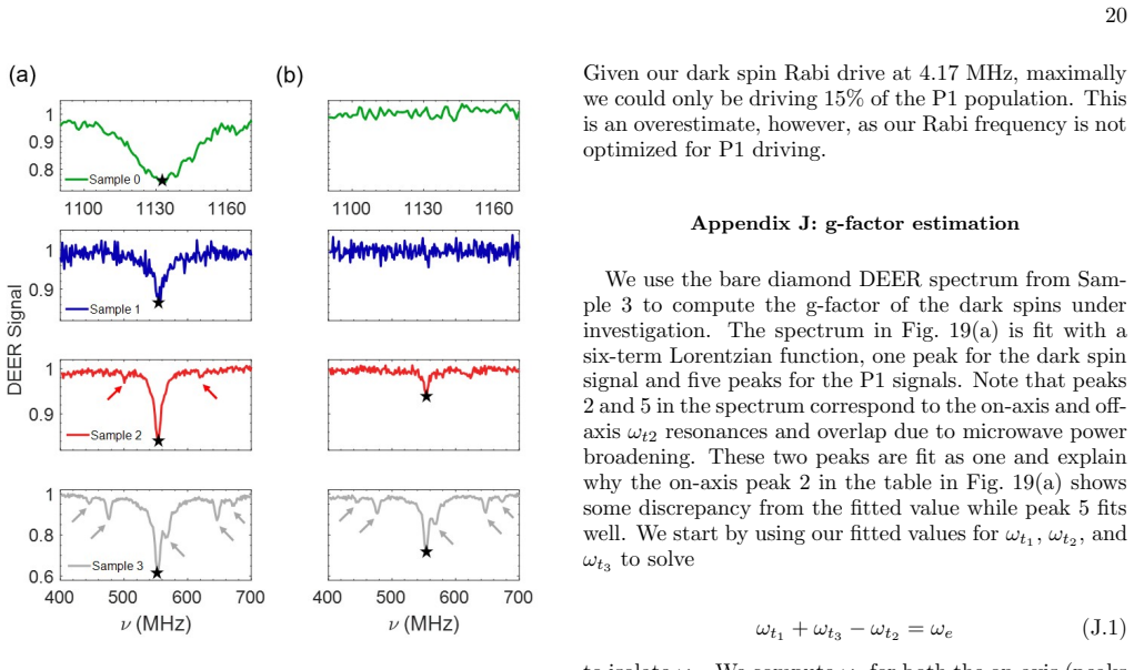
In Fig. 19(a), Sample 3 has on-axis (green - peaks 20 FIG. 18. DEER profile comparison between bare (a) and 300 ALD cycles (b). The four samples show smaller reductions in the dark spin resonance (black star) as the nitrogen implan- tation dose in increased from Sample 0 (green) to Sample 3 (gray). The appearance of P1 resonances (arrows) becomes apparent...
- [8]
-
[9]
R.-A. Eichel, Structural and dynamic properties of oxy- gen vacancies in perovskite oxides—analysis of defect chemistry by modern multi-frequency and pulsed epr techniques, Phys. Chem. Chem. Phys. 13, 368 (2011)
work page 2011
-
[10]
K. J. Reszka, P. Bilski, and C. F. Chignell, Spin trapping of nitric oxide by aci anions of nitroalkanes, Nitric Oxide 10, 53 (2004)
work page 2004
-
[11]
Y. Woldman, V. Khramtsov, I. Grigorev, I. Kiriljuk, and D. Utepbergenov, Spin trapping of nitric oxide by nitronylnitroxides: Measurement of the activity of no synthase from rat cerebellum, Biochemical and Biophys- ical Research Communications 202, 195 (1994)
work page 1994
-
[12]
D. M. Bailey, B. Davies, I. S. Young, M. J. Jackson, G. W. Davison, R. Isaacson, and R. S. Richardson, Epr spectroscopic detection of free radical outflow from an isolated muscle bed in exercising humans, Journal of Ap- plied Physiology 94, 1714 (2003), pMID: 12626489
work page 2003
-
[13]
L. Hofmann and S. Ruthstein, Epr spectroscopy pro- vides new insights into complex biological reaction mech- anisms, The Journal of Physical Chemistry B 126, 7486 (2022)
work page 2022
-
[14]
P. Kuppusamy, M. Chzhan, K. Vij, M. Shteynbuk, D. J. Lefer, E. Giannella, and J. L. Zweier, Three-dimensional spectral-spatial epr imaging of free radicals in the heart: a technique for imaging tissue metabolism and oxygena- tion., Proceedings of the National Academy of Sciences 91, 3388 (1994)
work page 1994
-
[15]
H. M. Swartz, N. Khan, J. Buckey, R. Comi, L. Gould, O. Grinberg, A. Hartford, H. Hopf, H. Hou, E. Hug, A. Iwasaki, P. Lesniewski, I. Salikhov, and T. Walczak, Clinical applications of epr: overview and perspectives, NMR in Biomedicine 17, 335 (2004)
work page 2004
-
[16]
G. C. Rex and S. Schlick, Study of polymer gels using paramagnetic probes: e.s.r. spectra of cu2+ in reversible polyacrylamide gels, Polymer 28, 2134 (1987)
work page 1987
-
[17]
R. D. Harvey and S. Schlick, Study of morphology in interpenetrating polymer networks using paramagnetic spin probes, Polymer 30, 11 (1989)
work page 1989
- [18]
- [19]
-
[20]
S. A. Goldman, G. V. Bruno, and J. H. Freed, Esr studies of anisotropic rotational reorientation and slow tumbling in liquid and frozen media. ii. saturation and nonsecular effects, The Journal of Chemical Physics59, 3071 (1973)
work page 1973
-
[21]
W. K. Schenken, Y. Liu, L. Krishna, A. A. A. Majid, C. A. Koh, P. C. Taylor, and R. T. Collins, Electron paramagnetic resonance study of type-ii silicon clathrate with low sodium guest concentration, Phys. Rev. B 101, 245204 (2020)
work page 2020
-
[22]
D. Schmalbein, G. Maresch, A. Kamlowski, and P. Hofer, The bruker high-frequency-epr system, Applied Magnetic Resonance 16, 185 (1999)
work page 1999
-
[23]
F. Shi, Q. Zhang, P. Wang, H. Sun, J. Wang, X. Rong, M. Chen, C. Ju, F. Reinhard, H. Chen, J. Wrachtrup, J. Wang, and J. Du, Single-protein spin resonance spec- troscopy under ambient conditions, Science 347, 1135 (2015)
work page 2015
-
[24]
F. Shi, F. Kong, P. Zhao, X. Zhang, M. Chen, S. Chen, Q. Zhang, M. Wang, X. Ye, Z. Wang, Z. Qin, X. Rong, J. Su, P. Wang, P. Z. Qin, and J. Du, Single-dna electron spin resonance spectroscopy in aqueous solutions, Nat. Methods 15, 697 (2018)
work page 2018
-
[25]
M. S. Grinolds, M. Warner, K. D. Greve, Y. Dovzhenko, L. Thiel, R. L. Walsworth, S. Hong, P. Maletinsky, and A. Yacoby, Subnanometre resolution in three- dimensional magnetic resonance imaging of individual dark spins, Nat. Nanotech. 9, 279 (2014)
work page 2014
-
[26]
B. L. Dwyer, L. V. Rodgers, E. K. Urbach, D. Bluvstein, S. Sangtawesin, H. Zhou, Y. Nassab, M. Fitzpatrick, Z. Yuan, K. De Greve, E. L. Peterson, H. Knowles, T. Sumarac, J.-P. Chou, A. Gali, V. Dobrovitski, M. D. Lukin, and N. P. de Leon, Probing spin dynamics on diamond surfaces using a single quantum sensor, PRX Quantum 3, 040328 (2022)
work page 2022
-
[27]
S. Sangtawesin, B. L. Dwyer, S. Srinivasan, J. J. Allred, L. V. H. Rodgers, K. De Greve, A. Stacey, N. Dontschuk, K. M. O’Donnell, D. Hu, D. A. Evans, C. Jaye, D. A. Fis- cher, M. L. Markham, D. J. Twitchen, H. Park, M. D. Lukin, and N. P. de Leon, Origins of diamond surface noise probed by correlating single-spin measurements with surface spectroscopy, P...
work page 2019
-
[28]
M. Joos, D. Bluvstein, Y. Lyu, D. Weld, and A. B. Jayich, Protecting qubit coherence by spectrally engineered driv- ing of the spin environment, npj Quantum Inf. 8, 47 (2022)
work page 2022
-
[29]
Z. Peng, T. Biktagirov, F. H. Cho, U. Gerstmann, and S. Takahashi, Investigation of near-surface defects of nan- odiamonds by high-frequency EPR and DFT calculation, The Journal of Chemical Physics 150, 134702 (2019)
work page 2019
- [30]
- [31]
-
[32]
W. K. Lo, Y. Zhang, H. Y. Chow, J. Wu, M. Y. Leung, K. O. Ho, X. Du, Y. Chen, Y. Shen, D. Pan, et al. , En- 27 hancement of quantum coherence in solid-state qubits via interface engineering, Nature Communications 16, 5984 (2025)
work page 2025
-
[33]
K. S. Liu, A. Henning, M. W. Heindl, R. D. Allert, J. D. Bartl, I. D. Sharp, R. Rizzato, and D. B. Bucher, Sur- face nmr using quantum sensors in diamond, Proceedings of the National Academy of Sciences 119, e2111607119 (2022)
work page 2022
- [34]
-
[35]
H. J. Mamin, M. H. Sherwood, and D. Rugar, Detecting external electron spins using nitrogen-vacancy centers, Phys. Rev. B 86, 195422 (2012)
work page 2012
-
[36]
U. Zvi, D. R. Candido, A. Weiss, A. R. Jones, L. Chen, I. Golovina, X. Yu, S. Wang, D. V. Talapin, M. E. Flatt´ e, A. P. Esser-Kahn, and P. C. Maurer, Engineering spin coherence in core-shell diamond nanocrystals, Pro- ceedings of the National Academy of Sciences 122.21, e2422542122 (2025)
work page 2025
- [37]
-
[38]
E. J. Davis, B. Ye, F. Machado, S. A. Meynell, W. Wu, T. Mittiga, W. Schenken, M. Joos, B. Kobrin, Y. Lyu, et al., Probing many-body dynamics in a two-dimensional dipolar spin ensemble, Nature Physics 19, 836 (2023)
work page 2023
-
[39]
G. N. Parsons, Functional model for analysis of ald nu- cleation and quantificaiton of area-selective deposition, Journal of Vacuum Science & Technology A 37, 020911 (2019)
work page 2019
- [40]
-
[41]
J. C. Jones, N. Delegan, F. J. Heremans, and A. B. F. Martinson, Nucleation dependence of atomic layer de- position on diamond surface termination, Carbon 213, 118276 (2023)
work page 2023
-
[42]
R. Vidrio, D. Vincent, B. Bachman, C. Saucedo, M. Za- hedian, Z. Xu, J. Lai, T. A. Grotjohn, S. Kolkowitz, J.-H. Seo, R. J. Hamers, K. G. Ray, Z. Ma, and J. T. Choy, Xps anlaysis of molecular contamination and sp2 amor- phous carbon on oxidized (100) diamond, Materials of Quantum Technology 4, 025201 (2024)
work page 2024
-
[43]
M. Xie, X. Yu, L. V. H. Rodgers, D. Xu, I. Chi-Duran, A. Toros, N. Quack, N. P. de Leon, and P. C. Maurer, Biocompatible surface functionalization architecture for a diamond quantum sensor, Proceedings of the National Academy of Sciences 119.8, e2114186119 (2022)
work page 2022
-
[44]
M. E. Dufond, M. W. Diouf, C. Badie, C. Laffon, P. Par- ent, D. Ferry, D. Gross, J. C. Kools, S. D. Elliot, and L. Santinacci, Quantifying the extent of ligand incorpo- ration and the effect on properties of TiO 2 thin films grown by atomic layer deposition using an alkoxide or an alkylamide, Chem. Mater. 32, 1393 (2020)
work page 2020
-
[45]
A. O. Sushkov, I. Lovchinsky, N. Chisholm, R. L. Walsworth, H. Park, and M. D. Lukin, Magnetic reso- nance detection of individual proton spins using quantum reporters, Phys. Rev. Lett. 113, 197601 (2014)
work page 2014
-
[46]
G. De Lange, T. Van Der Sar, M. Blok, Z.-H. Wang, V. Dobrovitski, and R. Hanson, Controlling the quantum dynamics of a mesoscopic spin bath in diamond, Scientific reports 2, 382 (2012)
work page 2012
-
[47]
R. Goldblatt, A. Martin, and A. Wood, Sensing coherent nuclear spin dynamics with an ensemble of paramagnetic nitrogen spins, PRX Quantum 5, 020334 (2024)
work page 2024
-
[48]
A. Laraoui, F. Dolde, C. Burk, F. Reinhard, J. Wrachtrup, and C. A. Meriles, High-resolution corre- lation spectroscopy of 13c spins near a nitrogen-vacancy centre in diamond, Nature communications 4, 1651 (2013)
work page 2013
-
[49]
M. Schlipf and F. Gygi, Optimization algorithm for the generation of oncv pseudopotentials, Computer Physics Communications 196, 36 (2015)
work page 2015
-
[50]
P. Giannozzi, S. Baroni, N. Bonini, M. Calandra, R. Car, C. Cavazzoni, D. Ceresoli, G. L. Chiarotti, M. Cococ- cioni, I. Dabo, et al. , Quantum espresso: a modular and open-source software project for quantum simulations of materials, Journal of Physics: Condensed Matter 21, 395502 (2009)
work page 2009
-
[51]
J. P. Perdew, K. Burke, and M. Ernzerhof, Generalized gradient approximation made simple, Phys. Rev. Lett. 77, 3865 (1996)
work page 1996
-
[52]
J. Heyd, G. E. Scuseria, and M. Ernzerhof, Hybrid func- tionals based on a screened coulomb potential, The Jour- nal of chemical physics 118, 8207 (2003)
work page 2003
-
[53]
J.-P. Chou, P. Udvarhelyi, N. P. de Leon, and A. Gali, Ab initio study of (100) diamond surface spins, Phys. Rev. Appl. 20, 014040 (2023)
work page 2023
-
[54]
C. G. Van de Walle and R. M. Martin, Theoretical study of band offsets at semiconductor interfaces, Phys. Rev. B 35, 8154 (1987)
work page 1987
- [55]
-
[56]
M. Schaffry, E. M. Gauger, J. J. Morton, and S. C. Ben- jamin, Proposed spin amplification for magnetic sensors employing crystal defects, Physical review letters 107, 207210 (2011)
work page 2011
-
[57]
D. B. Bucher, D. R. Glenn, H. Park, M. D. Lukin, and R. L. Walsworth, Hyperpolarization-enhanced nmr spec- troscopy with femtomole sensitivity using quantum de- fects in diamond, Physical Review X 10, 021053 (2020)
work page 2020
- [58]
- [59]
-
[60]
J. G. Bartholomew, K. de Oliveira Lima, A. Ferrier, and P. Goldner, Optical line width broadening mechanisms at the 10 khz level in eu3+: Y2o3 nanoparticles, Nano letters 17, 778 (2017)
work page 2017
-
[61]
S. Liu, A. Fossati, D. Serrano, A. Tallaire, A. Ferrier, and P. Goldner, Defect engineering for quantum grade rare-earth nanocrystals, ACS nano 14, 9953 (2020)
work page 2020
-
[62]
M. Bal, A. A. Murthy, S. Zhu, F. Crisa, X. You, Z. Huang, T. Roy, J. Lee, D. v. Zanten, R. Pilipenko, et al. , Systematic improvements in transmon qubit co- herence enabled by niobium surface encapsulation, npj 28 Quantum Information 10, 43 (2024)
work page 2024
-
[63]
R. D. Chang, N. Shumiya, R. A. McLellan, Y. Zhang, M. P. Bland, F. Bahrami, J. Mun, C. Zhou, K. Kisslinger, G. Cheng, et al. , Eliminating surface oxides of supercon- ducting circuits with noble metal encapsulation, Physical Review Letters 134, 097001 (2025)
work page 2025
-
[64]
F. A. Stevie and C. L. Donley, Introduction to x-ray pho- toelectron spectroscopy, J. Vac. Sci. Technology. A 38 (2020)
work page 2020
-
[65]
G. G. Fuentes, E. Elizalde, F. Yubero, and J. M. Sanz, Electron inelastic mean free path for Ti, TiC, TiN and TiO2 as determined by quantititative reflection electron energy-loss spectroscopy, Surf. Interface. Anal. 43, 230 (2002)
work page 2002
- [66]
-
[67]
C. G. Baldwin, J. E. Downes, C. J. McMahon, C. Bradac, and R. P. Mildren, Nanostructuing and oxidation of dia- mond by two-photon ultraviolet surface excitation: And XPS and NEXAFS study, Physical Review B 89, 195422 (2014)
work page 2014
- [68]
-
[69]
R. Pandiyan, N. Delegan, A. Dirany, P. Drogui, and M. A. E. Khakani, Probing the electronic surface proper- ties and bandgap narrowing of in situ N, W, and (W,N) doped magnetron sputtered TiO 2 films intended for elctro-photocatalyic applications, The Journal of Phys- ical Chemistry C 120, 631 (2016)
work page 2016
-
[70]
M. C. Biesinger, L. W. M. Lau, A. R. Gerson, and R. S. C. Smart, Resolving surface chemical states in XPS analysis of first row transition metals, oxides and hydrox- ides: Sc, Ti, V, Cu and Zn, Applied Surface Science 257, 887 (2010)
work page 2010
-
[71]
D. B. Bucher, D. P. L. Aude Craik, M. P. Backlund, M. J. Turner, O. Ben Dor, D. R. Glenn, and R. L. Walsworth, Quantum diamond spectrometer for nanoscale nmr and esr spectroscopy, Nature Protocols 14, 2707 (2019)
work page 2019
-
[72]
E. B. Fel’dman and S. Lacelle, Configurational averaging of dipolar interactions in magnetically diluted spin net- works, The Journal of chemical physics104, 2000 (1996)
work page 2000
-
[73]
L. M. Pham, S. J. DeVience, F. Casola, I. Lovchinsky, A. O. Sushkov, E. Bersin, J. Lee, E. Urbach, P. Cappel- laro, H. Park, et al. , Nmr technique for determining the depth of shallow nitrogen-vacancy centers in diamond, Physical Review B 93, 045425 (2016)
work page 2016
- [74]
- [75]
-
[76]
W. Yang, W.-L. Ma, and R.-B. Liu, Quantum many-body theory for electron spin decoherence in nanoscale nuclear spin baths, Reports on Progress in Physics 80, 016001 (2016)
work page 2016
-
[77]
J. D. A. Wood, D. A. Broadway, L. T. Hall, A. Stacey, D. A. Simpson, J.-P. Tetienne, and L. C. L. Hollenberg, Wide-band nanoscale magnetic resonance spectroscopy using quantum relaxation of a single spin in diamond, Phys. Rev. B 94, 155402 (2016)
work page 2016
-
[78]
K. Siegrist, Non-homogeneous poisson processes, Online; accessed 14-Feb-2025 (2022), in Probability, Mathemati- cal Statistics, and Stochastic Processes , LibreTexts
work page 2025
-
[79]
L. T. Hall, J. H. Cole, and L. C. Hollenberg, Ana- lytic solutions to the central-spin problem for nitrogen- vacancy centers in diamond, Physical Review B 90, 075201 (2014)
work page 2014
discussion (0)
Sign in with ORCID, Apple, or X to comment. Anyone can read and Pith papers without signing in.