Membrane-Associated Self-Assembly for Cellular Decision Making
Pith reviewed 2026-05-22 02:34 UTC · model grok-4.3
The pith
Spontaneous self-assembly of 3D subunits on 2D membranes acts as a sensitive switch for detecting receptors at physiological concentrations.
A machine-rendered reading of the paper's core claim, the machinery that carries it, and where it could break.
Core claim
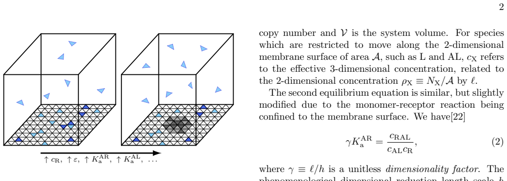
We show that spontaneous self-assembly of native 3D subunits on a two-dimensional substrate can act as a tunable and robust switch for detecting receptors at physiological concentrations, much more sensitive than other passive mechanisms. Analytical expressions for the critical receptor density driving stable subunit assembly agree closely with stochastic reaction-diffusion simulations, providing testable predictions for control by lipids, subunits, and receptors.
What carries the argument
Receptor-triggered spontaneous self-assembly of 3D subunits onto a 2D membrane, serving as a passive switch that produces a detectable response at low receptor densities.
If this is right
- Analytical expressions allow calculation of the critical receptor density required for assembly.
- The assembly mechanism is tunable by lipids, subunit properties, and receptor numbers.
- It produces a more sensitive response than alternative passive receptor detection processes.
- The switch-like behavior occurs without energy input from active cellular processes.
Where Pith is reading between the lines
- If this mechanism operates in cells, it could provide an energy-efficient way to amplify weak signals from sparse receptors.
- The assembly process might interact with known membrane curvature or lipid effects in real cells.
- Similar principles could be tested in artificial membrane systems to engineer simple sensors.
Load-bearing premise
That 3D subunits will spontaneously self-assemble on the 2D membrane when receptors are present, without needing energy or other active help, and that this assembly alone creates a clear switch-like detection.
What would settle it
Direct observation of whether subunit assemblies form at the predicted critical receptor density in a controlled membrane experiment; significant deviation from the analytical prediction would falsify the model.
Figures


read the original abstract
Cellular decision-making based on information received from the external environment is frequently initiated by transmembrane receptors. These receptors are known to propagate such information by triggering a series of irreversible, energy-consuming reactions. While this active mechanism ensures switch-like responses, here we show how spontaneous self-assembly of native 3D subunits on a two-dimensional substrate can similarly act as a tunable and robust switch for detecting receptors at physiological concentrations. This mechanism is much more sensitive than other passive mechanisms for receptor detection. We derive analytical expressions for the critical receptor density driving stable subunit assembly, in close agreement with stochastic reaction-diffusion simulations. The theory provides testable predictions for how lipids, subunits, and receptors each can control decision boundaries and magnitude of response.
Editorial analysis
A structured set of objections, weighed in public.
Referee Report
Summary. The manuscript claims that spontaneous self-assembly of native 3D subunits on a two-dimensional membrane, triggered by transmembrane receptors, can serve as a tunable and robust passive switch for cellular decision-making at physiological receptor concentrations. This mechanism is asserted to be more sensitive than other passive receptor-detection processes. Analytical expressions for the critical receptor density are derived from equilibrium considerations and stated to agree closely with stochastic reaction-diffusion simulations; the theory yields testable predictions for how lipids, subunits, and receptors modulate decision boundaries and response magnitude.
Significance. If the central result holds, the work identifies a passive, energy-independent route to switch-like behavior in membrane-associated self-assembly that could complement or explain aspects of receptor-mediated signaling. The combination of closed-form critical-density expressions with direct stochastic simulation validation constitutes a clear strength, as does the provision of concrete, falsifiable predictions for experimental control via lipid composition and subunit properties.
major comments (2)
- [§3.2, Eq. (7)] §3.2, Eq. (7): the equilibrium derivation of the critical receptor density omits an explicit nucleation barrier whose height scales with subunit interaction energy and local receptor number; near the predicted threshold the mean waiting time for a stable cluster may exceed cellular timescales even when the equilibrium state favors assembly, which would undermine the claims of spontaneous and robust switching. The reported agreement with stochastic reaction-diffusion simulations does not resolve this unless the runs explicitly document system size, receptor copy number (typically 10–100 per cell), and sampling of rare nucleation events.
- [§4.1] §4.1: the quantitative comparison asserting greater sensitivity than other passive mechanisms is presented without tabulated thresholds or direct numerical benchmarks against the cited passive alternatives, making it difficult to assess the magnitude of the claimed advantage.
minor comments (2)
- [§2.3] The parameter table in §2.3 lists interaction energies but does not specify the precise values or ranges used for the physiological receptor densities in the main figures.
- [Figure 3] Figure 3 caption should explicitly state the number of independent simulation trajectories and the criterion used to declare a 'stable cluster' for the purpose of comparing to the analytical threshold.
Simulated Author's Rebuttal
We thank the referee for their careful and constructive review. The comments highlight important distinctions between equilibrium stability and nucleation kinetics, as well as the need for clearer quantitative benchmarks. We have revised the manuscript to address both points directly while preserving the core claims supported by our derivations and simulations.
read point-by-point responses
-
Referee: [§3.2, Eq. (7)] §3.2, Eq. (7): the equilibrium derivation of the critical receptor density omits an explicit nucleation barrier whose height scales with subunit interaction energy and local receptor number; near the predicted threshold the mean waiting time for a stable cluster may exceed cellular timescales even when the equilibrium state favors assembly, which would undermine the claims of spontaneous and robust switching. The reported agreement with stochastic reaction-diffusion simulations does not resolve this unless the runs explicitly document system size, receptor copy number (typically 10–100 per cell), and sampling of rare nucleation events.
Authors: We agree that the equilibrium analysis in §3.2 identifies the thermodynamic threshold but does not explicitly quantify the nucleation barrier or associated waiting times. The stochastic reaction-diffusion simulations already include the full kinetic pathway and show spontaneous assembly within the simulated durations for the reported parameters. To strengthen the presentation, we have added a new subsection in §3.2 that estimates the nucleation barrier height using classical nucleation theory and shows that, for subunit interaction energies and receptor densities near the critical value, mean waiting times remain below one minute. We have also expanded the simulation methods to document a 2 μm × 2 μm periodic domain, receptor copy numbers of 10–150 per simulation (corresponding to physiological surface densities), and results averaged over 50 independent trajectories per condition to capture nucleation statistics. revision: partial
-
Referee: [§4.1] §4.1: the quantitative comparison asserting greater sensitivity than other passive mechanisms is presented without tabulated thresholds or direct numerical benchmarks against the cited passive alternatives, making it difficult to assess the magnitude of the claimed advantage.
Authors: We accept that the sensitivity comparison in §4.1 would benefit from explicit numerical benchmarks. In the revised manuscript we have inserted a new table that reports the critical receptor density (or equivalent threshold) for our self-assembly mechanism alongside the corresponding values for the passive alternatives discussed in the text (simple monovalent binding, receptor clustering without self-assembly, and lipid-phase separation). The table shows that our predicted critical density is 5–50 fold lower than the alternatives under comparable subunit and lipid parameters, providing a direct, quantitative basis for the sensitivity claim. revision: yes
Circularity Check
Analytical equilibrium derivation of critical receptor density validated by independent simulations
full rationale
The paper derives analytical expressions for the critical receptor density from equilibrium considerations of spontaneous 3D subunit self-assembly on a 2D membrane, then reports close agreement with separate stochastic reaction-diffusion simulations. This is a standard prediction-validation workflow rather than any reduction of the result to a fitted parameter, self-definition, or self-citation chain. No load-bearing step is shown to be equivalent to its inputs by construction; the central claim remains independent of the simulation data used only for comparison. The derivation is therefore self-contained against external benchmarks.
Axiom & Free-Parameter Ledger
axioms (1)
- domain assumption Native 3D subunits undergo spontaneous receptor-triggered self-assembly on a 2D substrate that produces a switch-like response at physiological concentrations.
Lean theorems connected to this paper
-
IndisputableMonolith/Cost/FunctionalEquation.leanwashburn_uniqueness_aczel unclear?
unclearRelation between the paper passage and the cited Recognition theorem.
We derive analytical expressions for the critical receptor density driving stable subunit assembly... free energy per surface monomer f = ½(È−1)Zε + eb(ϕ,È) − ϕȲ + (2ϕ−1)È + È ln(ϕÈ)
-
IndisputableMonolith/Foundation/AlexanderDuality.leanalexander_duality_circle_linking unclear?
unclearRelation between the paper passage and the cited Recognition theorem.
Z = 3 shown... hexagonal lattice
What do these tags mean?
- matches
- The paper's claim is directly supported by a theorem in the formal canon.
- supports
- The theorem supports part of the paper's argument, but the paper may add assumptions or extra steps.
- extends
- The paper goes beyond the formal theorem; the theorem is a base layer rather than the whole result.
- uses
- The paper appears to rely on the theorem as machinery.
- contradicts
- The paper's claim conflicts with a theorem or certificate in the canon.
- unclear
- Pith found a possible connection, but the passage is too broad, indirect, or ambiguous to say the theorem truly supports the claim.
Reference graph
Works this paper leans on
-
[1]
4, where assembly is guaranteed regardless of cR. Below this critical strength, the shape of the sensing decision boundary is directly tunable by K AR a andh via Eqn. (7). As monomer-receptor interactions are strengthened by either increasing K AR a or decreasing h, c∗ R decreases. This also changes the sensitivity of the threshold c∗ R to variations in a...
-
[2]
01 µM−1, and 0 . 033 µM−1 respectively. onal lattice, and they contain distinct sites to bind the lipids and receptors (Fig. 3a). Lipids and receptors have a single binding site. We include the one allosteric effect imposed in the theory: subunits bind receptors only after binding to the membrane. By simulating each system to equilibrium, we quantify the s...
-
[3]
C. Barata-Antunes, R. Alves, G. Talaia, M. Casal, H. Ger´ os, R. Mans, and S. Paiva, Computational and Structural Biotechnology Journal 19, 1713 (2021)
work page 2021
- [4]
-
[5]
C. M. Cadwell, W. Su, and A. P. Kowalczyk, Traffic 17, 1262 (2016)
work page 2016
-
[6]
D. A. Lauffenburger and J. J. Linderman, Receptors: models for binding, trafficking, and signaling (Oxford University Press, 1996)
work page 1996
-
[7]
J. E. Ferrell and S. H. Ha, Trends in biochemical sciences 39, 612 (2014)
work page 2014
-
[8]
G. Tkaˇ cik and W. Bialek, Annual Review of Condensed Matter Physics 7, 89 (2016)
work page 2016
-
[9]
P. Fran¸ cois and G. Altan-Bonnet, Journal of Statistical Physics 162, 1130 (2016)
work page 2016
-
[10]
G. Tkaˇ cik and P. R. t. Wolde, Annual Review of Bio- physics 54 (2025)
work page 2025
-
[11]
M. Mettlen, P.-H. Chen, S. Srinivasan, G. Danuser, and S. L. Schmid, Annual review of biochemistry 87, 871 (2018)
work page 2018
-
[12]
H. J. Green and N. H. Brown, Experimental Cell Re- search 378, 226 (2019)
work page 2019
-
[13]
C. Malinverno, S. Corallino, F. Giavazzi, M. Bergert, Q. Li, M. Leoni, A. Disanza, E. Frittoli, A. Oldani, E. Martini, et al. , Nature materials 16, 587 (2017)
work page 2017
-
[14]
A. Jhaveri, S. Loggia, Y. Qian, and M. E. Johnson, Proceedings of the National Academy of Sciences 121, e2403384121 (2024)
work page 2024
-
[15]
Ercolani, Journal of the American Chemical Society 125, 16097 (2003)
G. Ercolani, Journal of the American Chemical Society 125, 16097 (2003)
work page 2003
-
[16]
Zlotnick, Journal of molecular biology 241, 59 (1994)
A. Zlotnick, Journal of molecular biology 241, 59 (1994)
work page 1994
-
[17]
W. M. Jacobs, A. Reinhardt, and D. Frenkel, Proceedings of the National Academy of Sciences 112, 6313 (2015)
work page 2015
-
[18]
W. M. Jacobs, Physical review letters 126, 258101 (2021)
work page 2021
- [19]
-
[20]
C. G. Evans, J. O’Brien, E. Winfree, and A. Murugan, Nature 625, 500 (2024)
work page 2024
-
[21]
G. Adam and M. Delbr¨ uck, Structural chemistry and molecular biology 198, 198 (1968)
work page 1968
-
[22]
L. W¨ urthner, F. Brauns, G. Pawlik, J. Halatek, J. Kersse- makers, C. Dekker, and E. Frey, Proceedings of the Na- tional Academy of Sciences 119, e2206888119 (2022)
work page 2022
-
[23]
G.-K. Xu, J. Hu, R. Lipowsky, and T. R. Weikl, The Journal of chemical physics 143 (2015)
work page 2015
-
[24]
O. N. Yogurtcu and M. E. Johnson, PLoS computational biology 14, e1006031 (2018)
work page 2018
-
[25]
S.-K. Guo, A. J. Sodt, and M. E. Johnson, PLoS Com- putational Biology 18 (2022)
work page 2022
-
[26]
Z. Toprakcioglu, A. Kamada, T. C. Michaels, M. Xie, J. Krausser, J. Wei, A. Saric, M. Vendruscolo, and T. P. Knowles, Proceedings of the National Academy of Sci- ences 119, e2109718119 (2022)
work page 2022
- [27]
-
[28]
N. A. Ara´ ujo, L. M. Janssen, T. Barois, G. Boffetta, I. Cohen, A. Corbetta, O. Dauchot, M. Dijkstra, W. M. Durham, A. Dussutour, et al. , Soft matter 19, 1695 (2023)
work page 2023
-
[29]
M. Rouches, S. L. Veatch, and B. B. Machta, Proceedings of the National Academy of Sciences 118, e2103401118 (2021)
work page 2021
-
[30]
X. Zhao, G. Bartolucci, A. Honigmann, F. J¨ ulicher, and C. A. Weber, New Journal of Physics 23, 123003 (2021)
work page 2021
-
[31]
W. D. Kaplan, D. Chatain, P. Wynblatt, and W. C. Carter, Journal of Materials Science 48, 5681 (2013)
work page 2013
-
[32]
J. W. Cahn, The Journal of Chemical Physics 66, 3667 (1977)
work page 1977
-
[33]
Goldenfeld, Lectures on phase transitions and the renormalization group (CRC Press, 2018)
N. Goldenfeld, Lectures on phase transitions and the renormalization group (CRC Press, 2018)
work page 2018
-
[34]
L. M. Traub, Journal of Cell Biology 163, 203 (2003)
work page 2003
-
[35]
Y. Wu, J. Vendome, L. Shapiro, A. Ben-Shaul, and B. Honig, Nature 475, 510 (2011)
work page 2011
- [36]
-
[37]
M. J. Varga, Y. Fu, S. Loggia, O. N. Yogurtcu, and M. E. Johnson, Biophysical journal 118, 3026 (2020)
work page 2020
- [38]
-
[39]
M. E. Johnson and G. Hummer, Physical Review X 4, 031037 (2014)
work page 2014
-
[40]
M. Y. Hein, N. C. Hubner, I. Poser, J. Cox, N. Na- garaj, Y. Toyoda, I. A. Gak, I. Weisswange, J. Mansfeld, F. Buchholz, et al. , Cell 163, 712 (2015)
work page 2015
-
[41]
D. Duan, M. Hanson, D. O. Holland, and M. E. Johnson, Scientific Reports 12, 5413 (2022)
work page 2022
-
[42]
S. Sigismund, L. Lanzetti, G. Scita, and P. P. Di Fiore, Nature Reviews Molecular Cell Biology 22, 625 (2021)
work page 2021
-
[43]
B. Pearse and R. Crowther, Annual review of biophysics and biophysical chemistry 16, 49 (1987)
work page 1987
-
[44]
B. T. Kelly, S. C. Graham, N. Liska, P. N. Dannhauser, S. H¨ oning, E. J. Ungewickell, and D. J. Owen, Science 345, 459 (2014)
work page 2014
-
[45]
W. F. Zeno, J. B. Hochfelder, A. S. Thatte, L. Wang, A. K. Gadok, C. C. Hayden, E. M. Lafer, and J. C. Sta- chowiak, Biophysical Journal 120, 818 (2021)
work page 2021
-
[46]
S. J. Bryant and B. B. Machta, Physical review letters 131, 068401 (2023)
work page 2023
-
[47]
W. S. Hlavacek, A. Redondo, H. Metzger, C. Wofsy, and B. Goldstein, Proceedings of the National Academy of Sciences 98, 7295 (2001)
work page 2001
-
[48]
I. Levental and S. L. Veatch, Journal of molecular biolog y 428, 4749 (2016)
work page 2016
-
[49]
T. Baumgart, A. T. Hammond, P. Sengupta, S. T. Hess, D. A. Holowka, B. A. Baird, and W. W. Webb, Pro- ceedings of the National Academy of Sciences 104, 3165 (2007)
work page 2007
- [50]
-
[51]
R. D. Cadena-Nava, M. Comas-Garcia, R. F. Garmann, A. Rao, C. M. Knobler, and W. M. Gelbart, Journal of virology 86, 3318 (2012)
work page 2012
- [52]
-
[53]
X.-S. Wu, B. D. McNeil, J. Xu, J. Fan, L. Xue, E. Meli- coff, R. Adachi, L. Bai, and L.-G. Wu, Nature neuro- science 12, 1003 (2009)
work page 2009
-
[54]
W. Bialek and S. Setayeshgar, Proceedings of the Na- tional Academy of Sciences 102, 10040 (2005)
work page 2005
-
[55]
P. R. ten Wolde, N. B. Becker, T. E. Ouldridge, and A. Mugler, Journal of Statistical Physics 162, 1395 (2016). 7
work page 2016
- [56]
-
[57]
F. Yuan, S. Gollapudi, K. J. Day, G. Ashby, A. Sangani, B. T. Malady, L. Wang, E. M. Lafer, J. M. Huibregtse, and J. C. Stachowiak, PNAS nexus 3, pgae342 (2024)
work page 2024
-
[58]
S. Mondal, K. Narayan, S. Botterbusch, I. Powers, J. Zheng, H. P. James, R. Jin, and T. Baumgart, Na- ture communications 13, 5017 (2022). Supplementary Information for: Membrane-Associated Self-Assembly for Cellular Decision Ma king Samuel L. Foley and Margaret E. Johnson 1 Surface Free Energy Consider a 2-dimensional lattice with M total sites that can ...
work page 2022
discussion (0)
Sign in with ORCID, Apple, or X to comment. Anyone can read and Pith papers without signing in.