REVIEW 5 major objections 5 minor 22 references
Brain Tumor Detection through Thermal Imaging and MobileNET
T0 review · 5 major / 5 minor · reviewed 2026-08-06 · deepseek-v4-flash
Pith's one-line read This paper claims that a lightweight MobileNet can classify brain MRIs as tumor or no-tumor at about 98.8% validation accuracy using a jet color map as a stand-in for thermal imaging.
desk verdict The 'thermal imaging' is just a JET colormap, the claimed superiority over GoogLeNet contradicts the paper's own references, and the result is a smaller-scale replication of prior work. read the letter →
The pith
A machine-rendered reading of the paper's core claim, the machinery that carries it, and where it could break.
The reading
What carries the argument
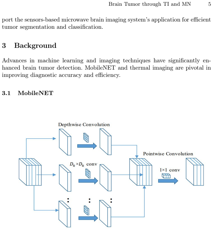
The mechanism that carries the argument is MobileNet's depthwise separable convolution, which splits each convolution into a depthwise pass with one filter per channel and a pointwise $1\times1$ pass, cutting computational cost from $K \times K \times C \times D \times H \times W$ to $K \times K \times C \times H \times W$. On the data side, the load-bearing preprocessing step is a standard computer-vision library's color-map function with the COLORMAP_JET palette, which maps grayscale MRI intensities to a blue-cyan-green-yellow-red scale; the paper treats this pseudocoloring as thermal imaging. Gaussian blur (a smoothing convolution) and Canny edge detection (a gradient-based boundary finder) are applied around the tumor region to complete the pipeline.
What would settle it
Train the same model once on original grayscale MRI slices and once on jet-colored versions, using the same data split; if the two accuracies are within random variation, the color map contributes no thermal signal. A stronger check is to run the identical model on true infrared thermal images of brain tissue and compare the resulting accuracy.
Extended reading notes
Core claim
The central claim, as the authors put it, is that a MobileNet with frozen feature-extraction layers and retrained final dense layers, fed $128\times128$ RGB versions of brain MRI slices recolored with a jet color map they call thermal imaging, classifies the scans with a validation accuracy of 98.8% (593 of 600 test images), a precision of 0.994, a recall of 0.984, and an F1 score of 0.989. The paper also reports average accuracy of 98.5% over five training runs and low validation loss and runtime, and it frames this as surpassing heavier models such as DenseNet and GoogleNet while using fewer computational resources. In plain terms, the paper is trying to show that inexpensive, lightweight deep learning plus simple image preprocessing can do automated brain-tumor screening without specialized thermal cameras.
Load-bearing premise
The paper assumes that recoloring grayscale MRI pixels with a color palette is the same as thermal imaging, so no actual infrared temperature measurements are needed for the method to be called thermal.
Editorial extensions
If this is right
- The reported 98.8% validation accuracy on 600 test images implies that a frozen-feature MobileNet with only its final dense layers retrained can reach roughly that accuracy on a moderate-size MRI dataset.
- Because the preprocessing steps are simple color mapping, blur, and edge detection, the full pipeline could run on a phone or laptop without a thermal camera or hospital-grade GPU.
- The 0.994 precision and 0.984 recall imply that, in this test set, the model misses 5 of every 311 tumor images and flags 2 of every 289 normal images as tumor.
- An average of 98.5% over five training runs suggests the headline accuracy is not a lucky single run, though only the average is reported and no variance or confidence interval is given.
Reading between the lines
- The paper does not compare the jet-colored input against the original grayscale input; running that ablation would separate MobileNet's contribution from the color map's contribution.
- If the color map matters at all, different palettes should change accuracy; measuring that sensitivity would test whether the thermal preprocessing carries diagnostic signal or merely changes the model's input distribution.
- Because the dataset is a single public source and the authors themselves flag cross-hospital variability as a limitation, the method's real-world value depends on validation on multi-scanner, multi-protocol MRIs or on actual infrared images.
Signed reviews
Editorial analysis
A structured set of objections, weighed in public.
Referee Report
Summary. This paper proposes a binary brain-tumor classifier that combines a MobileNet CNN with OpenCV image-processing techniques, including Gaussian blur, Canny edge detection, and a 'thermal imaging' step implemented as COLORMAP_JET pseudocoloring. The authors report a validation accuracy of 98.8% (Section 5.4) or an average accuracy of 98.5% (abstract) on a 600-image held-out test set from a Kaggle MRI dataset, and claim superiority over DenseNet and GoogleNet as well as low computational cost. The central claim is that the use of simulated thermal imaging via a pseudocolor map provides a novel, cost-effective detection approach.
Significance. If the result held as stated, the paper would provide a lightweight and inexpensive brain-tumor screening tool with roughly 98.8% accuracy, which could be relevant for resource-limited settings. The authors do take reasonable steps in using a standard held-out split (80/20), reporting a confusion matrix, and using transfer learning from ImageNet-pretrained MobileNet. However, the paper's stated novelty—thermal imaging—is not supported: the method described in Section 3.2 is a fixed pseudocolor lookup table on grayscale MRIs, not a thermal measurement. The accuracy claim also rests on an evaluation protocol that is insufficiently documented and on comparison claims that are both unsubstantiated and contradictory to the paper's own literature review. As a result, the contribution, if stripped of the thermal-imaging framing, reduces to a routine MobileNet MRI classification experiment with standard preprocessing, and the quantitative claims as presented cannot be accepted.
major comments (5)
- [Section 3.2, Section 1 novelty bullets] The method described in Section 3.2 applies OpenCV's COLORMAP_JET to grayscale MRI images; this is a fixed pointwise pseudocolor lookup table, not a measurement or model of temperature. The novelty bullets in Section 1 ('Integration of lightweight models like MobileNET with thermal imaging techniques' and 'Development of an OpenCV-based solution to emulate thermal imaging') rest on this identification, and the title itself claims thermal imaging. No infrared data are acquired, no bioheat model is used, and the reference [19] is not applied anywhere. Consequently, the central novelty and the associated cost argument collapse if the word 'thermal' is removed, because the experiments report classification of pseudocolored MRIs, not thermal imaging.
- [Section 1, Section 2, Section 5.4] Section 1 claims that the proposed model reaches 98.8% validation accuracy, 'exceeding cutting-edge models like DenseNET and GoogleNET,' but Section 2 cites [18] reporting GoogleNet at 99.45% accuracy. The paper contains no experiments with DenseNet or GoogLeNet, so the superiority claim is both contradicted by the cited literature and unsupported by any direct comparison. The 98.8% figure in Section 5.4 also conflicts with the abstract's stated 98.5% average accuracy.
- [Section 4, Section 5.2] Section 4 states that data visualization with thermal imaging, Gaussian blur, and Canny edge detection is used to set parameters such as kernel size, standard deviation, and colormap for later steps; this is done on the full dataset before the 80/20 split is described. Tuning parameters on the full dataset, including the eventual test portion, can inflate the reported test performance. In addition, Section 5.2 reports early stopping based on validation loss, but no separate validation set is described after the 80/20 split; it is unclear whether the test set double-serves as the validation set. This ambiguity undermines the reliability of the 98.8% accuracy figure.
- [Section 5.2, Section 5.4, Abstract] The paper states that results from five compilations were averaged (Section 5.2) and the abstract reports an average accuracy of 98.5%, yet Section 5.4 shows only a single confusion matrix (593 correct out of 600 test images) and provides no per-run accuracies, variance, or standard deviation. The claimed average and the implied stability cannot be independently verified from the reported material. The discrepancy between the abstract's 98.5% and Section 5.4's 98.8% further obscures the headline result.
- [Section 1, Section 5.4] The paper claims low runtime and low computational resource usage as a key advantage, but no runtime, inference latency, model size, or FLOP measurements are reported, and no comparison is made with DenseNet or GoogLeNet on these axes. The accessibility argument in Section 1 is therefore not supported by any quantitative evidence within the manuscript.
minor comments (5)
- [Section 5.4] The sentence 'Fig. displays the corresponding confusion matrix' is missing the figure number; please correct the reference.
- [Section 4, Section 5.4] There are several typographical errors, including 'graysacle' in Section 4 and 'weather truly there is any tumor' in Section 5.4, and the model name is rendered inconsistently as both 'MobileNET' and 'MobileNet'.
- [Section 5.2] The statement 'we have set the upper and lower threshold as 20 for edge detection' is unclear because Canny edge detection normally uses two distinct thresholds; please specify the exact threshold values and explain why a single value is used.
- [Section 5.3] The system configuration section should clarify whether timing or resource measurements, if any, were taken on Google Colab with a T4 GPU or on a local machine, since the claimed efficiency depends on this.
- [General] The paper does not provide a reproducibility statement or a link to code; adding such details would help readers verify the results and reuse the pipeline.
Circularity Check
The tumor-classification accuracy is a genuine held-out result, but the paper's central 'thermal imaging' novelty reduces to a COLORMAP_JET relabeling of grayscale MRI.
-
renaming known result
[Section 3.2 'Thermal Imaging' and Section 5.2 'Parameters'; cf. Section 1 novelty bullets]
"Thermal imaging identifies temperature variations caused by tumors' metabolic activity.Infraredcamerascapturethesedifferences,aidingearlydiagnosis.OpenCV's applyColorMap function was used withCOLORMAP_JET to visualize grayscale images with pseudocolor for better interpretation. ... We used COLORMAP_JET from OpenCV for thermal imaging purpose."
The paper's claimed novelty and cost argument rest on 'thermal imaging,' but by its own definition the thermal modality is just OpenCV's COLORMAP_JET applied pointwise to grayscale MRI. No infrared data are collected, no temperature is measured, and no bioheat model is run. Consequently, the statement 'thermal-imaging-based detection achieves 98.8%' reduces by construction to 'pseudocolored-MRI-based detection achieves 98.8%.' The word 'thermal' is a relabeling of a standard visualization colormap, so the cost/novelty conclusion follows from the label, not from any thermal measurement.
full rationale
The core classification evaluation is not circular: the MobileNet model is trained on an 80/20 split, and the 98.8% validation accuracy is computed from the confusion matrix on the held-out test portion (593/600 correct out of 600). No fitted parameter is relabeled as a prediction, and no self-citation chain is load-bearing. The accuracy is therefore an independent empirical result for the MRI classification task. However, the paper's central novelty and accessibility claim depend on the term 'thermal imaging,' which Section 3.2 defines as OpenCV's applyColorMap with COLORMAP_JET. Because COLORMAP_JET is a fixed pseudocolor lookup table and no thermal measurement is involved, the 'thermal' contribution is a rename rather than a derived modality; the claim that the system performs thermal imaging is true only by stipulative definition. This is partial circularity in the novelty framing, not in the model's predictive derivation. Separately, preprocessing parameters are selected after visualizing the full dataset and before the split is described, and the paper's claim of exceeding GoogleNet conflicts with the 99.45% GoogleNet accuracy cited in its own literature review; these are correctness/leakage concerns rather than circularity steps.
Assumptions & free parameters
free parameters (6)
- Input image resolution =
128x128
- Gaussian blur kernel size and sigma =
5x5, sigma=1
- Canny edge thresholds =
20 (both)
- Adam learning rate =
0.0001
- Dense layer width =
256 neurons
- Early stopping patience =
5 epochs
assumptions (5)
- domain assumption A JET color map applied to grayscale MRI is a valid simulation of thermal imaging for tumor detection.
- domain assumption The Kaggle subset is representative of clinical brain MRIs and the labels are correct.
- domain assumption ImageNet-pretrained MobileNet features transfer to 128x128 brain MRIs.
- domain assumption Image processing parameters selected during visualization generalize to the test split.
- domain assumption Binary tumor/no-tumor classification is the clinically relevant task for the claimed application.
Cite this review
Pith. "Pith review of Brain Tumor Detection through Thermal Imaging and MobileNET." pith.science (2026). https://pith.science/paper/GA3A3ZVW
@misc{pith2026250623627,
  author       = {Pith},
  title        = {Pith review of: Brain Tumor Detection through Thermal Imaging and MobileNET},
  year         = {2026},
  howpublished = {\url{https://pith.science/paper/GA3A3ZVW}},
  note         = {Machine review of arXiv:2506.23627}
}
read the original abstract
Brain plays a crucial role in regulating body functions and cognitive processes, with brain tumors posing significant risks to human health. Precise and prompt detection is a key factor in proper treatment and better patient outcomes. Traditional methods for detecting brain tumors, that include biopsies, MRI, and CT scans often face challenges due to their high costs and the need for specialized medical expertise. Recent developments in machine learning (ML) and deep learning (DL) has exhibited strong capabilities in automating the identification and categorization of brain tumors from medical images, especially MRI scans. However, these classical ML models have limitations, such as high computational demands, the need for large datasets, and long training times, which hinder their accessibility and efficiency. Our research uses MobileNET model for efficient detection of these tumors. The novelty of this project lies in building an accurate tumor detection model which use less computing re-sources and runs in less time followed by efficient decision making through the use of image processing technique for accurate results. The suggested method attained an average accuracy of 98.5%.
Figures


Figures from the paper (2 more)


Reference graph
Works this paper leans on
-
[19]
Brain tumor temperature effect extraction from MRI imaging using bioheat equation
Abdelmajid Bousselham et al. “Brain tumor temperature effect extraction from MRI imaging using bioheat equation”. In:Procedia Computer Science 127 (2018), pp. 336–343
work page 2018
-
[18]
Brain tumor detection using CNN, AlexNet and GoogLeNetensemblinglearningapproaches
Chetan Swarup et al. “Brain tumor detection using CNN, AlexNet and GoogLeNetensemblinglearningapproaches.” In: Electronic Research Archive 31.5 (2023)
work page 2023
-
[16]
Improving Brain Tumor MRI Image Classification Prediction based on Fine-tuned Mo- bileNet
Quy Thanh Lu, Triet Minh Nguyen, and Huan Le Lam. “Improving Brain Tumor MRI Image Classification Prediction based on Fine-tuned Mo- bileNet.” In: International Journal of Advanced Computer Science and Applications 15.1 (2024)
work page 2024
-
[1]
Ramazan İncir and Ferhat Bozkurt. “Improving brain tumor classification with combined convolutional neural networks and transfer learning”. In: Knowledge-Based Systems299 (2024), p. 111981
work page 2024
-
[2]
THE BRITISH NEUROPATHOLOGICAL SOCIETY AT THE UNIVERSITY OF LIVERPOOL, 15-17 JULY 1976
J Miles. “THE BRITISH NEUROPATHOLOGICAL SOCIETY AT THE UNIVERSITY OF LIVERPOOL, 15-17 JULY 1976”. In:Neuropathology and Applied Neurobiology2 (1976), pp. 489–495
work page 1976
-
[3]
The 2007 WHO classification of tumours of the central nervous system
David N Louis et al. “The 2007 WHO classification of tumours of the central nervous system”. In:Acta neuropathologica114 (2007), pp. 97–109
work page 2007
-
[4]
Brain Tumor: An overview of the basic clinical man- ifestations and treatment
AAK Abolanle et al. “Brain Tumor: An overview of the basic clinical man- ifestations and treatment”. In:Global J. Cancer Therapy6 (2020), pp. 38– 41
work page 2020
-
[5]
2016 updates to the WHO brain tumor classifica- tion system: what the radiologist needs to know
Derek R Johnson et al. “2016 updates to the WHO brain tumor classifica- tion system: what the radiologist needs to know”. In:Radiographics 37.7 (2017), pp. 2164–2180
work page 2017
Show all 22 references
-
[6]
Epidemiology of tumors of the brain and central nervous system: review of incidence and patterns among histological subtypes
Dimitris Vovoras, Keshav P Pokhrel, Chris P Tsokos, et al. “Epidemiology of tumors of the brain and central nervous system: review of incidence and patterns among histological subtypes”. In:open Journal of epidemiology 4.04 (2014), p. 224
2014
-
[7]
Liquid biopsy for brain tumors
Ganesh M Shankar et al. “Liquid biopsy for brain tumors”. In: Expert review of molecular diagnostics17.10 (2017), pp. 943–947
2017
-
[8]
Stereotactic biopsy of brain tumors
ChB Ostertag, HD Mennel, and M Kiessling. “Stereotactic biopsy of brain tumors.” In: Surgical neurology14.4 (1980), pp. 275–283
1980
-
[9]
Clas- sification of brain tumours types based on MRI images using mobilenet
TsamaraHanifaArfan,MardhiyaHayaty,andArifiyantoHadinegoro.“Clas- sification of brain tumours types based on MRI images using mobilenet”. In: 2021 2nd International Conference on Innovative and Creative Infor- mation Technology (ICITech). IEEE. 2021, pp. 69–73
2021
-
[10]
Brain tumor detection and multi-classification using advanced deep learning techniques
Tariq Sadad et al. “Brain tumor detection and multi-classification using advanced deep learning techniques”. In:Microscopy research and technique 84.6 (2021), pp. 1296–1308. Brain Tumor through TI and MN 13
2021
-
[11]
Brain tumor detection based on deep learning approaches and magnetic resonance imaging
Akmalbek Bobomirzaevich Abdusalomov, Mukhriddin Mukhiddinov, and Taeg Keun Whangbo. “Brain tumor detection based on deep learning approaches and magnetic resonance imaging”. In:Cancers 15.16 (2023), p. 4172
2023
-
[12]
Infrared thermal imaging: A review of the liter- ature and case report
Babak Kateb et al. “Infrared thermal imaging: A review of the liter- ature and case report”. In: NeuroImage 47 (2009). International Brain Mapping and Intraoperative Surgical Planning Society (IBMISPS), T154– T162. issn: 1053-8119. doi: https://doi.org/10.1016/j.neuroimage. 20...
2009 doi
-
[13]
Neural Network Based Brain Tumor Detec- tion Using Wireless Infrared Imaging Sensor
P. Mohamed Shakeel et al. “Neural Network Based Brain Tumor Detec- tion Using Wireless Infrared Imaging Sensor”. In:IEEE Access7 (2019), pp. 5577–5588. doi: 10.1109/ACCESS.2018.2883957
2019
-
[14]
A survey on brain tumor detection using image processing techniques
Luxit Kapoor and Sanjeev Thakur. “A survey on brain tumor detection using image processing techniques”. In:2017 7th International Conference on Cloud Computing, Data Science and Engineering - Confluence. 2017, pp. 582–585. doi: 10.1109/CONFLUENCE.2017.7943218
2017
-
[15]
A Novel Fragmentation-based Approach for Accurate Segmentation of Small-sized Brain Tumors in MRI Images
Mohd Anjum et al. “A Novel Fragmentation-based Approach for Accurate Segmentation of Small-sized Brain Tumors in MRI Images”. In:Current Medical Imaging21.1 (2025), E15734056305784
2025
-
[17]
MRI-basedbraintumordetectionusingconvolutional deep learning methods and chosen machine learning techniques
SoheilaSaeedietal. “MRI-basedbraintumordetectionusingconvolutional deep learning methods and chosen machine learning techniques”. In:BMC Medical Informatics and Decision Making23.1 (2023), p. 16
2023
-
[20]
Development of a real-time tactile sensing system for brain tumor diagnosis
Yoshihiro Tanaka et al. “Development of a real-time tactile sensing system for brain tumor diagnosis”. In:International journal of computer assisted radiology and surgery5 (2010), pp. 359–367
2010
-
[21]
Brain tumor segmentation and classification from sensor-based portable microwave brain imaging system using lightweight deep learning models
Amran Hossain et al. “Brain tumor segmentation and classification from sensor-based portable microwave brain imaging system using lightweight deep learning models”. In:Biosensors 13.3 (2023), p. 302
2023
-
[22]
Brain Tumor MRI Dataset
Msoud Nickparvar. Brain Tumor MRI Dataset. 2021. doi: 10 . 34740 / KAGGLE/DSV/2645886. url: https://www.kaggle.com/dsv/2645886
2021
Reviewed August 6, 2026 · model on record in the stance chip above.
Discussion (0). Continue with ORCID to comment.