REVIEW 4 major objections 6 minor 30 references
Automated surgical planning with nnU-Net: delineation of the anatomy in hepatobiliary phase MRI
T0 review · 4 major / 6 minor · reviewed 2026-08-15 · deepseek-v4-flash
Pith's one-line read A single automated MRI segmentation model can produce clinically usable 3D liver models in about 15 minutes per patient.
desk verdict Solid single-center clinical integration study for HBP-MRI liver segmentation, with real workflow gains, but the quantitative evaluation is thinner than the standard-of-care conclusion suggests. read the letter →
The pith
A machine-rendered reading of the paper's core claim, the machinery that carries it, and where it could break.
The reading
What carries the argument
The mechanism that carries the argument is an nnU-Net v1 segmentation network trained with a composite loss: clDice plus bootstrapped cross-entropy. clDice is a topology-preserving loss that skeletonizes the ground-truth and predicted vessel trees and penalizes disconnected or missing tubular structures; it is what keeps thin portal and hepatic vein branches and small bile ducts attached to the main tree. Bootstrapped cross-entropy selects only the hardest voxels for the loss, after a 400-epoch warm-up with ordinary cross-entropy, with the top-K fraction growing from 15% to 50% over the final 100 epochs. The input is the 20-minute hepatobiliary phase of a Gd-EOB-DTPA-enhanced 3T MRI, a sequence chosen because it shows both vascular anatomy and bile excretion. Manual segmentations made by two experienced technical physicians and confirmed by a hepatobiliary surgeon provide the reference standard for both training and evaluation.
What would settle it
Segment the same 18 test scans with two or more independent expert annotators and compute their pairwise overlap scores for portal vein, hepatic vein, biliary tree, and tumors; if expert-to-expert agreement is no better than the model's scores on those structures, the model is operating at the limit of the reference standard rather than demonstrating true anatomical accuracy.
Extended reading notes
Core claim
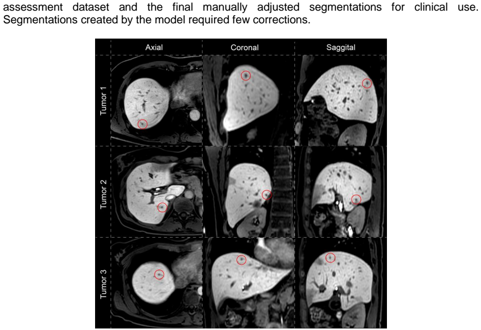
The central claim is that the hepatobiliary phase of gadoxetic acid-enhanced MRI carries enough contrast information for a self-configuring nnU-Net to delineate all five structures that matter for liver surgery planning, and that the resulting segmentations are clinically usable with only light manual touch-up. On the 18-patient test set, mean Dice similarity coefficients (0-to-1 overlap scores) were 0.97 for liver parenchyma, 0.80 for the hepatic vein, 0.79 for the biliary tree, 0.77 for tumors, and 0.74 for the portal vein; the average tumor detection rate was 76.6%, with a median of one false positive per patient. In the 10-patient assessment dataset collected after integration into clinical practice, the parenchyma reached 1.00, portal vein 0.98, hepatic vein 0.95, and tumor 0.80, with the largest manual corrections needed for tumors in patients with prior liver interventions, high tumor burden, or low scan quality. The workflow time for producing a 3D model fell from several hours to roughly 15 minutes per patient, and the network identified three sub-centimeter malignant lesions that radiologists had not initially reported.
Load-bearing premise
The load-bearing premise is that the manual outlines used for training and evaluation are correct for every structure; if those outlines are noisy, especially for thin vessels and small tumors, the reported overlap scores overstate how precisely the model matches true anatomy.
Editorial extensions
If this is right
- If these results generalize, 3D liver models can be produced for every patient scheduled for liver surgery, not only those treated at centers that can afford hours of manual segmentation.
- Preserving vessel-tree topology means the segmentations can serve as a map for image-guided procedures, where vessel bifurcations are used as registration landmarks.
- Cutting the segmentation step from several hours to about 15 minutes makes 3D planning compatible with normal clinical scheduling on a per-patient basis.
- Automated tumor outlining adds a safety check: in this cohort it surfaced three sub-centimeter lesions initially missed by the radiologist.
- The network handles common anatomical variations such as an absent gallbladder after cholecystectomy and regenerated anatomy after prior resection, so it does not fail at the edges of a typical liver-surgery population.
Reading between the lines
- A testable extension is external validation: running the trained network on multi-center MRI data with different scanners and protocols would show whether the reported vessel accuracy holds across imaging setups or is tied to one protocol.
- Since inter-observer variability of the manual reference was not reported, computing pairwise expert-vs-expert Dice on the same test scans would reveal how much of the model's apparent error is actually reference noise, especially for thin vessels and small tumors.
- The three incidentally detected sub-centimeter tumors suggest a second-reader role for the model, but establishing that requires a blinded prospective reader study comparing radiologists with and without the model's output.
- A natural next step is adding diffusion-weighted imaging or arterial-phase input to raise tumor detection beyond the current 76.6%; the trade-off in false positives is directly testable with the same evaluation protocol.
Signed reviews
Editorial analysis
A structured set of objections, weighed in public.
Referee Report
Summary. This manuscript reports the development and clinical integration of an nnU-Net-based automated segmentation method for hepatic anatomy—liver parenchyma, tumors, portal vein, hepatic vein, and biliary tree—from the hepatobiliary phase of gadoxetic acid-enhanced MRI. Manual segmentations from 90 patients were used to train (n=72) and test (n=18) the model, with an additional assessment cohort of 10 patients where model outputs were manually refined for clinical use. The authors report test-set Dice similarity coefficients of 0.97 for parenchyma, 0.80 for hepatic vein, 0.79 for biliary tree, 0.77 for tumors, and 0.74 for portal vein, together with a mean tumor detection rate of 76.6% and a median of one false positive per patient. In the assessment set, high DSC values are reported for parenchyma, portal vein, and hepatic vein after manual refinement, and the authors state that segmentation time decreased from several hours to about 15 minutes per patient. The paper concludes that the method enables accurate automated delineation and supports broader adoption of 3D planning in liver surgery.
Significance. If the reported performance holds under independent verification, the work has clear translational value: it addresses a real clinical bottleneck in liver surgery planning, uses a clinically relevant MRI phase, and includes a prospective assessment of workflow impact. The comparison with prior MRI-based liver segmentation studies (Zbinden et al., Ivashchenko et al., Oh et al.) is useful positioning. The strengths include a held-out test set, a consecutively collected single-center cohort, clinical integration with manual refinement, and reporting of tumor detection rate and false positives. However, the central quantitative claims rest on three points that are not currently established: the reliability of the manual ground truth, the contribution of the customized loss function, and the interpretation of the assessment-set DSC as evidence of accuracy. These are load-bearing because the weakest structures (portal vein, biliary tree, tumors) are exactly those where annotation variability and loss-function choices are most consequential.
major comments (4)
- [Methods, Manual segmentations; Results, Quantitative evaluation] The manuscript uses manual segmentations by two technical physicians, confirmed by a hepatobiliary surgeon, as ground truth for all five structures, but reports no inter-observer variability, no repeat-segmentation study, and no independent second contour for any structure. This is load-bearing because the structures with the lowest DSCs—portal vein (0.74±0.06), biliary tree (0.79±0.07), hepatic vein (0.80±0.04), and tumors (0.77±0.17)—are precisely the structures where boundary definition and lesion identification are most subjective. Without a human-human DSC baseline, the reader cannot separate model error from annotation noise, and the conclusion that the model provides 'accurate delineation' is not supported. Please provide an inter-observer variability measurement (e.g., DSC between independent manual segmentations) on a representative subset, or alternatively report the uncertainty this introduces into all quoted DSC values.
- [Methods, Automated segmentation model] The methodological claim that the combination of clDice and bootstrapped cross-entropy improves thin-structure delineation and topology preservation is not tested. The paper reports no ablation against a standard nnU-Net baseline (e.g., default cross-entropy plus Dice loss) on the same training and test splits. Since nnU-Net v1 with default settings is a strong baseline, the observed DSC values could be attributable to the architecture and data rather than to the customized loss. Moreover, the bootstrapped cross-entropy schedule (K growing from 15% to 50% over the final 100 epochs after a 400-epoch warm-up) introduces several free hyperparameters without sensitivity analysis. Please add an ablation study comparing (i) default nnU-Net, (ii) default nnU-Net plus clDice, and (iii) the full proposed loss combination, reporting DSC and topology metrics for at least the vascular and biliary structures.
- [Results, Performance of the network in prospective use; Discussion] The assessment-set evaluation compares the automated segmentations to the manually refined versions of those same automated segmentations. A high DSC in this comparison primarily measures the amount of editing performed, not the anatomical accuracy of the model, and the manual refinement process is likely biased toward minimal changes rather than independent re-delineation. The statement that 'minor adjustments were required for clinical use' is therefore not equivalent to anatomical accuracy. Please either compare assessment-set outputs to an independent manual segmentation of the assessment scans, or reframe this analysis explicitly as a measure of editing effort, supported by quantitative edit-distance or time metrics rather than DSC.
- [Discussion, Limitations; Results, Prospective tumor detection] The paper claims that 'the model detected three additional tumors initially missed by radiologists,' but this is an anecdotal observation from prospective clinical use without a systematic reference standard or an evaluation protocol for detection sensitivity in the assessment cohort. Given that the test-set tumor detection rate is only 76.6% with a median of one false positive per patient, this claim should be contextualized as a case observation, not a quantitative result. The external-validation limitation is explicitly acknowledged in the Discussion, and I agree it is important; however, given the conclusion advocating standard-of-care use, the absence of external or multi-center validation should also be reflected in the abstract and conclusion as a qualification of the generalizability claim.
minor comments (6)
- [Results, Quantitative evaluation; Figure 2] The text reports a combined DSC of 0.84±0.06 for 'gallbladder (central biliary tree) and bile ducts (peripheral biliary tree),' while the overall biliary tree DSC is 0.79±0.07. Please clarify whether these are separate structures or pooled in the figure, and define how cholecystectomy cases are handled in the calculation of the combined value.
- [Table 1] The pixel-spacing row reads '-1.5 1' and the footnote explains that negative pixel spacing indicates partially overlapping slices, but this formatting is confusing. Please present the acquisition and reconstruction voxel sizes more clearly, and specify which spacing value is used by nnU-Net for resampling.
- [Statistical analysis] The statistical analysis section states that DSC scores were compared using an unpaired Mann-Whitney test, but it does not state which groups were compared. Please specify the comparisons (e.g., central vs. peripheral vessels, training vs. test demographics) and correct the Python/SciPy version and the SPSS version if needed.
- [Introduction and Discussion] Reference 29 (Strahler, 1957) is listed in the references but does not appear to be cited in the main text. If it is used for the notion of vessel topology or branching order, please cite it explicitly where that concept is introduced.
- [Throughout] The framework is referred to inconsistently as 'nnUNet' and 'nnU-Net'; please use a single consistent spelling, preferably 'nnU-Net' as in the reference to Isensee et al.
- [Discussion, comparison with prior work] The comparison with Oh et al. reports higher DSC values for the proposed method across all structures, but no statistical comparison or matching of annotation protocols is provided. Please state whether the comparison is descriptive only, and note any differences in tumor inclusion criteria, vessel definition, or evaluation methodology that could affect the comparison.
Circularity Check
No significant circularity: test-set evaluation is held out, the loss design is not fitted to the test set, and the assessment-set DSCs are transparently framed as editing-effort measurements.
full rationale
The paper's central claim is an empirical performance evaluation rather than a derivation, so the main circularity patterns do not apply. Test-set DSCs (Figure 2) are computed on an 18-patient held-out split against manual segmentations that were not used to fit the network; the clDice + bootstrapped cross-entropy schedule was fixed before evaluation and was not tuned on the test set. The clDice citation (ref. 25) includes a co-author of this paper, but it is used as a published, externally implemented loss function, and no uniqueness or forced-choice argument rests on it. The assessment dataset compares the network output with a manual refinement of that same output; the manuscript explicitly labels this as quantifying required manual adjustments/editing effort ("quantify required adjustments using DSC"), not as an independent anatomical validation, so it does not constitute a prediction smuggled in as accuracy. Absence of inter-observer variability and of external validation are correctness/robustness limitations rather than circularity. No step in the paper reduces by construction to its own inputs.
Assumptions & free parameters
free parameters (4)
- Bootstrapped cross-entropy K schedule =
K grows linearly from 15% to 50% over the final 100 of 500 epochs
- Warm-up duration for bootstrapped cross-entropy =
400 epochs with K=100%
- Total training length =
500 epochs
- Loss combination weights =
not specified
assumptions (4)
- domain assumption Manual segmentations are accurate ground truth for all structures.
- domain assumption The 90-patient single-center cohort represents the clinical population for standard-of-care deployment.
- domain assumption clDice and bootstrapped cross-entropy improve topologically accurate vessel segmentation on this task.
- domain assumption Spatial resolution of 2.0x2.0x3.0 mm is adequate for delineating the target structures.
Cite this review
Pith. "Pith review of Automated surgical planning with nnU-Net: delineation of the anatomy in hepatobiliary phase MRI." pith.science (2026). https://pith.science/paper/G476NGVM
@misc{pith2026250814133,
  author       = {Pith},
  title        = {Pith review of: Automated surgical planning with nnU-Net: delineation of the anatomy in hepatobiliary phase MRI},
  year         = {2026},
  howpublished = {\url{https://pith.science/paper/G476NGVM}},
  note         = {Machine review of arXiv:2508.14133}
}
read the original abstract
Background: The aim of this study was to develop and evaluate a deep learning-based automated segmentation method for hepatic anatomy (i.e., parenchyma, tumors, portal vein, hepatic vein and biliary tree) from the hepatobiliary phase of gadoxetic acid-enhanced MRI. This method should ease the clinical workflow of preoperative planning. Methods: Manual segmentation was performed on hepatobiliary phase MRI scans from 90 consecutive patients who underwent liver surgery between January 2020 and October 2023. A deep learning network (nnU-Net v1) was trained on 72 patients with an extra focus on thin structures and topography preservation. Performance was evaluated on an 18-patient test set by comparing automated and manual segmentations using Dice similarity coefficient (DSC). Following clinical integration, 10 segmentations (assessment dataset) were generated using the network and manually refined for clinical use to quantify required adjustments using DSC. Results: In the test set, DSCs were 0.97+/-0.01 for liver parenchyma, 0.80+/-0.04 for hepatic vein, 0.79+/-0.07 for biliary tree, 0.77+/-0.17 for tumors, and 0.74+/-0.06 for portal vein. Average tumor detection rate was 76.6+/-24.1%, with a median of one false-positive per patient. The assessment dataset showed minor adjustments were required for clinical use of the 3D models, with high DSCs for parenchyma (1.00+/-0.00), portal vein (0.98+/-0.01) and hepatic vein (0.95+/-0.07). Tumor segmentation exhibited greater variability (DSC 0.80+/-0.27). During prospective clinical use, the model detected three additional tumors initially missed by radiologists. Conclusions: The proposed nnU-Net-based segmentation method enables accurate and automated delineation of hepatic anatomy. This enables 3D planning to be applied efficiently as a standard-of-care for every patient undergoing liver surgery.
Figures


Figures from the paper (3 more)


Reference graph
Works this paper leans on
-
[1]
Expanding Criteria for Resectability of Colorectal Liver Metastases
Pawlik TM, Schulick RD, Choti MA. Expanding Criteria for Resectability of Colorectal Liver Metastases. Oncologist. 2008;13(1):51–64
work page 2008
-
[2]
Evrard S, Torzilli G, Caballero C, Bonhomme B. Parenchymal sparing surgery brings treatment of colorectal liver metastases into the precision medicine era. Eur J Cancer. 2018 Nov 1;104:195–200
work page 2018
-
[3]
Boedecker C, Huettl F, Saalfeld P, Paschold M, Kneist W, Baumgart J, et al. Using virtual 3D-models in surgical planning: workflow of an immersive virtual reality application in liver surgery. Langenbeck’s Arch Surg. 2021;406(3):911–5
work page 2021
-
[4]
Fang C, An J, Bruno A, Cai X, Fan J, Fujimoto J, et al. Consensus recommendations of three-dimensional visualization for diagnosis and management of liver diseases. Hepatol Int. 2020 Jul 1;14(4):437–53
work page 2020
-
[5]
Zeng X, Tao H, Dong Y, Zhang Y, Yang J, Xuan F, et al. Impact of three-dimensional reconstruction visualization technology on short-term and long-term outcomes after hepatectomy in patients with hepatocellular carcinoma: a propensity-score-matched and inverse probability of treatment-weighted multicenter stud. Int J Surg. 2024;110(3):1663– 76
work page 2024
-
[6]
Ultrasound-based navigation for open liver surgery using active liver tracking
Smit JN, Kuhlmann KFD, Ivashchenko O V., Thomson BR, Langø T, Kok NFM, et al. Ultrasound-based navigation for open liver surgery using active liver tracking. Int J Comput Assist Radiol Surg. 2022 Oct 1;17(10):1765–73
work page 2022
-
[7]
A surgical navigation system to aid the ablation of vanished colorectal liver metastases
Olthof K, Smit J, Fusaglia M, Kok N, Ruers T, Kuhlmann K. A surgical navigation system to aid the ablation of vanished colorectal liver metastases. Br J Surg. 2024 May 3;111(5)
work page 2024
-
[8]
Deep learning for image-based liver analysis — A comprehensive review focusing on malignant lesions
Survarachakan S, Prasad PJR, Naseem R, Pérez de Frutos J, Kumar RP, Langø T, et al. Deep learning for image-based liver analysis — A comprehensive review focusing on malignant lesions. Vol. 130, Artificial Intelligence in Medicine. 2022. p. 102331
work page 2022
Show all 30 references
-
[9]
Deep learning techniques for liver and liver tumor segmentation: A review
Gul S, Khan MS, Bibi A, Khandakar A, Ayari MA, Chowdhury MEH. Deep learning techniques for liver and liver tumor segmentation: A review. Comput Biol Med. 2022 Aug 1;147:105620
2022
-
[10]
Jhaveri KS, Fischer SE, Hosseini-Nik H, Sreeharsha B, Menezes RJ, Gallinger S, et al. Prospective comparison of gadoxetic acid-enhanced liver MRI and contrast-enhanced CT with histopathological correlation for preoperative detection of colorectal liver metastases following che...
2017
-
[11]
Vreugdenburg TD, Ma N, Duncan JK, Riitano D, Cameron AL, Maddern GJ. Comparative diagnostic accuracy of hepatocyte-specific gadoxetic acid (Gd-EOB-DTPA) enhanced MR imaging and contrast enhanced CT for the detection of liver metastases: a systematic review and meta-analysis. V...
2016
-
[12]
Colorectal liver metastases: CT, MR imaging, and PET for diagnosis - Meta-analysis
Bipat S, Van Leeuwen MS, Comans EFI, Pijl MEJ, Bossuyt PMM, Zwinderman AH, et al. Colorectal liver metastases: CT, MR imaging, and PET for diagnosis - Meta-analysis. Radiology. 2005 Oct;237(1):123–31
2005
-
[13]
Automated MRI liver segmentation for anatomical segmentation, liver volumetry, and the extraction of radiomics
Gross M, Huber S, Arora S, Ze’evi T, Haider SP, Kucukkaya AS, et al. Automated MRI liver segmentation for anatomical segmentation, liver volumetry, and the extraction of radiomics. Eur Radiol. 2024 Aug 1;34(8):5056–65
2024
-
[14]
Automated segmentation of liver segment on portal venous phase MR images using a 3D convolutional neural network
Han X, Wu X, Wang S, Xu L, Xu H, Zheng D, et al. Automated segmentation of liver segment on portal venous phase MR images using a 3D convolutional neural network. Insights Imaging. 2022 Dec 1;13(1)
2022
-
[15]
Deep Learning Framework for Liver Segmentation from T1-Weighted MRI Images
Hossain MSA, Gul S, Chowdhury MEH, Khan MS, Sumon MSI, Bhuiyan EH, et al. Deep Learning Framework for Liver Segmentation from T1-Weighted MRI Images. Sensors (Basel). 2023 Nov 1;23(21):8890
2023
-
[16]
Training robust T1-weighted magnetic resonance imaging liver segmentation models using ensembles of datasets with different contrast protocols and liver disease etiologies
Patel N, Celaya A, Eltaher M, Glenn R, Savannah KB, Brock KK, et al. Training robust T1-weighted magnetic resonance imaging liver segmentation models using ensembles of datasets with different contrast protocols and liver disease etiologies. Sci Rep. 2024;14(1):20988
2024
-
[17]
Convolutional neural network for automated segmentation of the liver and its vessels on non-contrast T1 vibe Dixon acquisitions
Zbinden L, Catucci D, Suter Y, Berzigotti A, Ebner L, Christe A, et al. Convolutional neural network for automated segmentation of the liver and its vessels on non-contrast T1 vibe Dixon acquisitions. Sci Rep. 2022 Dec 21;12(1):1–11
2022
-
[18]
Automated 3D liver segmentation from hepatobiliary phase MRI for enhanced preoperative planning
Oh N, Kim JH, Rhu J, Jeong WK, Choi G seong, Kim JM, et al. Automated 3D liver segmentation from hepatobiliary phase MRI for enhanced preoperative planning. Sci Rep. 2023;13(1):17605
2023
-
[19]
Liver vessel MRI image segmentation based on dual-path diffusion model
Wu R, Peng Y, Wu S, Peng Z, Li Y, Zhou M, et al. Liver vessel MRI image segmentation based on dual-path diffusion model. J Radiat Res Appl Sci. 2024 Sep 1;17(3):101025
2024
-
[20]
A workflow for automated segmentation of the liver surface, hepatic vasculature and biliary tree anatomy from multiphase MR images
Ivashchenko O V., Rijkhorst EJ, ter Beek LC, Hoetjes NJ, Pouw B, Nijkamp J, et al. A workflow for automated segmentation of the liver surface, hepatic vasculature and biliary tree anatomy from multiphase MR images. Magn Reson Imaging. 2020 May 1;68:53–65
2020
-
[21]
3D Slicer as an image computing platform for the Quantitative Imaging Network
Fedorov A, Beichel R, Kalpathy-Cramer J, Finet J, Fillion-Robin JC, Pujol S, et al. 3D Slicer as an image computing platform for the Quantitative Imaging Network. Magn Reson Imaging. 2012 Nov;30(9):1323–41
2012
-
[22]
nnU-Net: a self-configuring method for deep learning-based biomedical image segmentation
Isensee F, Jaeger PF, Kohl SAA, Petersen J, Maier-Hein KH. nnU-Net: a self-configuring method for deep learning-based biomedical image segmentation. Nat Methods. 2021 Dec 7;18(2):203–11
2021
-
[23]
Deep interactive thin object selection
Liew JH, Cohen S, Price B, Mai L, Feng J. Deep interactive thin object selection. In: Proceedings - 2021 IEEE Winter Conference on Applications of Computer Vision, WACV
2021
-
[24]
Full-resolution residual networks for semantic segmentation in street scenes
Pohlen T, Hermans A, Mathias M, Leibe B. Full-resolution residual networks for semantic segmentation in street scenes. In: Proceedings - 30th IEEE Conference on Computer Vision and Pattern Recognition, CVPR 2017. Institute of Electrical and Electronics Engineers Inc.; 2017. p. 3309–18
2017
-
[25]
CLDICE - A novel topology-preserving loss function for tubular structure segmentation
Shit S, Paetzold JC, Sekuboyina A, Ezhov I, Unger A, Zhylka A, et al. CLDICE - A novel topology-preserving loss function for tubular structure segmentation. In: Proceedings of the IEEE Computer Society Conference on Computer Vision and Pattern Recognition. IEEE Computer Societ...
2021
-
[26]
Optimization of hepatic vasculature segmentation from contrast-enhanced MRI, exploring two 3D Unet modifications and various loss functions
Ivashchenko O V., Zhai Z, Stoel B, Ruers TJM. Optimization of hepatic vasculature segmentation from contrast-enhanced MRI, exploring two 3D Unet modifications and various loss functions. In SPIE-Intl Soc Optical Eng; 2021. p. 16
2021
-
[27]
Liver segmentation and metastases detection in MR images using convolutional neural networks
Jansen MJA, Kuijf HJ, Niekel M, Veldhuis WB, Wessels FJ, Viergever MA, et al. Liver segmentation and metastases detection in MR images using convolutional neural networks. J Med Imaging. 2019 Oct 15;6(04):1
2019
-
[28]
Clinical Implementation of In-House Developed MR-Based Patient-Specific 3D Models of Liver Anatomy
Ivashchenko O V., Smit JN, Nijkamp J, Ter Beek LC, Rijkhorst EJ, Kok NFM, et al. Clinical Implementation of In-House Developed MR-Based Patient-Specific 3D Models of Liver Anatomy. Eur Surg Res. 2021 Feb 16;61(4–5):143–52
2021
-
[29]
Quantitative analysis of watershed geomorphology
Strahler AN. Quantitative analysis of watershed geomorphology. Eos, Trans Am Geophys Union. 1957 Dec 1;38(6):913–20
1957
-
[2021]
Institute of Electrical and Electronics Engineers Inc.; 2021. p. 305–14
2021
Reviewed August 15, 2026 · model on record in the stance chip above.
Discussion (0). Continue with ORCID to comment.