REVIEW 4 major objections 6 minor 22 references
MRI-only landmark extraction for spinal digital twins achieves subpixel accuracy by estimating each vertebra's orientation instead of assuming image alignment.
Reviewed by Pith at T0; open to challenge. T0 means a machine referee read the full paper against a public rubric. the ladder, T0–T4 →
A rule-based pipeline estimates vertebral orientation and extracts landmark points from MRI for biomechanical spine models.
T0 review reviewed 2026-08-05 challenge →
load-bearing objection Useful incremental MRI adaptation of the authors' CT landmark pipeline with a clever orientation fix; the 'subpixel-accurate' claim is not measured and the hand-picked raycasting directions are a real gap. the 4 major comments →
Rule-based Key-Point Extraction for MR-Guided Biomechanical Digital Twins of the Spine
The pith
A machine-rendered reading of the paper's core claim, the machinery that carries it, and where it could break.
The reading
Core claim
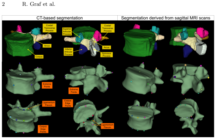
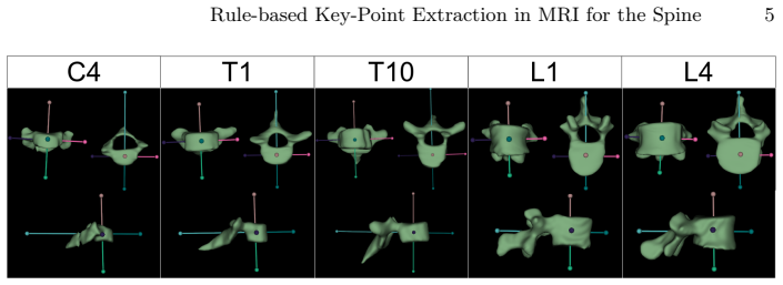
The paper's central claim is that a segmentation-only, rule-based pipeline can extract subpixel-accurate key points from low-resolution T2-weighted MRI by replacing image-axis assumptions with a per-vertebra local coordinate system and by locating boundaries through interpolated bisection. The front/back axis is computed by projecting the arcus and spinous process masks onto the plane orthogonal to the spline-defined up/down axis and joining their 2D center of mass to the vertebral corpus center; left/right follows as the cross product. Process endpoints are then obtained by raycasting along local directions that were empirically chosen to match anatomy, while vertebral body corners and liga
What carries the argument
The load-bearing mechanism is a per-vertebra orthonormal local coordinate system combined with raycasting and sub-voxel bisection. The up/down axis comes from a spline through vertebral body centers of mass; the front/back axis comes from projecting arcus and spinous process masks onto the plane orthogonal to that spline and connecting their 2D center of mass to the corpus center; left/right is the cross product. All landmarks are then defined by raycasting along local axes, or empirically chosen combinations of them, with corpus corners and ligamentum flavum points located by a 2D bisection search that interpolates at subvoxel positions.
Load-bearing premise
The load-bearing premise is that the hand-picked raycasting directions—0.5 lateral plus 0.5 posterior for transverse processes and down plus 0.2 posterior for the spinous process—match real anatomy across patients; if those direction coefficients are wrong for unseen populations or pathologies, every process endpoint shifts systematically even when the orientation estimate is correct.
What would settle it
Scan a cohort of patients with both CT and sagittal T2 MRI, compute POIs from the CT pipeline and from this MRI pipeline in the same world coordinate system, and measure the Euclidean distance between matched landmarks; systematic deviations that grow with scoliotic curvature or with transverse-process morphology would show the empirical raycasting directions do not generalize. A simpler check: rotate a high-resolution volume by known angles and verify that the computed landmarks rotate with the vertebra rather than drifting relative to it.
If this is right
- Multibody simulations of the spine can be built from routine MRI alone, avoiding radiation exposure and adding soft-tissue context that CT-based models lack.
- Rotated or scoliotic spines no longer need manual alignment before landmark extraction, since the pipeline derives a local coordinate system for every vertebra.
- Because the method runs on segmentation masks and completes in under a minute per spine, it scales to large cohorts and can feed subject-specific simulation studies.
- The landmark outputs can be corrected and used as training data for learning-based predictors, which the paper identifies as the natural next step beyond accumulated rule exceptions.
Where Pith is reading between the lines
- The fixed raycasting coefficients (0.5 lateral + 0.5 posterior for transverse processes, 0.2 posterior offset for the spinous process) are anatomical heuristics, not derived quantities; a matched CT-MRI comparison in the same subjects would reveal whether they generalize to scoliotic or morphologically unusual spines.
- Since the pipeline's accuracy ceiling is set by the segmentation, improving the effective resolution of the left-right axis, for example through super-resolution or better segmentation, should directly improve landmark placement.
- The large intervertebral forces at corner points that the authors attribute to the ligament model, not the landmarks, suggest MRI's soft-tissue contrast could eventually support extracting the actual ligament path instead of heuristic attachment offsets.
Editorial analysis
A structured set of objections, weighed in public.
Referee Report
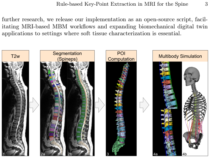
Summary. The paper adapts a rule-based CT key-point extraction pipeline to MRI for generating points of interest (POIs) for multibody spine models. The method takes SPINEPS segmentations from sagittal T2-weighted MRI, estimates a local vertebral coordinate system using a 2D projection of posterior structures, computes process endpoints by raycasting along hand-specified direction vectors, and localizes vertebral body corners and ligamentum flavum points via subvoxel bisection. The authors evaluate the orientation estimate on 90 CT vertebrae (Table 1) and report a qualitative expert assessment on 37 full-spine segmentations, arguing that the method is robust to low resolution and spinal curvature. The central claim is subpixel-accurate key-point extraction from MRI for biomechanical digital twins.
Significance. If fully validated, the contribution is valuable: it extends an existing CT-based biomechanical modeling pipeline to MRI, which is radiation-free and better suited for soft-tissue characterization; it is open-source and builds on publicly available segmentation tools; and it directly addresses vertebral orientation in the presence of deformities such as scoliosis. The orientation estimation results (mean error 1.72±1.76°, 80% below 3°, 100% below 10°) are promising. However, the headline claim of 'subpixel-accurate key-point extraction' is not yet supported by the evidence: the only quantitative evaluation concerns vertebral orientation, not key-point position, and the downstream MBS validation is anecdotal. The hand-picked raycasting directions are a potential source of systematic, reproducible error that the current validation cannot detect.
major comments (4)
- [Section 2.2] The raycasting directions for the transverse and spinous processes (a = 0.5l + 0.5p and a = d + 0.2p) are stated to be 'empirically chosen to best match the observed anatomical trajectories,' but no sensitivity analysis or independent validation of these coefficients is provided. These endpoints define muscle and ligament attachment sites in the MBS, so a systematic error here directly propagates into the biomechanical model. The coefficients are not stratified by vertebral level, despite known differences between cervical, thoracic, and lumbar process orientations, nor tested on pathological spines. Because the pipeline is deterministic, an incorrect direction produces a reproducible bias that the subvoxel bisection cannot correct. I recommend a quantitative comparison of the computed process endpoints against expert-annotated landmarks or CT-derived POIs on the same subjects, with per-
- [Section 2.3 and Abstract] The claim of 'subpixel-accurate' or 'subvoxel-accurate' landmark placement conflates numerical precision with measurement accuracy. The bisection search with interpolation guarantees that the reported coordinate can be placed at subvoxel resolution, but it does not establish that this coordinate is anatomically correct. Given the low left–right resolution of clinical sagittal T2-weighted MRI (3–4 mm), the segmentation boundary itself is uncertain, and no ground-truth comparison of key-point positions on MRI is presented. The phrase 'subpixel-accurate' in the Abstract should be replaced by a more precise statement, such as 'subvoxel-precise extraction,' unless a direct accuracy evaluation against independent reference landmarks is added.
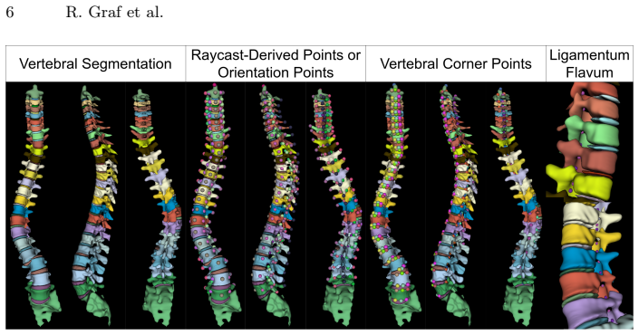
- [Section 3.2] The validation for the final key-point output is anecdotal. The authors state that two experts assessed 37 full-spine segmentations and that failures occurred only when the segmentation was flawed, but no quantitative scoring, inter-observer variability, error metrics, or case-level statistics are reported. The statement that 'we validated our point placement using an existing MBS framework [13]' is also weak evidence, because the framework originates from the same group and no quantitative comparison of MBS outputs (e.g., joint angles, loads, or ligament forces) is given. This section does not substantiate the reliability of the POIs for biomechanical simulation. A quantitative landmark error study, or at minimum a structured expert rating with defined criteria and agreement statistics, is needed.
- [Section 3.1 and Table 1] The orientation evaluation is the only quantitative experiment and it is limited in important ways: it is performed on CT, not on the target MRI modality; the manual angular measurements lack inter-observer variability; and the 90 vertebrae appear to come from non-pathological or mixed VerSe2020 subjects, with no explicit inclusion of scoliotic cases. The paper's motivation emphasizes scoliosis, but the only scoliosis evidence is the qualitative Figure 4. While the orientation method is segmentation-based and may transfer across modalities, the reported numbers do not directly support the MRI-specific claim. Please clarify how representative the evaluated vertebrae are of the target population and report measurement repeatability.
minor comments (6)
- [Section 2.1] Heading typo: 'V ertebra Orientation' should be 'Vertebra Orientation'.
- [Section 2.3] Heading typo: 'Sub-V oxel' should be 'Sub-Voxel'.
- [Abstract and Section 2.3] The terms 'subpixel' and 'subvoxel' are used interchangeably; please standardize, as 'pixel' and 'voxel' have different meanings in 2D and 3D.
- [Section 3.2] Please report the acquisition parameters of the 37 MRI scans, the number of subjects, and any inclusion/exclusion criteria, as this is important for reproducibility and for judging generalizability.
- [Section 2.3] The predefined precision threshold for the bisection search is not stated. Please give the actual value used in the experiments.
- [Table 1] The table would benefit from confidence intervals or a statistical test between the three methods; as presented, the standard deviations are large and the reader cannot assess the significance of the improvement.
Circularity Check
Validation leans on the authors' own CT/MBS pipeline; the core geometry derivation is otherwise self-contained.
specific steps
-
self citation load bearing
[Section 3.2, 'Points for Multi-Body Simulation']
"We validated our point placement using an existing MBS framework [13]. Despite operating at a lower resolution, we observed no large discrepancy for straight spines, compared to existing CT-based point extraction."
The MRI pipeline is explicitly 'the POI generation code developed in Lerchl et. al. [13]' (Sec. 2), and [13] is the same group's CT-based method. The validation therefore checks that the adapted MRI points agree with points produced by their own source code. Since both share the same raycasting rules and coordinate logic, 'no large discrepancy' is a self-consistency check, not an independent anatomical ground-truth test. The claim of reliability for MBS use is thus partially supported by a self-citation chain. This does not invalidate the orientation estimation, which is tested against manual measurements on VerSe2020, but the downstream POI validation is circular in the narrow sense of comparing a method to its own progenitor.
full rationale
The derivation of the POIs is rule-based and not claimed to follow from a theorem; the orientation estimation is an independent algorithm evaluated against manual measurements on an external CT dataset (VerSe2020), so the central novelty has non-circular support. The empirically chosen raycast directions (Sec. 2.2) are transparent free parameters rather than fitted predictions, and the subvoxel bisection (Sec. 2.3) is a definitional property of the interpolation, not a claim that the segmentation boundary equals the anatomical landmark. The main circularity-adjacent element is in Sec. 3.2: the MRI points are validated by comparing them to 'existing CT-based point extraction' and an 'existing MBS framework [13]', both from the same group and the same software lineage being adapted. This makes the observed agreement a consistency check with a self-citation, and it is load-bearing for the claim that the points are usable for MBS. Because the orientation evaluation and the expert qualitative check provide partially independent evidence, the paper is not wholly circular; but the downstream validation should not be read as independent confirmation.
Axiom & Free-Parameter Ledger
free parameters (4)
- Transverse process raycast blend =
0.5 * lateral + 0.5 * posterior
- Spinous process raycast blend =
0.2 * posterior added to inferior
- Vertebra-dependent shift factor =
f = (12 - vid)/11 + 1 for vid <= 11
- Bisection precision threshold =
not specified
axioms (3)
- domain assumption SPINEPS segmentation provides accurate vertebra subregion masks
- domain assumption Spline through vertebral body centers of mass defines the superior-inferior direction
- ad hoc to paper Projection of posterior structures onto a plane orthogonal to the superior-inferior axis removes out-of-plane asymmetry
Cite this review
Pith. "Pith review of Rule-based Key-Point Extraction for MR-Guided Biomechanical Digital Twins of the Spine." pith.science (2026). https://pith.science/paper/X2EHCSEF
@misc{pith2026250814708,
  author       = {Pith},
  title        = {Pith review of: Rule-based Key-Point Extraction for MR-Guided Biomechanical Digital Twins of the Spine},
  year         = {2026},
  howpublished = {\url{https://pith.science/paper/X2EHCSEF}},
  note         = {Machine review of arXiv:2508.14708}
}
read the original abstract
Digital twins offer a powerful framework for subject-specific simulation and clinical decision support, yet their development often hinges on accurate, individualized anatomical modeling. In this work, we present a rule-based approach for subpixel-accurate key-point extraction from MRI, adapted from prior CT-based methods. Our approach incorporates robust image alignment and vertebra-specific orientation estimation to generate anatomically meaningful landmarks that serve as boundary conditions and force application points, like muscle and ligament insertions in biomechanical models. These models enable the simulation of spinal mechanics considering the subject's individual anatomy, and thus support the development of tailored approaches in clinical diagnostics and treatment planning. By leveraging MR imaging, our method is radiation-free and well-suited for large-scale studies and use in underrepresented populations. This work contributes to the digital twin ecosystem by bridging the gap between precise medical image analysis with biomechanical simulation, and aligns with key themes in personalized modeling for healthcare.
Figures
Reference graph
Works this paper leans on
-
[1]
Journal of biomechanical engineering 144(10), 101007 (2022) 10 R
Balasubramanian, S., D’Andrea, C.R., Viraraghavan, G., Cahill, P.J.: Development of a finite element model of the pediatric thoracic and lumbar spine, ribcage, and pelvis with orthotropic region-specific vertebral growth. Journal of biomechanical engineering 144(10), 101007 (2022) 10 R. Graf et al
work page 2022
-
[2]
In: International Conference on Medical Image Computing and Computer-Assisted Intervention
Bürgin, V., Prevost, R., Stollenga, M.F.: Robust vertebra identification using si- multaneous node and edge predicting graph neural networks. In: International Conference on Medical Image Computing and Computer-Assisted Intervention. pp. 483–493. Springer (2023)
work page 2023
-
[3]
Multibody System Dynamics 30, 413–432 (2013)
Christophy, M., Curtin, M., Faruk Senan, N.A., Lotz, J.C., O’Reilly, O.M.: On the modeling of the intervertebral joint in multibody models for the spine. Multibody System Dynamics 30, 413–432 (2013)
work page 2013
-
[4]
Couvertier, M., Germaneau, A., Saget, M., Dupré, J.C., Doumalin, P., Brémand, F., Hesser, F., Brèque, C., Roulaud, M., Monlezun, O., et al.: Biomechanical anal- ysis of the thoracolumbar spine under physiological loadings: Experimental motion data corridors for validation of finite element models. Proceedings of the Institu- tion of Mechanical Engineers, ...
work page 2017
-
[5]
Scientific Reports10(1), 21519 (2020)
El Bojairami, I., El-Monajjed, K., Driscoll, M.: Development and validation of a timely and representative finite element human spine model for biomechanical simulations. Scientific Reports10(1), 21519 (2020)
work page 2020
-
[6]
Fürmetz, J., Daniel, T., Sass, J., Bergsträßer, M., Degen, N., Suero, E., Ahrens, P., Keppler, A., Böcker, W., Thaller, P., et al.: Three-dimensional assessment of patellofemoral anatomy: Reliability and reference ranges. The Knee29, 271–279 (2021)
work page 2021
-
[7]
Fürmetz, J., Sass, J., Ferreira, T., Jalali, J., Kovacs, L., Mück, F., Degen, N., Thaller, P.: Three-dimensional assessment of lower limb alignment: accuracy and reliability. The Knee26(1), 185–193 (2019)
work page 2019
-
[8]
Galbusera, F., Niemeyer, F., Bassani, T., Sconfienza, L.M., Wilke, H.J.: Estimating the three-dimensional vertebral orientation from a planar radiograph: Is it feasible? Journal of biomechanics102, 109328 (2020)
work page 2020
-
[9]
European Radiology Experimental7(1), 70 (2023)
Graf, R., Schmitt, J., Schlaeger, S., Möller, H.K., Sideri-Lampretsa, V., Sekuboy- ina, A., Krieg, S.M., Wiestler, B., Menze, B., Rueckert, D., et al.: Denoising diffusion-based MRI to CT image translation enables automated spinal segmenta- tion. European Radiology Experimental7(1), 70 (2023)
work page 2023
-
[10]
Computer methods in biomechanics and biomedical engineering 18(2), 175–184 (2015)
Huynh, K., Gibson, I., Jagdish, B., Lu, W.: Development and validation of a dis- cretised multi-body spine model in lifemod for biodynamic behaviour simulation. Computer methods in biomechanics and biomedical engineering 18(2), 175–184 (2015)
work page 2015
-
[11]
Khanal, B., Dahal, L., Adhikari, P., Khanal, B.: Automatic cobb angle detection using vertebra detector and vertebra corners regression. In: International work- shop and challenge on computational methods and clinical applications for spine imaging. pp. 81–87. Springer (2019)
work page 2019
-
[12]
Pattern Recognition and Image Analysis30, 512–522 (2020)
Lachinov, D., Getmanskaya, A., Turlapov, V.: Cephalometric landmark regression with convolutional neural networks on 3d computed tomography data. Pattern Recognition and Image Analysis30, 512–522 (2020)
work page 2020
-
[13]
Frontiers in bioengineering and biotechnology10, 862804 (2022)
Lerchl, T., El Husseini, M., Bayat, A., Sekuboyina, A., Hermann, L., Nispel, K., Baum, T., Löffler, M.T., Senner, V., Kirschke, J.S.: Validation of a patient-specific musculoskeletal model for lumbar load estimation generated by an automated pipeline from whole body ct. Frontiers in bioengineering and biotechnology10, 862804 (2022)
work page 2022
-
[14]
Bioengineering10(2), 202 (2023) Rule-based Key-Point Extraction in MRI for the Spine 11
Lerchl, T., Nispel, K., Baum, T., Bodden, J., Senner, V., Kirschke, J.S.: Multi- body models of the thoracolumbar spine: a review on applications, limitations, and challenges. Bioengineering10(2), 202 (2023) Rule-based Key-Point Extraction in MRI for the Spine 11
work page 2023
-
[15]
https://doi.org/10.1007/s00330-024-11155-y, https://doi.org/10
Möller, H., Graf, R., Schmitt, J., Keinert, B., Schön, H., Atad, M., Sekuboyina, A., Streckenbach, F., Kofler, F., Kroencke, T., Bette, S., Willich, S.N., Keil, T., Niendorf, T., Pischon, T., Endemann, B., Menze, B., Rueckert, D., Kirschke, J.S.: SPINEPS—automatic whole spine segmentation of t2-weighted MR im- ages using a two-phase approach to multi-clas...
-
[16]
Bioengineering10(3), 315 (2023)
Nispel, K., Lerchl, T., Senner, V., Kirschke, J.S.: Recent advances in coupled mbs and fem models of the spine—a review. Bioengineering10(3), 315 (2023)
work page 2023
-
[17]
In: Proceedings of the European conference on computer vision (ECCV) Workshops
O’Neil, A.Q., Kascenas, A., Henry, J., Wyeth, D., Shepherd, M., Beveridge, E., Clunie, L., Sansom, C., Seduikyte Keith Muir, E., Poole, I.: Attaining human-level performance with atlas location autocontext for anatomical landmark detection in 3d ct data. In: Proceedings of the European conference on computer vision (ECCV) Workshops. pp. 0–0 (2018)
work page 2018
-
[18]
Sekuboyina, A., Bayat, A., Husseini, M.E., Löffler, M., Rempfler, M., Kukačka, J., Tetteh, G., Valentinitsch, A., Payer, C., Urschler, M., et al.: Verse: a vertebrae labelling and segmentation benchmark (2020)
work page 2020
-
[19]
BMC oral health 23(1), 876 (2023)
Tao, L., Li, M., Zhang, X., Cheng, M., Yang, Y., Fu, Y., Zhang, R., Qian, D., Yu, H.: Automatic craniomaxillofacial landmarks detection in ct images of individuals with dentomaxillofacial deformities by a two-stage deep learning model. BMC oral health 23(1), 876 (2023)
work page 2023
-
[20]
Spine defor- mity 5(4), 225–230 (2017)
Wren, T.A., Ponrartana, S., Poorghasamians, E., Moreau, S., Aggabao, P.C., Za- slow, T.L., Edison, B.R., Gilsanz, V.: Biomechanical modeling of spine flexibility and its relationship to spinal range of motion and idiopathic scoliosis. Spine defor- mity 5(4), 225–230 (2017)
work page 2017
-
[21]
Bioengineering 11(1), 101 (2024)
Zhang, H., Chung, A.C.: A dual coordinate system vertebra landmark detection network with sparse-to-dense vertebral line interpolation. Bioengineering 11(1), 101 (2024)
work page 2024
-
[22]
Physics in Medicine & Biology68(18), 185011 (2023)
Zhang, Y., Liu, W., Zhao, J., Wang, D., Peng, F., Cui, S., Wang, B., Shi, Z., Liu, B., He, D., et al.: Improving pedicle screw path planning by vertebral posture estimation. Physics in Medicine & Biology68(18), 185011 (2023)
work page 2023
This paper was first reviewed by deepseek-v4-flash on August 5, 2026.
discussion (0)
Sign in with ORCID, Apple, or X to comment. Anyone can read and Pith papers without signing in.