EchoVLM: Dynamic Mixture-of-Experts Vision-Language Model for Universal Ultrasound Intelligence
Pith reviewed 2026-05-18 15:59 UTC · model grok-4.3
The pith
A mixture-of-experts vision-language model trained on seven anatomical regions improves performance on ultrasound report generation, diagnosis, and question answering.
A machine-rendered reading of the paper's core claim, the machinery that carries it, and where it could break.
Core claim
EchoVLM employs a Mixture of Experts architecture in a vision-language model trained on ultrasound data spanning seven anatomical regions. This enables the model to perform ultrasound report generation, diagnosis, and visual question-answering tasks. The design addresses limitations in general-purpose vision-language models, which show poor generalization in multi-organ lesion recognition and low efficiency in multi-task diagnostics.
What carries the argument
The Mixture of Experts architecture that dynamically activates specialized components based on input from different anatomical regions and tasks.
If this is right
- Ultrasound report generation achieves higher overlap with reference texts than general vision-language models.
- The same model supports diagnosis and visual question answering without task-specific retraining.
- Multi-region training reduces the need for separate models per organ or procedure.
- Clinical workflows gain a single system that processes images from varied body areas with consistent output quality.
Where Pith is reading between the lines
- Similar expert-routing designs could extend to other real-time imaging modalities where data variety across body regions is high.
- The approach may lower the expertise barrier for less experienced operators by standardizing output across tasks.
- If multi-region training proves robust, it could inform data collection strategies that prioritize breadth over volume in medical imaging datasets.
Load-bearing premise
Training on data from seven anatomical regions is enough to produce reliable generalization to new ultrasound images and clinical tasks.
What would settle it
Performance on a held-out ultrasound dataset from an eighth anatomical region or a new diagnostic workflow drops to levels comparable to or below general-purpose models.
Figures




read the original abstract
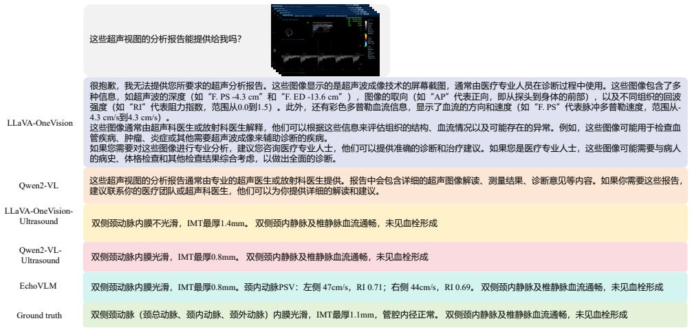
Ultrasound imaging has become the preferred imaging modality for early cancer screening due to its advantages of non-ionizing radiation, low cost, and real-time imaging capabilities. However, conventional ultrasound diagnosis heavily relies on physician expertise, presenting challenges of high subjectivity and low diagnostic efficiency. Vision-language models (VLMs) offer promising solutions for this issue, but existing general-purpose models demonstrate limited knowledge in ultrasound medical tasks, with poor generalization in multi-organ lesion recognition and low efficiency across multi-task diagnostics. To address these limitations, we propose EchoVLM, a vision-language model specifically designed for ultrasound medical imaging. The model employs a Mixture of Experts (MoE) architecture trained on data spanning seven anatomical regions. This design enables the model to perform multiple tasks, including ultrasound report generation, diagnosis and visual question-answering (VQA). The experimental results demonstrated that EchoVLM achieved significant improvements of 10.15 and 4.77 points in BLEU-1 scores and ROUGE-1 scores respectively compared to Qwen2-VL on the ultrasound report generation task. These findings suggest that EchoVLM has substantial potential to enhance diagnostic accuracy in ultrasound imaging, thereby providing a viable technical solution for future clinical applications. Source code and model weights are available at https://github.com/Asunatan/EchoVLM.
Editorial analysis
A structured set of objections, weighed in public.
Referee Report
Summary. The manuscript introduces EchoVLM, a dynamic Mixture-of-Experts vision-language model trained on ultrasound data spanning seven anatomical regions. It targets multiple tasks including report generation, diagnosis, and VQA, claiming to overcome limitations of general-purpose VLMs in ultrasound-specific knowledge and multi-organ generalization. The central empirical result is a 10.15-point BLEU-1 and 4.77-point ROUGE-1 improvement over Qwen2-VL on report generation, with the authors positioning the model as a step toward universal ultrasound intelligence. Code and model weights are released publicly.
Significance. If the reported gains are shown to be robust, the work could advance specialized medical VLMs by demonstrating that a dynamic MoE architecture can improve performance on ultrasound report generation across multiple regions. The public release of code and weights is a clear strength that enables reproducibility and external validation.
major comments (2)
- [Abstract and §4] Abstract and §4 (Experiments): The headline claim of 10.15 BLEU-1 and 4.77 ROUGE-1 gains is presented without any information on dataset size, train-test splits, statistical testing, baseline implementation details, or controls for image quality variation. These omissions are load-bearing because they prevent assessment of whether the improvements are attributable to the MoE design or to uncontrolled factors in the evaluation protocol.
- [Abstract and §5] Abstract and §5 (Discussion/Conclusion): The assertion of 'universal ultrasound intelligence' and reliable generalization to unseen images and clinical tasks rests on training across seven anatomical regions, yet no quantitative results on cross-region hold-out sets, external validation, or performance degradation on out-of-distribution anatomy are supplied. This is load-bearing for the central claim, as in-distribution fitting within the same seven regions could fully explain the reported metrics without requiring the dynamic MoE mechanism.
minor comments (2)
- [Abstract] The abstract would be strengthened by briefly stating the total number of images or patients and the specific metrics used for the diagnosis and VQA tasks.
- [§3] Notation for the dynamic routing mechanism in the MoE layers should be defined explicitly on first use to aid readers unfamiliar with the architecture.
Simulated Author's Rebuttal
We thank the referee for the constructive and detailed feedback. The comments highlight important aspects of experimental transparency and the scope of our generalization claims. We address each point below and have revised the manuscript to improve clarity and accuracy without overstating the results.
read point-by-point responses
-
Referee: [Abstract and §4] Abstract and §4 (Experiments): The headline claim of 10.15 BLEU-1 and 4.77 ROUGE-1 gains is presented without any information on dataset size, train-test splits, statistical testing, baseline implementation details, or controls for image quality variation. These omissions are load-bearing because they prevent assessment of whether the improvements are attributable to the MoE design or to uncontrolled factors in the evaluation protocol.
Authors: We agree that these details are essential for evaluating the robustness of the reported gains. In the revised manuscript, we have expanded Section 4 (Experiments) to include the full dataset composition (total ultrasound images and reports across the seven regions, along with patient counts), the exact train/validation/test split ratios used, and detailed baseline implementation information for Qwen2-VL, including prompt engineering and fine-tuning hyperparameters. We have also added results from paired statistical tests (t-tests with p-values) confirming the significance of the BLEU-1 and ROUGE-1 improvements. Regarding image quality variation, we note that the dataset consists of standard clinical acquisitions; we have added a brief analysis of quality metrics and how the model processes variable inputs. These additions make the evaluation protocol fully reproducible and support attribution to the dynamic MoE architecture. revision: yes
-
Referee: [Abstract and §5] Abstract and §5 (Discussion/Conclusion): The assertion of 'universal ultrasound intelligence' and reliable generalization to unseen images and clinical tasks rests on training across seven anatomical regions, yet no quantitative results on cross-region hold-out sets, external validation, or performance degradation on out-of-distribution anatomy are supplied. This is load-bearing for the central claim, as in-distribution fitting within the same seven regions could fully explain the reported metrics without requiring the dynamic MoE mechanism.
Authors: We acknowledge that the original phrasing of 'universal ultrasound intelligence' could be interpreted as claiming stronger out-of-distribution generalization than the experiments directly demonstrate. The current results show consistent gains across the seven in-distribution anatomical regions, with ablation studies indicating that the dynamic MoE enables region-specific expert activation that improves performance over a single dense model. However, we did not perform explicit leave-one-region-out hold-out tests or external validation on unseen anatomy. In the revised version, we have updated the abstract, title framing, and conclusion to describe the contribution as advancing 'multi-region ultrasound intelligence' and have added a limitations subsection that explicitly discusses the absence of cross-region hold-out results and the need for future external validation. We maintain that the MoE design provides measurable benefits for handling diversity within the trained distribution, but we have moderated the broader claims accordingly. revision: partial
Circularity Check
No significant circularity; empirical results on held-out test data
full rationale
The paper reports standard empirical metrics (BLEU-1 and ROUGE-1 gains on ultrasound report generation) after training a MoE VLM on data spanning seven anatomical regions. No derivation chain, first-principles equations, or algebraic predictions are presented that reduce by construction to fitted inputs or self-citations. The headline improvements are framed as test-set performance comparisons against Qwen2-VL, with no evidence of self-definitional loops, fitted parameters renamed as predictions, or load-bearing self-citations that would force the central claims. Generalization to unseen images is an empirical assumption open to external validation but does not constitute circularity under the specified criteria. The work is self-contained against external benchmarks.
Axiom & Free-Parameter Ledger
Lean theorems connected to this paper
-
IndisputableMonolith/Foundation/RealityFromDistinction.leanreality_from_one_distinction unclear?
unclearRelation between the paper passage and the cited Recognition theorem.
We have compiled a large-scale dataset from 15 hospitals, containing 208,941 cases and 1.47 million key imaging sections across seven major organ systems.
What do these tags mean?
- matches
- The paper's claim is directly supported by a theorem in the formal canon.
- supports
- The theorem supports part of the paper's argument, but the paper may add assumptions or extra steps.
- extends
- The paper goes beyond the formal theorem; the theorem is a base layer rather than the whole result.
- uses
- The paper appears to rely on the theorem as machinery.
- contradicts
- The paper's claim conflicts with a theorem or certificate in the canon.
- unclear
- Pith found a possible connection, but the passage is too broad, indirect, or ambiguous to say the theorem truly supports the claim.
Forward citations
Cited by 2 Pith papers
-
Look-Closer-Then-Diagnose: Confidence-Aware Ultrasound VQA via Active Zooming
A structured Zoom-then-Diagnose paradigm with uncertainty-aware GRPO rewards improves lesion localization by 39.3% on liver, breast, and thyroid ultrasound VQA datasets by encouraging caution under ambiguity.
-
Echo-{\alpha}: Large Agentic Multimodal Reasoning Model for Ultrasound Interpretation
Echo-α integrates organ-specific detectors with global visual context via an invoke-and-reason agentic loop, trained on a nine-task curriculum plus sequential RL, to achieve superior grounding (56.73%/43.78% F1@0.5) a...
Reference graph
Works this paper leans on
-
[1]
MouSi: Poly-Visual-Expert Vision-Language Models. arXiv:2401.17221. Guo, Q.; Mello, S. D.; Yin, H.; Byeon, W.; Cheung, K. C.; Yu, Y .; Luo, P.; and Liu, S. 2024. RegionGPT: Towards Region Understanding Vision Language Model. arXiv:2403.02330. Hong, W.; Wang, W.; Ding, M.; Yu, W.; Lv, Q.; Wang, Y .; Cheng, Y .; Huang, S.; Ji, J.; Xue, Z.; Zhao, L.; Yang, Z...
-
[2]
CogVLM2: Visual Language Models for Image and Video Understanding
CogVLM2: Visual Language Models for Image and Video Understanding. arXiv:2408.16500. Hu, E. J.; Shen, Y .; Wallis, P.; Allen-Zhu, Z.; Li, Y .; Wang, S.; Wang, L.; and Chen, W. 2021. LoRA: Low-Rank Adap- tation of Large Language Models. arXiv:2106.09685. Kar, O. F.; Tonioni, A.; Poklukar, P.; Kulshrestha, A.; Zamir, A.; and Tombari, F. 2024. BRA VE: Broade...
work page internal anchor Pith review Pith/arXiv arXiv 2021
-
[3]
LLaVA-OneVision: Easy Visual Task Transfer
LLaV A-OneVision: Easy Visual Task Transfer. arXiv:2408.03326. Li, C.; Wong, C.; Zhang, S.; Usuyama, N.; Liu, H.; Yang, J.; Naumann, T.; Poon, H.; and Gao, J. 2023a. LLaV A- Med: Training a Large Language-and-Vision Assistant for Biomedicine in One Day. arXiv:2306.00890. Li, J.; Li, D.; Savarese, S.; and Hoi, S. 2023b. BLIP-2: bootstrapping language-image...
work page internal anchor Pith review Pith/arXiv arXiv 2026
-
[4]
In2024 IEEE/CVF Conference on Computer Vision and Pattern Recognition (CVPR), 9568–
Eyes Wide Shut? Exploring the Visual Shortcomings of Multimodal LLMs . In2024 IEEE/CVF Conference on Computer Vision and Pattern Recognition (CVPR), 9568–
-
[5]
Los Alamitos, CA, USA: IEEE Computer Society. Vasu, P. K. A.; Pouransari, H.; Faghri, F.; Vemulapalli, R.; and Tuzel, O. 2024. MobileCLIP: Fast Image-Text Models through Multi-Modal Reinforced Training . In 2024 IEEE/CVF Conference on Computer Vision and Pat- tern Recognition (CVPR), 15963–15974. Los Alamitos, CA, USA: IEEE Computer Society. Wang, W.; Lv,...
work page internal anchor Pith review Pith/arXiv arXiv 2024
-
[6]
Patient information: Name, age, date of birth, etc
-
[7]
Clinical history: Reason for the ultrasound, symptoms, and medical history
-
[8]
Technical details: Type of ultrasound, settings used, and the probe frequency
-
[9]
Description of findings: Detailed observations of the ultrasound images, including any abnormalities or normal findings
-
[10]
Measurements: Quantitative data such as size, shape, and other dimensions of structures observed
-
[11]
Interpretation: Clinical significance of the findings, including any diagnoses or recommendations
-
[12]
Conclusion: Summary of the report and any follow-up recommendations. To provide a corresponding medical report, a healthcare professional would need to: - Review the ultrasound images in detail. - Compare the images to clinical findings and medical history. - Use their professional judgment to interpret the images and make a diagnosis. - Write a comprehen...
-
[13]
**Patient Information**: Name, age, date of the scan, and any relevant medical history
-
[14]
**Technical Details**: Date and time of the scan, machine settings, and any specific techniques used
-
[15]
**Findings**: Description of any abnormalities or normal findings in the pelvic organs, such as the uterus, ovaries, and surrounding structures
-
[16]
**Measurements**: Any measurements taken, such as the size of the uterus or ovaries
-
[17]
**Impressions**: Interpretation of the findings by the radiologist, including any recommendations for further testing or treatment
-
[18]
**Signatures**: Signatures of the radiologist and any other relevant healthcare providers. If you have access to the full report, it would provide a comprehensive overview of the findings and recommendations. Qwen2-VL Transvaginal, transabdominal, and color Doppler ultrasound examinations revealed a normal uterine size of 64 mm in length, 46 mm in anterop...
work page 2020
-
[19]
第一张图片显示了一个器官的超声波图像,可能是一个腹部器官,如肝脏或肾脏。
-
[20]
第二张图片显示了一个器官的超声波图像,可能是一个腹部器官,如肝脏或肾脏。
-
[21]
第三张图片显示了一个器官的超声波图像,可能是一个腹部器官,如肝脏或肾脏。
-
[22]
第四张图片显示了一个器官的超声波图像,可能是一个腹部器官,如肝脏或肾脏。
-
[23]
第五张图片显示了一个器官的超声波图像,可能是一个腹部器官,如肝脏或肾脏。
-
[24]
I request a clinical report for these ultrasound images
第六张图片显示了一个器官的超声波图像,可能是一个腹部器官,如肝脏或肾脏。 这些图像通常用于医学诊断,帮助医生观察内部器官的结构和功能。 Qwen2-VL 左肾大小11.7cm×5.6cm,实质厚度1.5cm。右肾大小10.0cm×4.0cm,实质厚度1.5cm。双肾实质回声正常,皮髓质分界清楚,集合系统正常,无局灶性病变, 肾门血流通畅。 膀胱壁不厚,内膜平整,腔内未见病变。双输尿管未见扩张。 EchoVLM 左肾大小12.9cm×5.3cm,实质厚薄不均匀,较厚处约0.6cm,集合形态不规则分离,呈调色板状,范围约11.7cm×5.6cm。 右肾大小10.0cm×4.0cm,实质厚 1.5cm,右肾实质回声正常,皮髓质分界清楚,集合系统正常,无局灶性病变,肾门血流通畅。 双输尿管未见扩张。 Ground...
-
[25]
**Anatomical Orientation**: The ultrasound images are oriented with the transducer (the device that emits the sound waves) at the top of the screen, and the patient's body parts are shown in a cross-sectional view
-
[26]
**Color Doppler**: The color Doppler feature in these images indicates the direction and speed of blood flow. Red areas represent blood flowing away from the transducer, while blue areas represent blood flowing towards the transducer. The presence of color Doppler suggests that the images are showing vascular structures, such as arteries and veins
-
[27]
Darker areas typically indicate denser tissues, while lighter areas indicate less dense tissues
**B-Mode Ultrasound**: The grayscale areas represent different densities of tissue. Darker areas typically indicate denser tissues, while lighter areas indicate less dense tissues
-
[28]
**Measurement Tools**: The images include measurement tools such as calipers and rulers to provide scale and to measure specific dimensions of the structures seen
-
[29]
**Annotations**: There are annotations such as "ID: [A unique identifier]" and "XG3: [Abdominal]" which indicate the patient's identification number and the specific area of the body being imaged (abdomen)
-
[30]
**Scale Indicators**: The scale indicators at the bottom of the images show the depth in centimeters, which helps in understanding the location within the body
-
[31]
**Image Quality**: The quality of the images appears to be high, with clear delineation of structures and good contrast between different tissues
-
[32]
**Clinical Context**: These images are likely part of a diagnostic study, possibly for conditions such as vascular disease, tumors, or other abnormalities within the abdominal region. To provide a detailed clinical report, a radiologist or a medical professional with expertise in ultrasound interpretation would need to analyze these images thoroughly. The...
-
[33]
Qwen2-VL birads 3EchoVLM birads 3Ground truth Figure 12: Breast Case Study
**Image 8**: 3 All images are rated as 3, which indicates a benign lesion or finding that requires further evaluation. Qwen2-VL birads 3EchoVLM birads 3Ground truth Figure 12: Breast Case Study. Please issue a detailed medical report based on the provided ultrasound images. The echogenicity of bilateral breast tissue was coarse and mixed, with uniformly d...
-
[34]
The nodule measures approximately 7.2 mm x 6.2 mm
**Image 1:** - **Description:** A well-defined, hypoechoic (dark) nodule is present in the right lobe of the thyroid gland. The nodule measures approximately 7.2 mm x 6.2 mm. - **TI-RADS Classification:** TI-RADS 4 - **Impression:** The nodule has characteristics consistent with a high risk of malignancy
-
[35]
The nodule measures approximately 6.0 mm x 5.0 mm
**Image 2:** - **Description:** A similar well-defined, hypoechoic nodule is present in the left lobe of the thyroid gland. The nodule measures approximately 6.0 mm x 5.0 mm. - **TI-RADS Classification:** TI-RADS 4 - **Impression:** The nodule has characteristics consistent with a high risk of malignancy
-
[36]
The nodule measures approximately 8.0 mm x 7.0 mm
**Image 3:** - **Description:** A well-defined, hypoechoic nodule is present in the right lobe of the thyroid gland. The nodule measures approximately 8.0 mm x 7.0 mm. - **TI-RADS Classification:** TI-RADS 4 - **Impression:** The nodule has characteristics consistent with a high risk of malignancy
-
[37]
The nodule measures approximately 10.0 mm x 8.0 mm
**Image 4:** - **Description:** A well-defined, hypoechoic nodule is present in the left lobe of the thyroid gland. The nodule measures approximately 10.0 mm x 8.0 mm. - **TI-RADS Classification:** TI-RADS 4 - **Impression:** The nodule has characteristics consistent with a high risk of malignancy. **Recommendations:** - **Further Evaluation:** Given the ...
discussion (0)
Sign in with ORCID, Apple, or X to comment. Anyone can read and Pith papers without signing in.