2D-RIXS: Resonant inelastic x-ray scattering microscopy with high energy and spatial resolutions
Pith reviewed 2026-05-17 06:53 UTC · model grok-4.3
The pith
A 2D-RIXS microscopy system delivers 1.0 micrometer vertical spatial resolution with ultrahigh energy resolution in soft x-rays.
A machine-rendered reading of the paper's core claim, the machinery that carries it, and where it could break.
Core claim
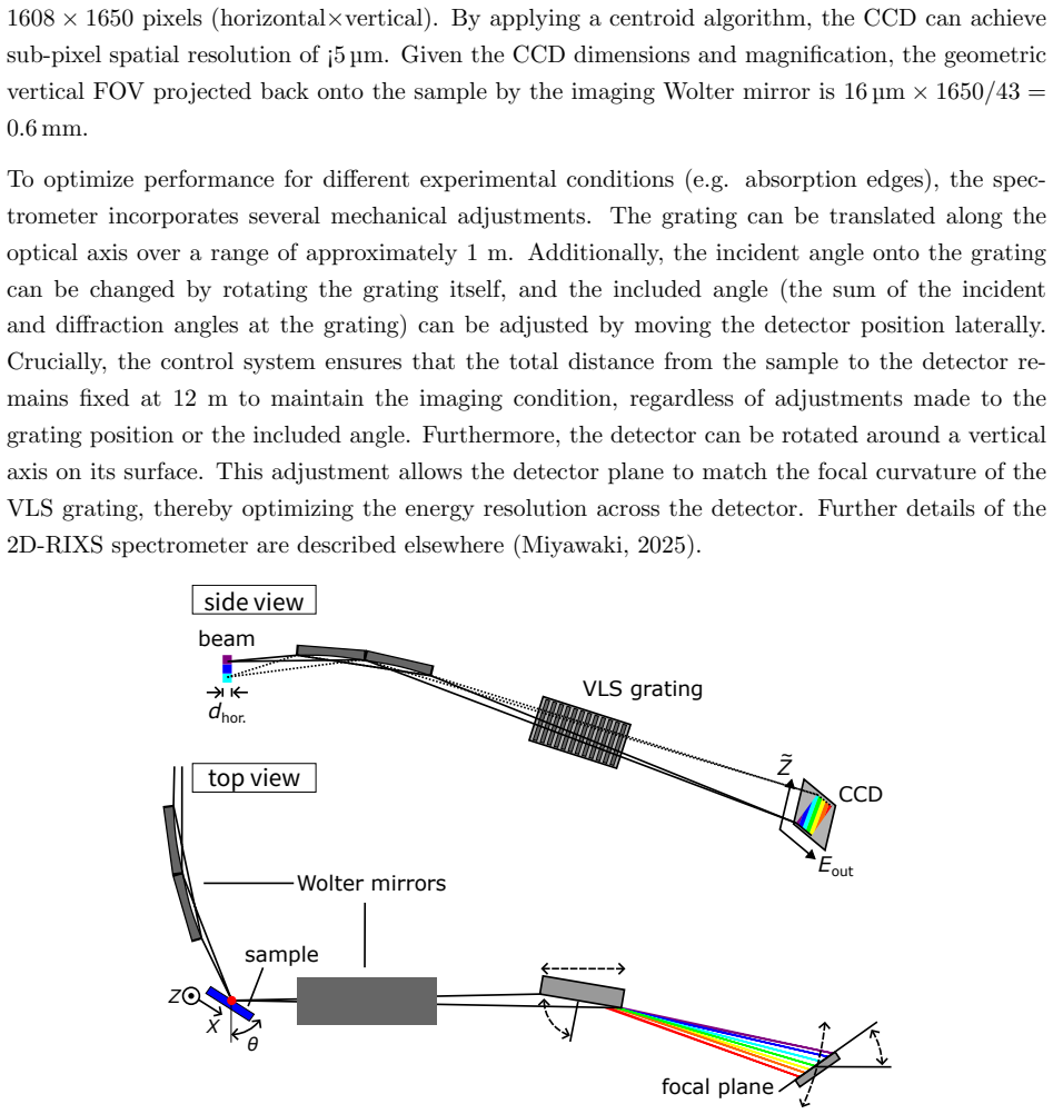
The central claim is that the new 2D-RIXS system, using a Wolter mirror and grating spectrometer, provides micrometer-scale spatial resolution alongside ultrahigh energy resolution, as verified by test chart measurements yielding 1.0 um vertical and 0.8 um horizontal resolutions, and demonstrated through imaging of inhomogeneous samples like exfoliated nanoflakes.
What carries the argument
The Wolter type-I mirror combined with the varied-line-spacing grating spectrometer, which enables simultaneous spatial imaging and high-resolution energy analysis of scattered x-rays.
Load-bearing premise
The spatial and energy resolutions achieved on test charts and selected samples will hold for arbitrary inhomogeneous samples without needing extra corrections or being affected by beamline instabilities.
What would settle it
Performing RIXS imaging on a highly complex, multi-phase sample and checking if the measured spatial resolution remains at 1.0 um or if energy resolution degrades due to sample inhomogeneity.
Figures


read the original abstract
A two-dimensional resonant inelastic x-ray scattering (2D-RIXS) microscopy system has been developed at the beamline BL02U of NanoTerasu. The instrument combines a Wolter type-I mirror for spatial imaging with a varied-line-spacing grating spectrometer, simultaneously achieving micrometer-scale spatial resolution and ultrahigh energy resolution in the soft x-ray regime. Test chart measurements confirm a vertical spatial resolution of 1.0 um near the field-of-view center, and the horizontal resolution determined by the incident beam footprint is 0.8 um. RIXS imaging capabilities have been demonstrated by the measurements of a patterned NanoTerasu logo and exfoliated NiPS${}_3$ nanoflakes, highlighting its efficiency in locating specific microscale regions within inhomogeneous samples. These results establish 2D-RIXS microscopy as a position-sensitive probe of elementary excitations in quantum materials and functional devices.
Editorial analysis
A structured set of objections, weighed in public.
Referee Report
Summary. The manuscript reports the development of a 2D-RIXS microscopy instrument at beamline BL02U of NanoTerasu. It combines a Wolter type-I mirror for spatial imaging with a varied-line-spacing grating spectrometer to achieve simultaneous micrometer-scale spatial resolution and ultrahigh energy resolution in the soft x-ray regime. Test-chart measurements establish a vertical spatial resolution of 1.0 μm near the field-of-view center and a horizontal resolution of 0.8 μm set by the incident beam footprint. The capabilities are demonstrated on a patterned NanoTerasu logo and exfoliated NiPS3 nanoflakes to illustrate utility for locating microscale regions in inhomogeneous samples.
Significance. If the reported spatial resolutions and energy performance are robustly validated with quantitative metrics and error analysis, the instrument would represent a meaningful advance for position-resolved studies of elementary excitations in quantum materials and devices. The direct demonstrations on inhomogeneous samples provide concrete evidence of practical utility beyond idealized test objects.
major comments (2)
- [Abstract and Results] Abstract and Results: the energy resolution is stated only as 'ultrahigh' without a numerical value (e.g., meV FWHM), error bars, or comparison to the spectrometer's design resolution. This quantitative detail is load-bearing for the central claim of simultaneous high spatial and energy performance and should be added with supporting spectra or fitting details.
- [Methods/Experimental] Methods/Experimental section: no description is given of the data processing steps used to extract the 1.0 μm and 0.8 μm spatial resolutions from the test-chart images, nor of any corrections applied for beamline instabilities or sample-specific effects. These steps are necessary to assess whether the quoted resolutions are reproducible across the demonstrated inhomogeneous samples.
minor comments (3)
- [Figures] Figure captions should explicitly state the photon energy, polarization, and acquisition time for each RIXS image to allow direct comparison with other soft-x-ray instruments.
- [Discussion] The manuscript would benefit from a brief table comparing the achieved spatial and energy resolutions with those of existing RIXS microscopes or spectrometers at comparable beamlines.
- [Throughout] Minor typographical inconsistencies appear in the NiPS3 chemical formula subscript formatting between the abstract and main text.
Simulated Author's Rebuttal
We thank the referee for the constructive review and the recommendation for minor revision. The comments highlight important areas for improving the quantitative presentation of the instrument performance and the transparency of the analysis methods. We address each point below and have revised the manuscript accordingly.
read point-by-point responses
-
Referee: [Abstract and Results] Abstract and Results: the energy resolution is stated only as 'ultrahigh' without a numerical value (e.g., meV FWHM), error bars, or comparison to the spectrometer's design resolution. This quantitative detail is load-bearing for the central claim of simultaneous high spatial and energy performance and should be added with supporting spectra or fitting details.
Authors: We agree that a numerical value for the energy resolution, including error bars and comparison to the design resolution, strengthens the central claim. In the revised manuscript we have updated both the Abstract and Results sections to report the measured energy resolution (with FWHM value, uncertainties from spectral fitting, and direct comparison to the spectrometer design resolution). Supporting spectra and fitting details have been added to the main text or supplementary material as appropriate. revision: yes
-
Referee: [Methods/Experimental] Methods/Experimental section: no description is given of the data processing steps used to extract the 1.0 μm and 0.8 μm spatial resolutions from the test-chart images, nor of any corrections applied for beamline instabilities or sample-specific effects. These steps are necessary to assess whether the quoted resolutions are reproducible across the demonstrated inhomogeneous samples.
Authors: We appreciate this comment on reproducibility. The revised Methods/Experimental section now includes a detailed description of the data-processing pipeline: line-profile extraction from the test-chart images, Gaussian fitting procedures used to determine the 1.0 μm vertical and 0.8 μm horizontal resolutions, and the specific corrections applied for beamline instabilities and sample-related effects. These steps were applied uniformly to both the test-chart data and the measurements on the inhomogeneous samples (patterned logo and NiPS3 nanoflakes). revision: yes
Circularity Check
No significant circularity
full rationale
The paper describes the development and characterization of a 2D-RIXS microscopy instrument at beamline BL02U. All load-bearing claims rest on direct empirical measurements: vertical spatial resolution of 1.0 um is confirmed via test chart data, horizontal resolution of 0.8 um follows from the incident beam footprint, and imaging utility is shown through explicit demonstrations on a patterned logo and NiPS3 nanoflakes. No equations, fitted parameters, derivations, or self-citation chains are present that could reduce any result to its own inputs by construction. The argument is self-contained through instrument performance data and sample applications, with no theoretical steps that invoke uniqueness theorems, ansatzes, or renamings of prior results.
Axiom & Free-Parameter Ledger
Forward citations
Cited by 1 Pith paper
-
Recent Progress in Ultrafast Dynamics of Transition-Metal Compounds Studied by Time-Resolved X-ray Techniques
A review of progress in femtosecond X-ray techniques for probing laser-induced demagnetization, spin transitions, and valence changes in transition-metal compounds.
Reference graph
Works this paper leans on
-
[1]
Ag˚ aker, M., S¨ oderstr¨ om, J., Baumann, T. M., Englund, C.-J., Kjellsson, L., Boll, R., De Fanis, A., Dold, S., Mazza, T., Monta˜ no, J., M¨ unnich, A., Mullins, T., Ovcharenko, Y., Rennhack, N., Schmidt, P., Senfftleben, B., Turcato, M., Usenko, S., Meyer, M., Nordgren, J. & Rubensson, J.-E. (2024).J. Synchrotron Rad.31(5), 1264–1275. Ament, L. J. P.,...
work page 2024
-
[2]
Dvorak, J., Jarrige, I., Bisogni, V., Coburn, S. & Leonhardt, W. (2016).Rev. Sci. Instrum.87(11), 115109. Imada, M., Fujimori, A. & Tokura, Y. (1998).Rev. Mod. Phys.70, 1039–1263. 10 Ishii, K., Tohyama, T. & Mizuki, J. (2013).J. Phys. Soc. Jpn.82(2), 021015. Marschall, F., Yin, Z., Rehanek, J., Beye, M., D¨ oring, F., Kubiˇ cek, K., Raiser, D., Veedu, S. ...
work page 2016
-
[3]
Mitrano, M., Johnston, S., Kim, Y.-J. & Dean, M. P. M. (2024).Phys. Rev. X,14, 040501. Miyawaki, J. (2025).in preparation. Miyawaki, J., Fujii, K., Imazono, T., Horiba, K., Ohtsubo, Y., Inami, N., Nakatani, T., Inaba, K., Agui, A., Kimura, H. & Takahasi, M. (2022).J. Phys.: Conf. Ser.2380(1), 012030. Pelliciari, J., Mejia, E., Woods, J. M., Gu, Y., Li, J....
work page 2024
-
[4]
Yamamoto, K., Ugalino, R., Fujii, K., Ohtsubo, Y., Iwasawa, H., Kitamura, M., Imazono, T., Inami, N., Nakatani, T., Inaba, K., Agui, A., Takeuchi, T., Kimura, H., Takahasi, M., Horiba, K. & Miyawaki, J. (2025).J. Phys.: Conf. Ser.3010(1), 012115. Yang, Z., Wang, L., Zhao, D., Luo, M., Laha, S., G¨ uth, A., Taniguchi, T., Watanabe, K., Lotsch, B. V., Smet,...
work page 2025
-
[5]
Zhou, K.-J., Matsuyama, S. & Strocov, V. N. (2020).J. Synchrotron Rad.27(5), 1235–1239. Zhou, K.-J., Walters, A., Garcia-Fernandez, M., Rice, T., Hand, M., Nag, A., Li, J., Agrestini, S., Garland, P., Wang, H., Alcock, S., Nistea, I., Nutter, B., Rubies, N., Knap, G., Gaughran, M., Yuan, F., Chang, P., Emmins, J. & Howell, G. (2022).J. Synchrotron Rad.29(...
work page 2020
discussion (0)
Sign in with ORCID, Apple, or X to comment. Anyone can read and Pith papers without signing in.