Progressive Deep Learning for Automated Spheno-Occipital Synchondrosis Maturation Assessment
Pith reviewed 2026-05-10 16:28 UTC · model grok-4.3
The pith
Sequentially activating deeper layers in a neural network improves accuracy for assessing spheno-occipital synchondrosis maturation stages from CBCT scans.
A machine-rendered reading of the paper's core claim, the machinery that carries it, and where it could break.
Core claim
The authors claim that training a network by progressively activating its deeper blocks over time creates a depth-based curriculum that aligns feature learning with the biological progression of synchondrosis fusion. This leads to more stable optimization and higher classification accuracy than training the full network at once, with the largest gains on ambiguous intermediate maturation stages. These improvements hold across convolutional and transformer models and require no changes to the architecture or loss function.
What carries the argument
The progressive representation-learning framework, which sequentially grows the model by activating deeper blocks over training time to encode coarse cranial base morphology first before specializing in subtle fusion patterns.
If this is right
- Higher accuracy particularly on transitional fusion stages
- More stable training optimization across different network types
- Effective without modifications to architecture or loss functions
- Applicable to modeling other continuous biological processes in imaging
Where Pith is reading between the lines
- This training strategy might generalize to other medical imaging tasks involving gradual changes, such as bone age assessment or tumor progression.
- It highlights that the order of feature learning can substitute for complex model designs in fine-grained anatomical classification.
- Future work could test if this method reduces the amount of training data needed for similar problems.
Load-bearing premise
That growing the model depth sequentially will naturally align with the biological stages of fusion and improve separation of close stages by mirroring how experts reason.
What would settle it
Running the same experiments but finding no accuracy gain or even a drop for the progressive method on a standard test set of CBCT images would disprove the main claim.
Figures


read the original abstract
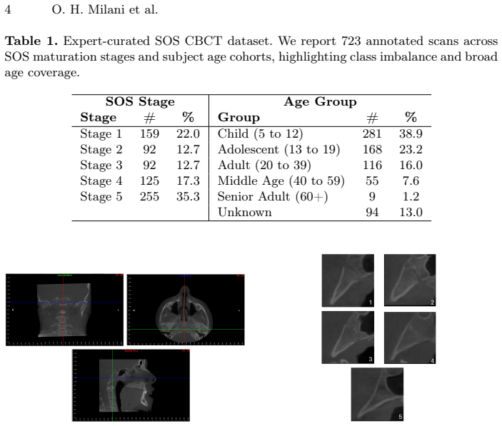
Accurate assessment of spheno-occipital synchondrosis (SOS) maturation is a key indicator of craniofacial growth and a critical determinant for orthodontic and surgical timing. However, SOS staging from cone-beam CT (CBCT) relies on subtle, continuously evolving morphological cues, leading to high inter-observer variability and poor reproducibility, especially at transitional fusion stages. We frame SOS assessment as a fine-grained visual recognition problem and propose a progressive representation-learning framework that explicitly mirrors how expert clinicians reason about synchondral fusion: from coarse anatomical structure to increasingly subtle patterns of closure. Rather than training a full-capacity network end-to-end, we sequentially grow the model by activating deeper blocks over time, allowing early layers to first encode stable cranial base morphology before higher-level layers specialize in discriminating adjacent maturation stages. This yields a curriculum over network depth that aligns deep feature learning with the biological continuum of SOS fusion. Extensive experiments across convolutional and transformer-based architectures show that this expert-inspired training strategy produces more stable optimization and consistently higher accuracy than standard training, particularly for ambiguous intermediate stages. Importantly, these gains are achieved without changing network architectures or loss functions, demonstrating that training dynamics alone can substantially improve anatomical representation learning. The proposed framework establishes a principled link between expert dental intuition and deep visual representations, enabling robust, data-efficient SOS staging from CBCT and offering a general strategy for modeling other continuous biological processes in medical imaging.
Editorial analysis
A structured set of objections, weighed in public.
Referee Report
Summary. The manuscript presents a progressive deep learning framework for automated assessment of spheno-occipital synchondrosis (SOS) maturation from cone-beam CT (CBCT) images. It proposes sequentially growing the model by activating deeper blocks over time to mirror expert clinician reasoning, from coarse cranial base morphology to subtle fusion patterns. The central claim is that this strategy yields more stable optimization and consistently higher accuracy than standard end-to-end training across convolutional and transformer architectures, particularly for ambiguous intermediate stages, without modifying network architectures or loss functions.
Significance. If substantiated with rigorous, controlled experiments, the significance lies in demonstrating that training dynamics—specifically a curriculum over network depth—can substantially improve performance on fine-grained anatomical classification tasks involving continuous biological processes. This could advance data-efficient learning in medical imaging and establish a principled connection between clinical expertise and deep feature learning, with potential applications beyond SOS staging to other staged or transitional anatomical assessments.
major comments (2)
- [Experiments] Experiments section: The comparison between the progressive activation strategy and standard end-to-end training does not specify whether total training epochs, gradient updates, or wall-clock time are matched. If each progressive stage trains for a fixed number of epochs before activating the next block, the cumulative optimization budget exceeds the baseline, providing an alternative explanation for reported gains in accuracy and stability on ambiguous intermediate stages rather than the curriculum itself.
- [Results] Results section: The manuscript asserts 'consistently higher accuracy' and 'more stable optimization' but provides no quantitative metrics (e.g., per-stage accuracies, confusion matrices focused on transitional fusion stages, standard deviations over multiple runs, or statistical significance tests such as McNemar's test or paired t-tests against baselines). This leaves the central empirical claim without verifiable support.
minor comments (2)
- [Abstract] Abstract: The description of the method and results is repetitive; the final two sentences restate the same benefits without adding new information.
- [Method] Method: The progressive activation schedule lacks pseudocode or a precise definition of 'deeper blocks' for both CNN and transformer architectures, hindering reproducibility.
Simulated Author's Rebuttal
We thank the referee for their constructive comments, which help improve the clarity and rigor of our manuscript. We address each major comment below and will make the necessary revisions to strengthen the experimental validation and reporting.
read point-by-point responses
-
Referee: [Experiments] Experiments section: The comparison between the progressive activation strategy and standard end-to-end training does not specify whether total training epochs, gradient updates, or wall-clock time are matched. If each progressive stage trains for a fixed number of epochs before activating the next block, the cumulative optimization budget exceeds the baseline, providing an alternative explanation for reported gains in accuracy and stability on ambiguous intermediate stages rather than the curriculum itself.
Authors: We appreciate this observation and acknowledge that the manuscript does not explicitly detail the matching of training budgets. Upon review, our experimental protocol was designed to match the total number of gradient updates and epochs across methods by proportionally allocating epochs to each progressive stage. However, to eliminate any ambiguity, we will revise the Experiments section to include a clear description of the training schedule, a table comparing total epochs and wall-clock time, and confirmation that the optimization budget is equivalent. This will demonstrate that the performance gains are attributable to the progressive strategy rather than increased training resources. revision: yes
-
Referee: [Results] Results section: The manuscript asserts 'consistently higher accuracy' and 'more stable optimization' but provides no quantitative metrics (e.g., per-stage accuracies, confusion matrices focused on transitional fusion stages, standard deviations over multiple runs, or statistical significance tests such as McNemar's test or paired t-tests against baselines). This leaves the central empirical claim without verifiable support.
Authors: We agree that providing these quantitative metrics would enhance the verifiability of our claims. While the manuscript reports overall accuracy improvements and qualitative observations on stability, we will expand the Results section to include per-stage accuracy breakdowns (with emphasis on intermediate stages), confusion matrices highlighting errors in transitional fusion stages, standard deviations from multiple independent runs, and results from statistical significance tests such as paired t-tests and McNemar's test. These additions will provide rigorous support for the reported benefits of the progressive framework. revision: yes
Circularity Check
No circularity: purely empirical curriculum with experimental comparisons
full rationale
The paper proposes a progressive training strategy for deep networks on SOS staging from CBCT, framing it as mirroring expert clinician reasoning via sequential block activation. No mathematical derivation, equations, or first-principles predictions are present that could reduce to inputs by construction. Claims rest on empirical accuracy gains versus standard end-to-end training across architectures, without self-citations, fitted parameters renamed as predictions, or uniqueness theorems. The curriculum is an explicit design choice, not a result derived from itself; any confounding in training budgets is an experimental validity issue, not circularity. The approach is self-contained as a standard empirical ML contribution.
Axiom & Free-Parameter Ledger
free parameters (1)
- progressive activation schedule
axioms (1)
- domain assumption Early layers encode stable cranial base morphology while deeper layers can specialize in subtle fusion patterns when activated sequentially
Reference graph
Works this paper leans on
-
[1]
American Journal of Orthodontics and Dentofacial Orthopedics122(4), 380–385 (2002)
Mito, T., Sato, K., Mitani, H.: Cervical vertebral bone age in girls. American Journal of Orthodontics and Dentofacial Orthopedics122(4), 380–385 (2002). https://doi.org/10.1067/mod.2002.126896
-
[2]
Journal of Children’s Orthopaedics4(5), 467–470 (2010)
Calfee, R.P., Sutter, M., Steffen, J.A., Goldfarb, C.A.: Skeletal and chronological ages in American adolescents: current findings in skeletal maturation. Journal of Children’s Orthopaedics4(5), 467–470 (2010)
work page 2010
-
[3]
Karlberg, J.: Secular trends in pubertal development. Hormone Research57(Suppl. 2), 19–30 (2002)
work page 2002
-
[4]
Indian Journal of Endocrinology and Metabolism18(Suppl 1), S63–S71 (2014)
De Sanctis, V., Di Maio, S., Soliman, A.T., Raiola, G., Elalaily, R., Millimaggi, G.: Hand X-ray in pediatric endocrinology: Skeletal age assessment and beyond. Indian Journal of Endocrinology and Metabolism18(Suppl 1), S63–S71 (2014)
work page 2014
-
[5]
Australian Dental Journal62, 33–50 (2017)
Scarfe, W.C., Azevedo, B., Toghyani, S., Farman, A.G.: Cone beam computed tomographic imaging in orthodontics. Australian Dental Journal62, 33–50 (2017)
work page 2017
-
[6]
Journal of Applied Clinical Medical Physics22(2), 85–89 (2021)
Olch, A.J., Alaei, P.: How low can you go? A CBCT dose reduction study. Journal of Applied Clinical Medical Physics22(2), 85–89 (2021)
work page 2021
-
[7]
The Angle Orthodontist 91(4), 538–543 (2021)
Alhazmi,A.,Aldossary,M.,Palomo,J.M.,Hans,M.,Latimer,B.,Simpson,S.:Cor- relation of spheno-occipital synchondrosis fusion stages with a hand-wrist skeletal maturity index: a cone beam computed tomography study. The Angle Orthodontist 91(4), 538–543 (2021)
work page 2021
-
[8]
Angle Orthodontist94(6), 641–647 (2024)
Booth, E., Viana, G., Shirazi, S., Miller, S., Sellke, T., Elnagar, M., Viana, M., At- sawasuwan, P.: Correlations of spheno-occipital synchondrosis, cervical vertebrae, midpalatal suture, and third molar maturation stages. Angle Orthodontist94(6), 641–647 (2024). https://doi.org/10.2319/041224-295.1
-
[9]
PLoS ONE11(8), e0161104 (2016)
Fernández-Pérez, M.J., Alarcón, J.A., McNamara Jr., J.A., Velasco-Torres, M., Benavides, E., Galindo-Moreno, P., Catena, A.: Spheno-occipital synchondrosis fusion correlates with cervical vertebrae maturation. PLoS ONE11(8), e0161104 (2016)
work page 2016
-
[10]
The Anatomical Record147(1), 15–23 (1963)
Powell, T.V., Brodie, A.G.: Closure of the spheno-occipital synchondrosis. The Anatomical Record147(1), 15–23 (1963)
work page 1963
-
[11]
American Journal of Physical Anthropology157(1), 42–57 (2015)
Lottering, N., MacGregor, D.M., Alston, C.L., Gregory, L.S.: Ontogeny of the spheno-occipital synchondrosis in a modern Queensland, Australian population using computed tomography. American Journal of Physical Anthropology157(1), 42–57 (2015)
work page 2015
-
[12]
Journal of Forensic Dental Sciences5(2), 72–76 (2013)
Krishan, K., Kanchan, T.: Evaluation of spheno-occipital synchondrosis: A review of literature and considerations from forensic anthropologic point of view. Journal of Forensic Dental Sciences5(2), 72–76 (2013)
work page 2013
-
[13]
Turkish Journal of Orthodontics36(3), 186 (2023)
Tashayyodi, N., Kajan, Z.D., Ostovarrad, F., Khosravifard, N.: Relationship of the Fusion Stage of Spheno-Occipital Synchondrosis with Midpalatal and Zygomatico- maxillary Sutures on Cone-Beam Computed Tomography Scans of Patients Aged Between 7 and 21 Years. Turkish Journal of Orthodontics36(3), 186 (2023)
work page 2023
-
[14]
PLoS ONE12(8), e0183305 (2017)
Alhazmi, A., Vargas, E., Palomo, J.M., Hans, M., Latimer, B., Simpson, S.: Timing and rate of spheno-occipital synchondrosis closure and its relationship to puberty. PLoS ONE12(8), e0183305 (2017)
work page 2017
-
[15]
The Angle Orthodontist80(1), 106–110 (2010)
Leonardi, R., Cutrera, A., Barbato, E.: Rapid maxillary expansion affects the spheno-occipital synchondrosis in youngsters: a study with low-dose computed to- mography. The Angle Orthodontist80(1), 106–110 (2010)
work page 2010
-
[16]
The Angle Orthodontist91(6), 822–829 (2021)
Leonardi, R., Ronsivalle, V., Lagravere, M.O., Barbato, E., Isola, G., Lo Giudice, A.: Three-dimensional assessment of the spheno-occipital synchondrosis and clivus Title Suppressed Due to Excessive Length 15 aftertooth-borneandbone-bornerapidmaxillaryexpansion:aretrospectiveCBCT study using voxel-based superimposition. The Angle Orthodontist91(6), 822–829 (2021)
work page 2021
-
[17]
Plastic and Reconstructive Surgery134(3), 504–510 (2014)
Goldstein, J.A., Paliga, J.T., Wink, J.D., Bartlett, S.P., Nah, H.-D., Taylor, J.A.: Earlier evidence of spheno-occipital synchondrosis fusion correlates with severity of midface hypoplasia in patients with syndromic craniosynostosis. Plastic and Reconstructive Surgery134(3), 504–510 (2014)
work page 2014
-
[18]
Journal of Oral and Maxillofacial Surgery72(6), 1173–1179 (2014)
Tahiri, Y., Paliga, J.T., Vossough, A., Bartlett, S.P., Taylor, J.A.: The spheno- occipital synchondrosis fuses prematurely in patients with Crouzon syndrome and midface hypoplasia compared with age- and gender-matched controls. Journal of Oral and Maxillofacial Surgery72(6), 1173–1179 (2014)
work page 2014
-
[19]
Dentomaxillofacial Radiology44(2015)
Kapila, S.D., Nervina, J.M.: CBCT in orthodontics: Assessment of treatment out- comes and indications for its use. Dentomaxillofacial Radiology44(2015). Preprint at https://doi.org/10.1259/dmfr.20140282
-
[20]
Korean Journal of Radiology18(4), 570–584 (2017)
Lee, J.-G., Jun, S., Cho, Y.-W., Lee, H., Kim, G.B., Seo, J.B., Kim, N.: Deep learning in medical imaging: general overview. Korean Journal of Radiology18(4), 570–584 (2017)
work page 2017
-
[21]
American Journal of Orthodontics and Dentofacial Orthopedics160(2), 170–192.e4 (2021)
Mohammad-Rahimi, H., Nadimi, M., Rohban, M.H., Shamsoddin, E., Lee, V.Y., Motamedian, S.R.: Machine learning and orthodontics, current trends and the future opportunities: A scoping review. American Journal of Orthodontics and Dentofacial Orthopedics160(2), 170–192.e4 (2021). https://doi.org/10.1016/j.ajodo.2021.02.013
-
[22]
Graphical Models109, 101071 (2020)
Zhang, J., Li, C., Song, Q., Gao, L., Lai, Y.-K.: Automatic 3D tooth segmenta- tion using convolutional neural networks in harmonic parameter space. Graphical Models109, 101071 (2020)
work page 2020
-
[23]
Thurzo, A., Kosnáčová, H.S., Kurilová, V., Kosmel’, S., Beňuš, R., Moravansk` y, N., Kováč, P., Kuracinová, K.M., Palkovič, M., Varga, I.: Use of advanced artificial intelligence in forensic medicine, forensic anthropology and clinical anatomy. In: Healthcare, vol. 9(11), p. 1545 (2021)
work page 2021
-
[24]
Journal of Pharmacy and Bioallied Sciences16(Suppl 3), S2585–S2587 (2024)
Kumar, N., Malik, B., Dubey, A., Kaur, H., Mujoo, S., Jugade, S.C., Gulia, S.K.: Long-Term Trends in the Utilization of Cone Beam Computed Tomography in Oral and Maxillofacial Radiology. Journal of Pharmacy and Bioallied Sciences16(Suppl 3), S2585–S2587 (2024). https://doi.org/10.4103/jpbs.jpbs_295_24
-
[25]
Bassed, R.B., Briggs, C., Drummer, O.H.: Analysis of time of closure of the spheno- occipitalsynchondrosisusingcomputedtomography.ForensicScienceInternational 200(1–3), 161–164 (2010)
work page 2010
-
[26]
PLOS ONE17(7), e0269198 (2022)
Atici, S.F., Ansari, R., Allareddy, V., Suhaym, O., Cetin, A.E., Elnagar, M.H.: Fully automated determination of the cervical vertebrae maturation stages using deep learning with directional filters. PLOS ONE17(7), e0269198 (2022)
work page 2022
discussion (0)
Sign in with ORCID, Apple, or X to comment. Anyone can read and Pith papers without signing in.