Predicting Metastatic Risk from Primary Tissue Architecture via Distance-Aware Spatial Modeling
Pith reviewed 2026-06-30 10:09 UTC · model grok-4.3
The pith
A multiple instance learning model using signed distance functions to tissue phenotypes captures spatial architecture to predict metastatic risk from primary tumor histology.
A machine-rendered reading of the paper's core claim, the machinery that carries it, and where it could break.
Core claim
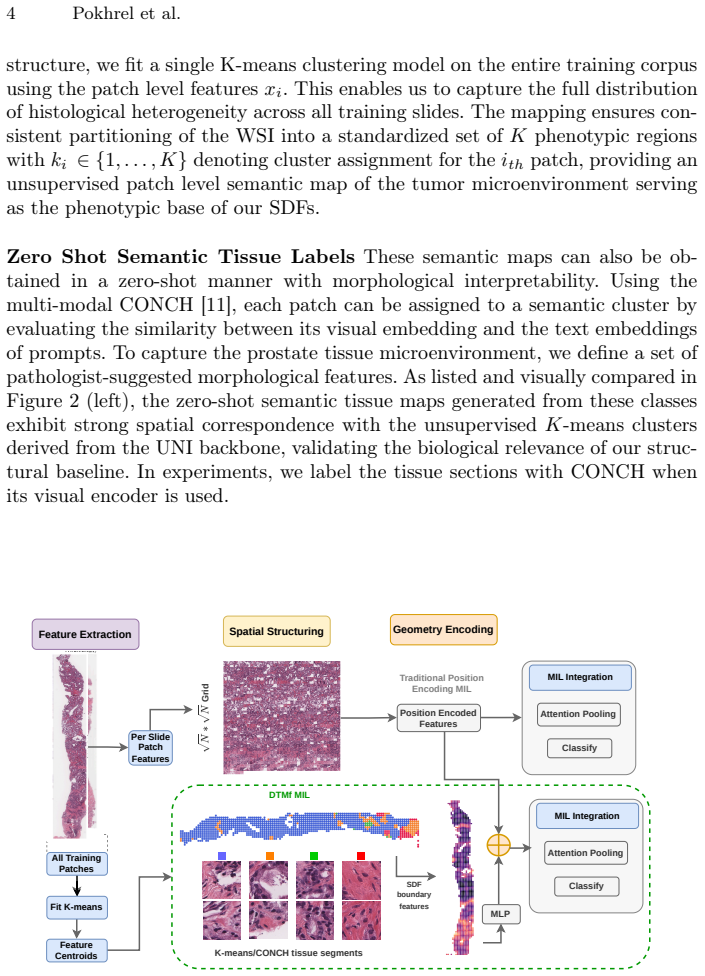
By computing signed distance functions relative to tissue phenotypes, DTMf-MIL learns structural signatures of metastatic risk that standard MIL models discard, resulting in superior performance on the task of predicting distant metastasis from primary tumor histology.
What carries the argument
Signed distance functions relative to tissue phenotypes (tumor cells, tumor-associated fibroblasts, infiltrating lymphocytes) that supply explicit spatial priors to a multiple instance learning model.
If this is right
- Explicit spatial modeling improves accuracy on metastasis prediction compared with methods that ignore tissue layout.
- The same spatial priors produce consistent gains in diagnostic accuracy on diverse clinical tasks beyond metastasis prediction.
- Structural signatures encoded by distance functions to cell-type interfaces are learnable and clinically actionable from primary tumor slides.
Where Pith is reading between the lines
- The same distance-based priors could be tested for predicting treatment response or recurrence risk in the same tissue samples.
- If the geometric interfaces prove decisive, simpler non-learned geometric features might achieve comparable gains with lower compute cost.
- The approach suggests that spatial modeling may transfer to other whole-slide imaging tasks where microenvironment geometry matters.
Load-bearing premise
Metastatic risk is inherently dictated by the geometric arrangement of the tumor microenvironment at the interface with tumor cells, and signed distance functions capture the relevant structural signatures.
What would settle it
If a standard MIL model that receives no signed distance information achieves the same or higher accuracy on the metastasis prediction task as DTMf-MIL, the necessity of the spatial priors would be falsified.
Figures
read the original abstract
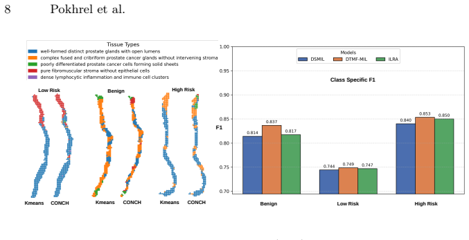
Predicting the risk of distant metastasis from primary tumor tissue histology is a critical yet challenging task in computational pathology. Multiple Instance Learning (MIL) approaches can attend to subdomains in tumor regions that harbor features of metastatic cancer progression. However MIL models treat tissue patches as unordered bags, discarding the spatial layout that defines the metastatic potential. We propose that metastatic risk is inherently dictated by the geometric arrangement of the tumor microenvironment at the interface with tumor cells. Our model is designed to explicitly capture the spatial relationships between tumor cells, tumor associated fibroblasts and infiltrating lymphocytes. For this purpose, we propose Distance aware Tissue Modeling for Multiple Instance Learning(DTMf-MIL), a novel method that reinforces visual features with explicit spatial priors. By computing signed distance functions (SDF) relative to tissue phenotypes, our model learns to recognize structural signatures of metastatic risk. This geometric awareness translates directly to superior clinical performance as DTMf-MIL significantly outperforms state-of-the-art methods that ignore spatial layout on metastasis prediction from tissue in the primary tumor. We further validate our approach on public benchmarks, demonstrating that spatial awareness consistently improves diagnostic accuracy across diverse clinical tasks.
Editorial analysis
A structured set of objections, weighed in public.
Referee Report
Summary. The paper proposes DTMf-MIL, a multiple instance learning model for predicting distant metastasis risk from primary tumor histology. It augments standard MIL with signed distance functions (SDFs) computed relative to tissue phenotypes (tumor cells, tumor-associated fibroblasts, and infiltrating lymphocytes) to explicitly encode spatial layout and geometric relationships at the tumor-microenvironment interface, claiming that this spatial awareness yields significant outperformance over spatial-ignoring MIL baselines on metastasis prediction and other diagnostic tasks across public benchmarks.
Significance. If the performance gains are attributable to the SDF spatial priors rather than ancillary modeling choices, the work would meaningfully advance computational pathology by providing a concrete mechanism to incorporate tissue architecture into MIL frameworks, addressing a recognized limitation of bag-based models and potentially improving risk stratification in clinical settings.
major comments (2)
- [Abstract] Abstract: The central claim that DTMf-MIL 'significantly outperforms state-of-the-art methods that ignore spatial layout' is asserted without any mention of the datasets used, statistical tests performed, baseline implementations, effect sizes, or ablation controls that isolate the SDF component. This absence makes it impossible to evaluate whether the reported gains derive from the geometric priors or from differences in model capacity, feature fusion, or training procedure, directly undermining attribution to the proposed spatial modeling.
- [Abstract] Abstract / Methods (implied): The paper states that 'metastatic risk is inherently dictated by the geometric arrangement' and that SDFs 'learn to recognize structural signatures,' yet provides no controlled experiment (e.g., replacing SDF inputs with non-spatial equivalents while holding architecture fixed) to test this assumption. Without such an ablation, the load-bearing hypothesis that signed distance functions capture the relevant metastatic signatures remains unverified.
Simulated Author's Rebuttal
We appreciate the referee's comments on the abstract and the need for clearer attribution of results to the SDF priors. We address each point below and will make revisions to strengthen the manuscript.
read point-by-point responses
-
Referee: [Abstract] Abstract: The central claim that DTMf-MIL 'significantly outperforms state-of-the-art methods that ignore spatial layout' is asserted without any mention of the datasets used, statistical tests performed, baseline implementations, effect sizes, or ablation controls that isolate the SDF component. This absence makes it impossible to evaluate whether the reported gains derive from the geometric priors or from differences in model capacity, feature fusion, or training procedure, directly undermining attribution to the proposed spatial modeling.
Authors: We agree that the abstract, being a concise summary, does not include these details. The full manuscript specifies the public benchmarks used, the statistical tests (e.g., paired t-tests or Wilcoxon), the baseline implementations (standard MIL variants), effect sizes (AUC improvements), and ablation studies. To address this, we will revise the abstract to briefly mention the evaluation setup and key results with statistical significance. revision: yes
-
Referee: [Abstract] Abstract / Methods (implied): The paper states that 'metastatic risk is inherently dictated by the geometric arrangement' and that SDFs 'learn to recognize structural signatures,' yet provides no controlled experiment (e.g., replacing SDF inputs with non-spatial equivalents while holding architecture fixed) to test this assumption. Without such an ablation, the load-bearing hypothesis that signed distance functions capture the relevant metastatic signatures remains unverified.
Authors: The manuscript includes comparisons to spatial-ignoring baselines, which serve as controls. However, we acknowledge that a more direct ablation—replacing SDF inputs with non-spatial equivalents (e.g., random or constant values) while keeping the model architecture fixed—would provide stronger evidence. We will add this controlled experiment to the revised version to isolate the contribution of the SDF priors. revision: yes
Circularity Check
No circularity: empirical model validated on external benchmarks
full rationale
The paper proposes DTMf-MIL, a MIL variant that augments visual features with signed distance functions computed from tissue phenotypes to encode spatial layout for metastasis prediction. The central claim is empirical outperformance versus spatial-ignoring baselines on clinical tasks and public benchmarks. No equations, parameter-fitting procedures, self-citations, or uniqueness theorems are described that reduce any prediction or result to the inputs by construction. The derivation chain consists of a modeling choice (SDF priors) plus data-driven validation, which remains independent of the target performance metric.
Axiom & Free-Parameter Ledger
Reference graph
Works this paper leans on
-
[1]
The Cancer Imaging Archive (2016)
Albertina, B., Watson, M., Holback, C., Jarosz, R., Kirk, S., Lee, Y., Rieger-Christ, K., Lemmerman, J.: The Cancer Genome Atlas Lung Adenocarcinoma Collection (TCGA-LUAD) (Version 4) [Data set]. The Cancer Imaging Archive (2016)
2016
-
[2]
Nature Medicine28(1), 154–163 (Jan 2022)
Bulten, W., Kartasalo, K., Chen, P.H.C., Ström, P., Pinckaers, H., Nagpal, K., Cai, Y., Steiner, D.F., van Boven, H., Vink, R., Hulsbergen-van de Kaa, C., van der Laak, J., Amin, M.B., Evans, A.J., van der Kwast, T., Allan, R., Humphrey, P.A., Grönberg, H., Samaratunga, H., Delahunt, B., Tsuzuki, T., Häkkinen, T., Egevad, L., Demkin, M., Dane, S., Tan, F....
2022
-
[3]
Nature Medicine30(3), 850–862 (Mar 2024)
Chen, R.J., Ding, T., Lu, M.Y., Williamson, D.F.K., Jaume, G., Song, A.H., Chen, B.,Zhang,A.,Shao,D.,Shaban,M.,Williams,M.,Oldenburg,L.,Weishaupt,L.L., Wang,J.J.,Vaidya,A.,Le,L.P.,Gerber,G.,Sahai,S.,Williams,W.,Mahmood,F.: Towards a general-purpose foundation model for computational pathology. Nature Medicine30(3), 850–862 (Mar 2024)
2024
- [4]
-
[5]
Dillekås, H., Rogers, M.S., Straume, O.: Are 90% of deaths from cancer caused by metastases? Cancer Med8(12), 5574–5576 (Aug 2019)
2019
-
[6]
He, K., Zhang, X., Ren, S., Sun, J.: Deep residual learning for image recognition (2015)
2015
-
[7]
Ilse, M., Tomczak, J.M., Welling, M.: Attention-based deep multiple instance learn- ing (2018)
2018
-
[8]
The Cancer Imaging Archive (2016)
Kirk,S.,Lee,Y.,Kumar,P.,Filippini,J.,Albertina,B.,Watson,M.,Rieger-Christ, K., Lemmerman, J.: The Cancer Genome Atlas Lung Squamous Cell Carcinoma Collection (TCGA-LUSC) (Version 4) [Data set]. The Cancer Imaging Archive (2016)
2016
-
[9]
Li, B., Li, Y., Eliceiri, K.W.: Dual-stream multiple instance learning network for whole slide image classification with self-supervised contrastive learning (2021) 10 Pokhrel et al
2021
-
[10]
GigaScience7(6), giy065 (05 2018)
Litjens, G., Bandi, P., Ehteshami Bejnordi, B., Geessink, O., Balkenhol, M., Bult, P., Halilovic, A., Hermsen, M., van de Loo, R., Vogels, R., Manson, Q.F., Stathonikos, N., Baidoshvili, A., van Diest, P., Wauters, C., van Dijk, M., van der Laak, J.: 1399 h&e-stained sentinel lymph node sections of breast cancer pa- tients: the camelyon dataset. GigaS...
2018
-
[11]
Lu, M.Y., Chen, B., Williamson, D.F.K., Chen, R.J., Liang, I., Ding, T., Jaume, G., Odintsov, I., Zhang, A., Le, L.P., Gerber, G., Parwani, A.V., Mahmood, F.: Towards a visual-language foundation model for computational pathology (2023)
2023
-
[12]
Lu, M.Y., Williamson, D.F.K., Chen, T.Y., Chen, R.J., Barbieri, M., Mahmood, F.: Data efficient and weakly supervised computational pathology on whole slide images (2020)
2020
-
[13]
Biol Open5(3), 289–299 (Feb 2016)
Lyons, S.M., Alizadeh, E., Mannheimer, J., Schuamberg, K., Castle, J., Schroder, B., Turk, P., Thamm, D., Prasad, A.: Changes in cell shape are correlated with metastatic potential in murine and human osteosarcomas. Biol Open5(3), 289–299 (Feb 2016)
2016
-
[14]
Nature529(7586), 298–306 (Jan 2016)
Massagué, J., Obenauf, A.C.: Metastatic colonization by circulating tumour cells. Nature529(7586), 298–306 (Jan 2016)
2016
-
[15]
Science314(5797), 298–300 (Oct 2006)
Nelson, C.M., Vanduijn, M.M., Inman, J.L., Fletcher, D.A., Bissell, M.J.: Tissue geometry determines sites of mammary branching morphogenesis in organotypic cultures. Science314(5797), 298–300 (Oct 2006)
2006
-
[16]
Front Cell Dev Biol10, 1048630 (Oct 2022)
Nevarez, A.J., Hao, N.: Quantitative cell imaging approaches to metastatic state profiling. Front Cell Dev Biol10, 1048630 (Oct 2022)
2022
-
[17]
Shao, Z., Bian, H., Chen, Y., Wang, Y., Zhang, J., Ji, X., Zhang, Y.: Transmil: Transformer based correlated multiple instance learning for whole slide image clas- sification (2021)
2021
-
[18]
Sci Rep8(1), 13692 (Sep 2018)
Sirinukunwattana, K., Snead, D., Epstein, D., Aftab, Z., Mujeeb, I., Tsang, Y.W., Cree, I., Rajpoot, N.: Novel digital signatures of tissue phenotypes for predicting distant metastasis in colorectal cancer. Sci Rep8(1), 13692 (Sep 2018)
2018
-
[19]
Tang, W., Zhou, F., Huang, S., Zhu, X., Zhang, Y., Liu, B.: Feature re-embedding: Towards foundation model-level performance in computational pathology (2024)
2024
-
[20]
Science Advances6(4), eaaw6938 (2020)
Wu, P.H., Gilkes, D.M., Phillip, J.M., Narkar, A., Cheng, T.W.T., Marchand, J., Lee, M.H., Li, R., Wirtz, D.: Single-cell morphology encodes metastatic potential. Science Advances6(4), eaaw6938 (2020)
2020
-
[21]
In: The Eleventh International Conference on Learning Representations (2023)
Xiang, J., Zhang, J.: Exploring low-rank property in multiple instance learning for whole slide image classification. In: The Eleventh International Conference on Learning Representations (2023)
2023
-
[22]
In: International Workshop on Efficient Medical Artificial Intelligence
Zhang, B., Manoochehri, H., Li, X., Knudsen, B.S., Tasdizen, T.: Weaksupcon: Weakly supervised contrastive learning for encoder pre-training. In: International Workshop on Efficient Medical Artificial Intelligence. pp. 310–319. Springer (2025)
2025
discussion (0)
Sign in with ORCID, Apple, or X to comment. Anyone can read and Pith papers without signing in.