REVIEW 1 major objections 1 minor 44 references
Automatic acute ischemic stroke lesion segmentation using semi-supervised learning
T0 review · 1 major / 1 minor · reviewed 2026-08-14 · deepseek-v4-flash
Pith's one-line read Semi-supervised stroke lesion segmentation with only five fully labeled subjects can match fully supervised accuracy.
desk verdict A plausible semi-supervised AIS segmentation pipeline with real but modest empirical claims; the headline Dice is a point estimate on a private dataset with no error bars, and the abstract/full-text subject-count mismatch needs fixing. read the letter →
The pith
A machine-rendered reading of the paper's core claim, the machinery that carries it, and where it could break.
The reading
What carries the argument
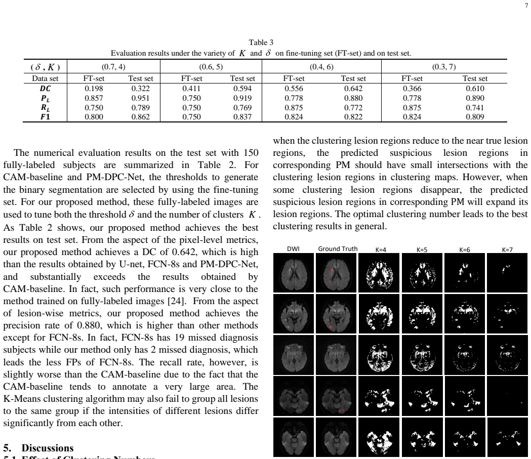
The load-bearing mechanism is a two-pathway fusion. The DPC-Net, a VGG-16 truncated before the third max-pooling layer with an extra global-average-pooling side branch at an earlier convolution block, is trained on slice-level labels and produces two class activation maps at different resolutions; thresholding the coarse map and multiplying it by the fine map yields a probability map of suspicious lesion regions. Independently, K-Means clustering on the DWI separates hyperintense pixels, using the clinical prior that AIS lesions appear bright on DWI, and connected components of the brightest cluster form candidate regions. A region-growing algorithm then starts from probability-map pixels above $\delta$ and, when they fall inside a K-Means component, grows to fill that component, so semantic information from the network and intensity information from clustering jointly decide the final boundary. The values $K$ and $\delta$ are selected by grid search on the five fully labeled subjects.
What would settle it
Repeat the grid search on a different set of five fully labeled subjects, for example with leave-one-out over the fine-tuning set, and measure Dice on the same 150-subject test set; if the mean Dice falls well below 0.642, the reported accuracy is tied to the particular choice of five subjects rather than to the method itself.
Extended reading notes
Core claim
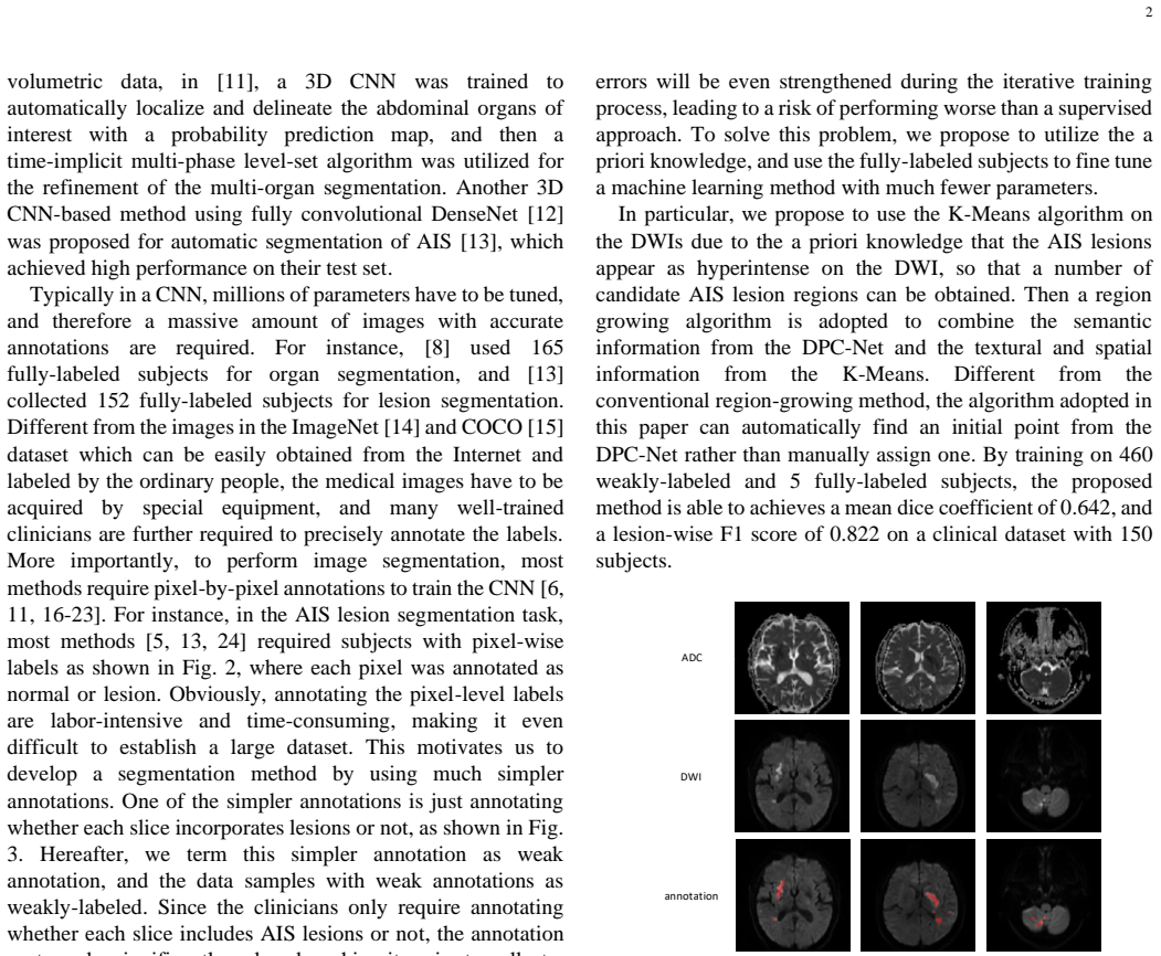
The central claim is that combining weak semantic supervision with unsupervised intensity clustering yields lesion segmentations comparable to fully supervised deep networks. Concretely, the paper reports that its semi-supervised pipeline, trained on 460 weakly labeled subjects and fine-tuned with five fully labeled subjects, achieves a mean Dice coefficient of 0.642 and a lesion-wise F1 score of 0.822 on a 150-subject clinical test set, with lesion-wise precision 0.880. The method deliberately avoids generating fake labels for weakly labeled data, instead using fully labeled subjects only to tune two parameters: the cluster count $K$ in K-Means and the threshold $\delta$ in region growing. The paper further claims this design is especially sensitive to small lesions, reporting a Dice of 0.708 on a 90-subject small-lesion subset.
Load-bearing premise
The load-bearing premise is that the two parameters tuned on just five fully labeled subjects, the number of K-Means clusters and the probability threshold, keep working on the 150-patient test set rather than reflecting only those five patients.
Editorial extensions
If this is right
- A hospital could build an AIS segmentation system from cheap slice-level screening labels plus a handful of detailed cases, reducing expert marking time.
- The reported Dice of 0.642 is close to the 0.67 of a fully supervised DWI method, suggesting weakly supervised training need not sacrifice much accuracy.
- Because the method preserves small lesions better than large ones (Dice 0.708 versus 0.543), it may be useful for lacunar infarctions, which are common but hard to spot.
- The high lesion-wise precision of 0.880 means few false lesion detections per patient, which matters if the output is used to flag suspected strokes.
Reading between the lines
- The same weak-label-plus-clustering recipe could be tried on other hyperintense-lesion tasks in MRI, such as multiple sclerosis plaques, where the intensity prior and slice-level labels are similarly cheap.
- A direct robustness test would vary which five subjects form the fine-tuning set; if $K$ and $\delta$ shift materially, a slightly larger fine-tuning set or a learned parameter predictor would be needed.
- The K-Means branch could be replaced by a learned intensity model trained on the five fully labeled slices, potentially removing the grid search while keeping the semi-supervised structure.
- A head-to-head evaluation on one shared public benchmark would show whether the annotation saving transfers across scanners, field strengths, and imaging protocols.
Signed reviews
Editorial analysis
A structured set of objections, weighed in public.
Referee Report
Summary. The paper proposes a semi-supervised pipeline for acute ischemic stroke (AIS) lesion segmentation on diffusion-weighted images (DWI) and apparent diffusion coefficient (ADC) maps. The method has three stages: a double-path classification network (DPC-Net) trained on 460 slice-level weakly labeled subjects to produce probability maps; K-Means clustering on DWI intensities to identify hyperintense candidate regions; and a region-growing step that combines the DPC-Net probability map with the K-Means clusters, using two parameters K (number of clusters) and delta (probability threshold) tuned on five fully labeled subjects. The authors report a Dice coefficient of 0.642 and a lesion-wise F1 of 0.822 on a 150-subject clinical test set, and compare with CAM-baseline, U-Net, and FCN-8s baselines trained on the same five fully labeled subjects. The central claim is that with very few pixel-level annotations, the proposed method approaches fully-supervised performance.
Significance. If the reported results are stable, the paper would be a meaningful practical contribution: it demonstrates a semi-supervised method that avoids the error-propagation of self-training, uses only slice-level weak labels plus five pixel-level annotations, and achieves Dice/F1 values in the range of fully-supervised AIS segmentation methods. The algorithmic combination of a weakly supervised CNN with an unsupervised intensity prior and a small fully labeled fine-tuning set is well motivated. The paper also reports lesion-wise metrics, which are clinically relevant given the importance of small lacunar lesions. However, the strength of this contribution rests almost entirely on the reliability of the point estimates from the 150-subject test set, since the method's two free parameters are selected on only five subjects without variance estimation.
major comments (1)
- [Section 4.1 and Table 3] In Table 4, the test set is split into large (60) and small (90) lesion sets, and the proposed method achieves higher DC on small lesions (0.708) than on large ones (0.543). The discussion explains that hyperintensity distribution is uneven in large lesions, but this size-dependent behavior is not analyzed with respect to the hyperparameter tuning; since 85% of AIS are lacunar, the authors should discuss whether the grid search on five subjects may be biased toward small lesions and how this affects the generalizability of the selected K and δ.
minor comments (1)
- [References] Reference [25] is an unpublished medRxiv preprint by the same group; the claim in Section 1 that 'the lesion-wise detection rate is high' for weak supervision relies on this preprint, and it would be preferable to cite a peer-reviewed source or describe the result in sufficient detail to be self-contained.
Circularity Check
No circularity: the reported Dice/F1 come from evaluation on a held-out test set; hyperparameter tuning on five subjects is model selection, not a circular prediction.
full rationale
This is an empirical segmentation paper, not a derivation from first principles. The central claim is that a semi-supervised pipeline (DPC-Net trained on 460 weakly-labeled subjects, K-Means clustering, and region growing with DPC-Net seeds) achieves DC 0.642 and lesion-wise F1 0.822 on a 150-subject test set. The pipeline's output is computed from DWI/ADC inputs via the network and Algorithm 1, and the evaluation uses ground-truth annotations that are not used to construct the output. The only tunable parameters, K and delta, are selected by grid search on five fully-labeled fine-tuning subjects and then applied to the separate 150-subject test set; this is standard model selection rather than fitting the evaluation metric by construction. The thresholds for comparison methods are also selected on the fine-tuning set, so the comparisons are not systematically favorable in a definitional way. The paper does cite prior work by overlapping authors, notably [25], but that citation supports a background claim about weakly-supervised detection rates and is not load-bearing for the numerical results or for the method's design choices. No equation defines the predicted segmentation in terms of the reported Dice coefficient, and no fitted parameter is renamed as a prediction. The main scientific weaknesses, such as the small fine-tuning set and lack of confidence intervals, are soundness/reproducibility concerns, not circularity.
Assumptions & free parameters
free parameters (3)
- K (number of clusters in K-Means) =
6
- delta (DPC-Net probability threshold for region-growing seeds) =
0.41
- CAM binarization threshold in Eq. (1) =
0.5
assumptions (4)
- domain assumption Acute ischemic stroke lesions appear hyperintense on DWI and hypointense on ADC.
- domain assumption Magnetic susceptibility artifacts are hyperintense on DWI but lack ADC abnormality, and can be rejected using semantic information from DPC-Net.
- domain assumption Weak slice-level labels are sufficient to train a classifier whose CAMs provide reliable lesion localization.
- ad hoc to paper The five fully-labeled subjects used for grid search are representative enough that the selected K and delta generalize to the test population.
Cite this review
Pith. "Pith review of Automatic acute ischemic stroke lesion segmentation using semi-supervised learning." pith.science (2026). https://pith.science/paper/HTRDQFY3
@misc{pith2026190803735,
  author       = {Pith},
  title        = {Pith review of: Automatic acute ischemic stroke lesion segmentation using semi-supervised learning},
  year         = {2026},
  howpublished = {\url{https://pith.science/paper/HTRDQFY3}},
  note         = {Machine review of arXiv:1908.03735}
}
read the original abstract
Ischemic stroke is a common disease in the elderly population, which can cause long-term disability and even death. However, the time window for treatment of ischemic stroke in its acute stage is very short. To fast localize and quantitively evaluate the acute ischemic stroke (AIS) lesions, many deep-learning-based lesion segmentation methods have been proposed in the literature, where a deep convolutional neural network (CNN) was trained on hundreds of fully labeled subjects with accurate annotations of AIS lesions. Despite that high segmentation accuracy can be achieved, the accurate labels should be annotated by experienced clinicians, and it is therefore very time-consuming to obtain a large number of fully labeled subjects. In this paper, we propose a semi-supervised method to automatically segment AIS lesions in diffusion weighted images and apparent diffusion coefficient maps. By using a large number of weakly labeled subjects and a small number of fully labeled subjects, our proposed method is able to accurately detect and segment the AIS lesions. In particular, our proposed method consists of three parts: 1) a double-path classification net (DPC-Net) trained in a weakly-supervised way is used to detect the suspicious regions of AIS lesions; 2) a pixel-level K-Means clustering algorithm is used to identify the hyperintensive regions on the DWIs; and 3) a region-growing algorithm combines the outputs of the DPC-Net and the K-Means to obtain the final precise lesion segmentation. In our experiment, we use 460 weakly labeled subjects and 15 fully labeled subjects to train and fine-tune the proposed method. By evaluating on a clinical dataset with 150 fully labeled subjects, our proposed method achieves a mean dice coefficient of 0.642, and a lesion-wise F1 score of 0.822.
Figures


Figures from the paper (3 more)


Reference graph
Works this paper leans on
-
[1]
Lopez, A. D., Mathers, C. D., Ezzati, M., Jamison, D. T. & Murray, C. J.,2006. Global and regional burden of disease and risk factors, 2001: systematic analysis of population health data. Lancet , 367(9524), pp. 1747-57
work page 2006
-
[2]
J., Muntner, P., Alonso, A., Bittencourt, M
Benjamin, E. J., Muntner, P., Alonso, A., Bittencourt, M. S., Callaway, C. W., Carson, A. P., Chamberlain, A. M., Chang, A. R., Cheng, S., Das, S. R., Delling, F. N., Djousse, L., Elkind, M. S. V ., Ferguson, J. F., Fornage, M., Jordan, L. C., Khan, S. S., Kissela, B. M., Knutson, K. L., Kwan, T. W., Lackland, D. T., Lewis, T. T., Lichtman, J. H., Longene...
work page 2019
-
[3]
Yang, J., Wong, A., Wang, Z., Liu, W., Au, L., Xiong, Y ., Chu, W. W., Leung, E. Y ., Chen, S., Lau, C., Chan, A. Y ., Lau, A. Y ., Fan, F., Ip, V ., Soo, Y ., Leung, T., Ho, C. L., Wong, L. K. & Mok, V . C.,2015. Risk factors for incident dementia after stroke and transient ischemic attack. Alzheimers Dement, 11(1), pp. 16-23
work page 2015
-
[4]
Dutil, F., Havaei, M., Pal, C., Larochelle, H. & Jodoin, P .,2015. A convolutional neural network approach to brain lesion segmentation. Ischemic Stroke Lesion Segmentation, pp. 51-56
work page 2015
-
[5]
Liu, L., Chen, S., Zhang, F., Wu, F. -X., Pan, Y . & Wang, J.,2019. Deep convolutional neural network for automatically segmenting acute i schemic stroke lesion in multi -modality MRI. Neural Computing and Applications
work page 2019
-
[6]
Kamnitsas, K., Chen, L., Ledig, C., Rueckert, D. & Glocker, B.,2015. Multi-scale 3D convolutional neural networks for lesion segmentation in brain MRI. Ischemic stroke lesi on segmentation, 13, p. 46
work page 2015
-
[7]
Roy, S., Butman, J. A., Reich, D. S., Calabresi, P . A. & Pham, D. L.,2018. Multiple sclerosis lesion segmentation from brain MRI via fully convolutional neural networks. arXiv preprint arXiv:1803.09172
arXiv 2018
-
[8]
Sharma, K., Ruppr echt, C., Caroli, A., Aparicio, M. C., Remuzzi, A., Baust, M. & Navab, N., 2017. Automatic segmentation of kidneys using deep learning for total kidney volume quantification in autosomal dominant polycystic kidney disease. Scientific reports, 7(1), p. 2049
work page 2017
Show all 44 references
-
[9]
Aslani, S., Dayan, M., Storelli, L., Filippi, M., Murino, V ., Rocca, M. A. & Sona, D.,2019. Multi-branch convolutional neural network for multiple sclerosis lesion segmentation. NeuroImage , 196, pp. 1-15
2019
-
[10]
M., Vu, C
Xu, B., Chai, Y ., Galarza, C. M., Vu, C. Q., Tamrazi, B., Gaonkar, B., Macyszyn, L., Coates, T. D., Lepore, N. & Wood, J. C., (2018). Orchestral fully convolutional networks for small lesion segmentation in brain MRI. 2018 IEEE 15th International Symposium on Biomedical Imagi...
2018
-
[11]
& Kong, D., 2017
Hu, P., Wu, F., Peng, J., Bao, Y ., Chen, F. & Kong, D., 2017. Automatic abdominal multi -organ segmentation using deep convolutional neural network and time -implicit level sets. International journal of computer assisted radiolog y and surgery , 12(3), pp. 399-411
2017
-
[12]
& Weinberger, K
Huang, G., Liu, Z., V an Der Maaten, L. & Weinberger, K. Q., (2017). Densely connected convolutional networks. Proceedings of the IEEE conference on computer vision and pattern recognition, pp. 4700-4708
2017
-
[13]
M., Mok, V
Zhang, R., Zhao, L., Lou, W., Abrigo, J. M., Mok, V . C., Chu, W. C., Wang, D. & Shi, L., 2018. Automatic segmentation of acute ischemic stroke from DWI using 3 -D fully convolutional DenseNets. IEEE transactions on medical imaging , 37(9), pp. 2149-2160
2018
-
[14]
& Bernstein, M., 2015
Russakovsky, O., Deng, J., Su, H., Krause, J., Satheesh, S., Ma, S., Huang, Z., Karpathy, A., Khosla, A. & Bernstein, M., 2015. Imagenet large scale visual recognition challenge. International journal of computer vision, 115(3), pp. 211-252
2015
-
[15]
& Zitnick, C
Lin, T.-Y ., Maire, M., Belongie, S., Hays, J., Perona, P ., Ramanan, D., Dollár, P. & Zitnick, C. L., ( 2014). Microsoft coco: Common objects in context. European conference on computer vision, pp. 740-755
2014
-
[16]
& Zhao, T., ( 2018)
Taha, A., Lo, P ., Li, J. & Zhao, T., ( 2018). Kid-Net: convolution networks for kidney vessels segmentation from CT -volumes. International Conference on Medical Image Computing and Computer-Assisted Intervention, pp. 463-471
2018
-
[17]
& Han, B., ( 2015)
Noh, H., Hong, S. & Han, B., ( 2015). Learning deconvolution network for semantic segm entation. Proceedings of the IEEE international conference on computer vision, pp. 1520-1528
2015
-
[18]
& Rahman, M
Ibtehaz, N. & Rahman, M. S.,2019. MultiResUNet: Rethinking the U-Net Architecture for Multimodal Biomedical Image Segmentation. arXiv preprint arXiv:1902.04049
2019 arXiv
-
[19]
& Liu, S.,2018
Liu, Z., Cao, C., Ding, S., Liu, Z., Han, T. & Liu, S.,2018. Towards Clinical Diagnosis: Automated Stroke Lesion Segmentation on Multi-Spectral MR Image Using Convolutional Neural Network. IEEE Access, 6, pp. 57006-57016
2018
-
[20]
& Darrell, T., ( 2015)
Long, J., Shelhamer, E. & Darrell, T., ( 2015). Fully convolutional networks for semantic segmentation. Proceedings of the IEEE 9 conference on computer vision and pattern recognition, pp. 3431-3440
2015
-
[21]
& Ahmadi, S
Milletari, F., Navab, N. & Ahmadi, S. -A., ( 2016). V -net: Fully convolutional neural networks for volumetric medical image segmentation. 2016 Fourth International Conference on 3D Vision (3DV), pp. 565-571
2016
-
[22]
& Y uille, A
Chen, L.-C., Papandreou, G., Kokkinos, I., Murphy, K. & Y uille, A. L.,2014. Semantic image segmentation wit h deep convolutional nets and fully connected crfs. arXiv preprint arXiv:1412.7062
2014 arXiv
-
[23]
& Y uille, A
Chen, L.-C., Papandreou, G., Kokkinos, I., Murphy, K. & Y uille, A. L.,2017. Deeplab: Semantic image segmentation with deep convolutional nets, atrous convolution, and fully connected crfs. IEEE transactions on pattern analysis and machine intelligence , 40(4), pp. 834-848
2017
-
[24]
& Rueckert, D., 2017
Chen, L., Bentley, P. & Rueckert, D., 2017. Fully automatic acute ischemic lesion segmentation in DWI using convolutional neural networks. NeuroImage: Clinical, 15, pp. 633-643
2017
-
[25]
& Xia, S.,2020
Cao, C., Liu, Z., Liu, G., Jin, S. & Xia, S.,2020. Suitability of Deep Weakly Supervised Learning to detect Acute Ischemic Stroke and Hemorrhagic Infarction Lesions Using Diffusion -weighted Imaging. medRxiv
2020
-
[26]
R., Kasprian, G., Donner, R., Brugger, P
Dittrich, E., Raviv, T. R., Kasprian, G., Donner, R., Brugger, P . C., Prayer, D. & Langs, G., 2014. A spatio -temporal latent atlas for semi-supervised learning of fetal brain segmentations and morphological age estimation. Medical image analysis , 18(1), pp. 9-21
2014
-
[27]
Bai, W., Oktay, O., Sinclair, M., Suzuki, H., Rajchl, M., Tarroni, G., Glocker, B., King, A., Matthews, P. M. & Rueckert, D., ( 2017). Semi-supervised learning for network -based cardiac MR image segmentation. International Conference on Medical Image Computing and Computer-As...
2017
-
[28]
& Ye, C., (2019)
Cui, W., Liu, Y ., Li, Y ., Guo, M., Li, Y ., Li, X., Wang, T., Zeng, X. & Ye, C., (2019). Semi-supervised brain lesion segmentation with an adapted mean teacher model. International Conference on Information Processing in Medical Imaging, pp. 554-565
2019
-
[29]
& Wright, C., ( 2013)
Nabizadeh, N., Kubat, M., John, N. & Wright, C., ( 2013). Automatic ischemic stroke lesion segmentation using single mr modality and gravitational histogram optimization based brain segmentation. Proceedings of the International Conference on Image Processing, Computer Vision,...
2013
-
[30]
& Carey, L.,2014
Mitra, J., Bourgeat, P ., Fripp, J., Ghose, S., Rose, S., Salvado, O., Connelly, A., Campbell, B., Palmer, S., Sharma, G., Christensen, S. & Carey, L.,2014. Lesion segmentation from multimodal MRI using random forest following ischemic stroke. Neuroimage , 98, pp. 324-35
2014
-
[31]
& Handels, H., ( 2014)
Maier, O., Wilms, M., von der Gablentz, J., Krämer, U. & Handels, H., ( 2014). Ischemic stroke lesion segme ntation in multi -spectral MR images with support vector machine classifiers. Medical Imaging 2014: Computer-Aided Diagnosis, p. 903504
2014
-
[32]
& Basit, A., (2015)
Mahmood, Q. & Basit, A., (2015). Automatic ischemic stroke lesion segmentation in multi -spectral MRI images using random forests classifier. BrainLes 2015, pp. 266-274
2015
-
[33]
H., von der Gablentz, J., Häni, L., Heinrich, M
Maier, O., Menze, B. H., von der Gablentz, J., Häni, L., Heinrich, M. P., Liebrand, M., Winzeck, S., Basit, A., Bentley, P. & Chen, L.,2017. ISLES 2015-A public evaluation benchmark for ischemic stroke lesion segmentation from multispectral MRI. Medical image analysis, 35, pp. 250-269
2017
-
[34]
F., Simpson, J
Kamnitsas, K., Ledig, C., Newcombe, V . F., Simpson, J. P., Kane, A. D., Menon, D. K., Rueckert, D. & Glocker, B., 2017. Efficient multi-scale 3D CNN with fully connected CRF for accurate brain lesion segmentation. Medical image analysis, 36, pp. 61-78
2017
-
[35]
Jia, Z., Huang, X., Chang, E. I. & Xu, Y .,2017. Constrained Deep Weak Supervision for Histopathology Image Segmentation. IEEE Transactions on Medical Imaging, 36(11), pp. 2376-2388
2017
-
[36]
& Tu, Z., 2014
Xu, Y ., Zhu, J.-Y ., Eric, I., Chang, C., Lai, M. & Tu, Z., 2014. Weakly supervised histopathology cancer image segmentation and classification. Medical image analysis, 18(3), pp. 591-604
2014
-
[37]
& Zisserman, A., 2014
Simonyan, K. & Zisserman, A., 2014. V ery Deep Convolutional Networks for Large-Scale Image Recognition. Computer Science
2014
-
[38]
& Torralba, A., (2016)
Zhou, B., Khosla, A., Lapedriza, A., Oliva, A. & Torralba, A., (2016). Learning deep features for discriminative localization. Proceedings of the IEEE conference on computer vision and pattern recognition, pp. 2921-2929
2016
-
[39]
& Brox, T., ( 2015)
Ronneberger, O., Fischer, P . & Brox, T., ( 2015). U -net: Convolutional networks for biomedical image segmentation. International Conference on Medical image computing and computer-assisted intervention, pp. 234-241
2015
-
[40]
& Bengio, Y ., (2010)
Glorot, X. & Bengio, Y ., (2010). Understanding the difficulty of training deep feedforward neural networks. Proceedings of the thirteenth international conference on artificial intelligence and statistics, pp. 249-256
2010
-
[41]
Kingma, D. P . & Ba, J., 2014. Adam: A method for stochastic optimization. arXiv preprint arXiv:1412.6980
2014 arXiv
-
[42]
& Blezek, D
Lowekamp, B., Chen, D., Ibanez, L. & Blezek, D. 2013. The design of simpleITK
2013
-
[43]
A., Piven, J., Hazlett, H
Yushkevich, P . A., Piven, J., Hazlett, H. C., Smith, R. G., Ho, S., Gee, J. C. & Gerig, G., 2006. User -guided 3D active contour segmentation of anatomical structures: significantly improved efficiency and reliability. Neuroimage, 31(3), pp. 1116-28
2006
-
[44]
Size criterion for lacunar infarction
Lodder, J., 2007. Size criterion for lacunar infarction. Cerebrovascular Diseases, 24(1), p. 156
2007
Reviewed August 14, 2026 · model on record in the stance chip above.
Discussion (0). Continue with ORCID to comment.