REVIEW 3 major objections 6 minor 13 references
Collaborative Multi-agent Learning for MR Knee Articular Cartilage Segmentation
T0 review · 3 major / 6 minor · reviewed 2026-08-14 · deepseek-v4-flash
Pith's one-line read Multi-agent adversarial learning segments knee cartilage in 3D MR scans with best scores
desk verdict A legitimate adversarial multi-agent framework that beats its own baseline, but the coarse-to-fine ROI dependency leaves a real gap for exactly the pathology the paper targets. read the letter →
The pith
A machine-rendered reading of the paper's core claim, the machinery that carries it, and where it could break.
The reading
What carries the argument
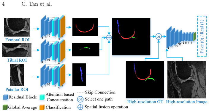
The ROI-fusion layer F(Af, At, Ap) is the load-bearing component: it takes the three agents' binary cartilage masks, uses the recorded ROI coordinates to fuse them back into the original knee joint space as a single multi-cartilage label, and then backpropagates the adversarial discriminator's loss to each individual agent. This lets the agents be trained both on their own fine-grained ROI (low class imbalance) and on the joint label (shape/spatial constraints) simultaneously. Attention-based skip connections in each agent (a learned single-channel mask weighting the low-level features before concatenation) support thin-structure, unclear-boundary segmentation.
What would settle it
Run the proposed pipeline on a set of knee MR volumes with large focal cartilage defects or severe atrophy and check whether the coarse segmentor (C0) still produces a mask that overlaps each GT cartilage; if any true-positive cartilage voxel is entirely outside the extracted ROI, the final segmentation cannot recover it and the claimed reliability fails on those cases.
Extended reading notes
Core claim
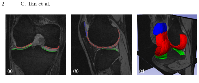
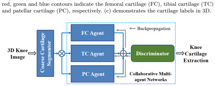
The paper claims that a collaborative multi-agent segmentation network, where each agent segments one cartilage within a tightly cropped ROI and an adversarial sub-network judges the fused multi-cartilage label against the ground truth, produces the most accurate delineation of all three knee cartilages compared to the evaluated alternatives. The ROI-fusion layer is the key mechanism: it reassembles the individual binary masks into a full multi-label volume and, during backpropagation, passes the discriminator's loss back to every agent, forcing the agents to jointly respect inter-cartilage position and shape priors. In the authors' experiments on 176 OAI MR knee volumes, this full pipeline (P2) achieves the best Dice similarity coefficient, volumetric overlap error, and average surface distance for each cartilage and overall, and the authors conclude it can provide reliable automatic cartilage assessments for knee osteoarthritis analysis.
Load-bearing premise
The coarse segmentor reliably localizes every cartilage into its predetermined ROI, so each agent only ever sees the correct tissue; if a severely damaged or atrophic cartilage is missed by the coarse mask, no agent can recover it.
Editorial extensions
If this is right
- Automatic, reliable cartilage segmentation becomes feasible on high-resolution 3D MR data with limited GPU memory, since each agent only processes a small ROI.
- The same collaborative multi-agent pattern—ROI-wise agents plus adversarial joint-label learning—can be transferred to other multi-organ or multi-structure segmentation tasks where inter-object spatial constraints matter.
- Comparing P2 with P1 shows that the adversarial collaboration step, not just the ROI cropping or the attention skips, is what brings the accuracy gain for all three cartilages.
- The reported low average surface distances suggest the method could support downstream quantitative biomarkers such as cartilage thickness and surface area in OA studies.
Reading between the lines
- The two-stage design makes the final mask's accuracy conditional on the coarse segmentor's recall: a cartilage so damaged or atrophied that the coarse mask misses it will never reach any agent, so the method's clinical value on severe OA cases hinges on the coarse stage never dropping a true positive.
- One could test the framework's generality by applying the same ROI-fusion-plus-adversarial collaboration to other paired thin structures (e.g., hip cartilage, intervertebral discs) where a fixed ROI size and location prior are easy to define.
- The attention-based skip connection is generic, so an ablation that swaps it for plain concatenation inside the same collaborative setup would isolate how much of the gain comes from attention versus from the adversarial collaborator.
Signed reviews
Editorial analysis
A structured set of objections, weighed in public.
Referee Report
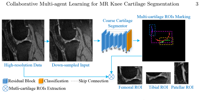
Summary. The paper proposes a coarse-to-fine framework for segmenting femoral, tibial, and patellar cartilage in high-resolution 3D MR knee volumes from the OAI dataset. A coarse VNet-like segmentor localizes each cartilage and extracts fixed-size ROIs; three fine segmentation agents then process the ROIs with attention-augmented skip connections. A ROI-fusion layer reassembles the agent outputs into a multi-label mask in the original volume, and an adversarial discriminator, conditioned on the input image, provides a joint shape and spatial-constraint loss to the agents. Experiments on 176 iMorphics volumes (120/26/30 train/validation/test split) compare the full method (P2), the same agents without the adversarial module (P1), the coarse segmentor (C0), and two variants in which DenseASPP blocks replace parts of the agent network (D1, D2). P2 reports the highest DSC, lowest VOE, and lowest ASD across all cartilage groups.
Significance. If the results hold, the main contribution is a practical and memory-efficient way to segment all three knee cartilages in high-resolution data, with a clear ablation (P2 vs P1) isolating the effect of adversarial collaborative learning. The use of a public dataset, the defined train/validation/test split, and the inclusion of mean/std metrics are strengths. The attention-augmented skip connections and the ROI-fusion design are reasonable and clearly described. However, the broader claims of 'superior performance' against state-of-the-art methods are not yet supported by the experimental design, and the method's dependence on coarse-stage ROI detection is not analyzed. The paper is a solid application contribution but needs revision before the claims match the evidence.
major comments (3)
- [Section 3, Experimental results, Table 1] The comparison against DenseASPP is not a comparison against the original DenseASPP network. D1 and D2 are defined as the proposed agent network with DenseASPP blocks inserted in place of residual blocks/skip connections at certain levels. Therefore the statement in the abstract and conclusions that the proposed method shows 'superior performance' over the state-of-the-art DenseASPP is not established. The authors should either evaluate the original DenseASPP architecture (or a standard VNet with DenseASPP) on the same data and protocol, or rephrase the claim as an ablation of two architectural variations within the proposed framework.
- [Section 2, ROI extraction; Section 3, Experimental settings] The method's upper bound depends on the coarse segmentor detecting each cartilage and producing a ROI that contains the cartilage. If the coarse mask misses a severely atrophic or damaged cartilage, the corresponding agent receives an empty or incomplete ROI and cannot recover the tissue. The paper reports C0 patellar DSC of 0.771 ± 0.132, indicating substantial case-to-case variability in the coarse stage, yet P2 reports patellar DSC of 0.880 ± 0.043. No analysis is given of how many test volumes have incomplete or empty coarse ROIs, whether any ground-truth voxels fall outside the extracted ROIs, or how the fixed ROI sizes were chosen beyond 'adequate evaluation on the training data'. Without this information, the claim that the method 'can be used to automatically generate reliable assessments of all important articular cartilages' is not supported for the pathological cases that matter most.
- [Table 1 and Section 3] The reported improvements of P2 over P1, D1, D2, and C0 are not accompanied by any statistical significance test. With 30 test volumes and standard deviations as large as 0.439 ASD for P1 patellar, the differences may not be statistically significant. The authors should add paired tests (e.g., Wilcoxon signed-rank or paired t-test) on DSC, VOE, and ASD across the 30 test subjects, or report confidence intervals, so that the central claim of improvement from collaborative learning is supported rigorously.
minor comments (6)
- [Section 3, Experimental settings] The downsizing of the input from 384×384×160 to 192×192×160 for the coarse segmentor is mentioned, but the interpolation method and whether the same resolution is used at test time are not specified.
- [Section 2, Collaborative multi-agent learning] The attention skip connection is described as 'o (α⊙Il,I_up_h)' with o denoting concatenation, but the notation could be made cleaner by explicitly writing concat(α⊙Il, I_up_h) and specifying the channel dimensions of α.
- [Equations (1) and (2)] The sums in Eq. (1) and (2) are over i without explicitly stating the domain; the authors should state that i ranges over the training samples in a mini-batch.
- [Table 1] The table layout places mean and standard deviation on separate rows without clear separation, making it hard to read. Consider using '0.900 ± 0.037' format in a single cell.
- [Section 3, Experimental results] The comparison with Xu et al. and Ambellan et al. is indirect because those methods were evaluated on different data splits and protocols. This is acknowledged in part, but the sentence 'the proposed method acquires comparable DSC and VOE scores' should explicitly state that this is an indirect comparison.
- [Section 3, Experimental settings] The fixed ROI sizes are not reported in physical or voxel units, and no justification is given for why these sizes are sufficient to cover the observed anatomical variability across the OAI population.
Circularity Check
No significant circularity: the central segmentation claim is evaluated against external manual labels in the OAI dataset, and no prediction or derived result reduces by construction to its own inputs.
full rationale
The paper's core claim is empirical: a multi-agent segmentation network with attention skip connections, ROI fusion, and adversarial training produces accurate cartilage labels on a held-out test set of the iMorphics/OAI data. The evaluation metrics (DSC, VOE, ASD) are computed against manual ground truth labels that are not used as inputs to the network at test time, so the headline performance comparison is externally grounded rather than self-referential. The only tuning choice tied to training data is the statement that 'Fixed ROI size of each type of cartilage is pre-defined based on adequate evaluation on the training data.' This is ordinary hyperparameter selection and does not by itself force any test-time segmentation result; the fine agents still learn to segment from image intensities within those ROIs. The coarse segmentor used for ROI extraction is also learned and evaluated, and the final P2 predictions are not algebraically or statistically dictated by C0 outputs. The paper cites some prior work by overlapping authors, notably Tan et al. ISBI 2018 and Uzunbas et al. MICCAI 2013, but those citations are contextual literature references rather than load-bearing justifications of the proposed architecture or its uniqueness. No fitted parameter is renamed as a prediction, no uniqueness theorem is imported from the authors' own prior work, and no known result is repackaged under new coordinates. Potential concerns about the coarse segmentor missing atrophic cartilage would be correctness or robustness issues, not circularity, because the claim of reliable assessment is not derived from the assumption that the coarse segmentor always succeeds. Overall, the derivation chain is self-contained with respect to the test evaluation and contains no circular step.
Assumptions & free parameters
free parameters (2)
- ROI sizes for femoral, tibial, and patellar cartilage =
not reported
- Training hyperparameters =
agent LR 0.001, discriminator LR 0.0002, batch size 1, LR decay 0.95 every 10 epochs
assumptions (3)
- domain assumption The manual cartilage labels in the OAI iMorphics dataset are accurate ground truth for metric computation.
- domain assumption The coarse segmentor localizes each cartilage within the fixed ROI.
- domain assumption Adversarial training of the fused mask improves anatomically meaningful segmentation rather than merely fooling the discriminator.
Cite this review
Pith. "Pith review of Collaborative Multi-agent Learning for MR Knee Articular Cartilage Segmentation." pith.science (2026). https://pith.science/paper/6F2FEBUM
@misc{pith2026190804469,
  author       = {Pith},
  title        = {Pith review of: Collaborative Multi-agent Learning for MR Knee Articular Cartilage Segmentation},
  year         = {2026},
  howpublished = {\url{https://pith.science/paper/6F2FEBUM}},
  note         = {Machine review of arXiv:1908.04469}
}
read the original abstract
The 3D morphology and quantitative assessment of knee articular cartilages (i.e., femoral, tibial, and patellar cartilage) in magnetic resonance (MR) imaging is of great importance for knee radiographic osteoarthritis (OA) diagnostic decision making. However, effective and efficient delineation of all the knee articular cartilages in large-sized and high-resolution 3D MR knee data is still an open challenge. In this paper, we propose a novel framework to solve the MR knee cartilage segmentation task. The key contribution is the adversarial learning based collaborative multi-agent segmentation network. In the proposed network, we use three parallel segmentation agents to label cartilages in their respective region of interest (ROI), and then fuse the three cartilages by a novel ROI-fusion layer. The collaborative learning is driven by an adversarial sub-network. The ROI-fusion layer not only fuses the individual cartilages from multiple agents, but also backpropagates the training loss from the adversarial sub-network to each agent to enable joint learning of shape and spatial constraints. Extensive evaluations are conducted on a dataset including hundreds of MR knee volumes with diverse populations, and the proposed method shows superior performance.
Figures
Figures from the paper (3 more)
Reference graph
Works this paper leans on
-
[1]
Ambellan, F., Tack, A., Ehlke, M., Zachow, S.: Automated segmentation of knee bone and cartilage combining statistical shape knowledge and convolutional neural networks: Data from the osteoarthritis initiative. Medical Image Analysis (2018)
work page 2018
-
[2]
Eckstein, F., Wirth, W.: Quantitative cartilage imaging in knee osteoarthritis. Arthritis 2011 (2010)
work page 2010
-
[3]
IEEE transactions on medical imaging 38(2), 585--595 (2019)
He, K., Cao, X., Shi, Y., Nie, D., Gao, Y., Shen, D.: Pelvic organ segmentation using distinctive curve guided fully convolutional networks. IEEE transactions on medical imaging 38(2), 585--595 (2019)
work page 2019
-
[4]
Osteoarthritis and Cartilage 19(8), 990--1002 (2011)
Hunter, D.J., Guermazi, A., Lo, G.H., Grainger, A.J., Conaghan, P.G., Boudreau, R.M., Roemer, F.W.: Evolution of semi-quantitative whole joint assessment of knee OA : MOAKS ( MRI O steoarthritis K nee S core). Osteoarthritis and Cartilage 19(8), 990--1002 (2011)
work page 2011
-
[5]
arXiv preprint arXiv:1804.02391 (2018)
Jetley, S., Lord, N.A., Lee, N., Torr, P.H.: Learn to pay attention. arXiv preprint arXiv:1804.02391 (2018)
arXiv 2018
-
[6]
In: 3D Vision (3DV), 2016 Fourth International Conference on
Milletari, F., Navab, N., Ahmadi, S.A.: V-net: Fully convolutional neural networks for volumetric medical image segmentation. In: 3D Vision (3DV), 2016 Fourth International Conference on. pp. 565--571. IEEE (2016)
work page 2016
- [7]
-
[8]
Uzunba s , M.G., Chen, C., Zhang, S., Pohl, K.M., Li, K., Metaxas, D.: Collaborative multi organ segmentation by integrating deformable and graphical models. In: MICCAI. pp. 157--164. Springer (2013)
work page 2013
Show all 13 references
-
[9]
In: MICCAI
Xu, C., Xu, L., Brahm, G., Zhang, H., Li, S.: Mutgan: Simultaneous segmentation and quantification of myocardial infarction without contrast agents via joint adversarial learning. In: MICCAI. pp. 525--534. Springer (2018)
2018
-
[10]
In: Deep Learning in Medical Image Analysis and Multimodal Learning for Clinical Decision Support, pp
Xu, Z., Shen, Z., Niethammer, M.: Contextual additive networks to efficiently boost 3d image segmentations. In: Deep Learning in Medical Image Analysis and Multimodal Learning for Clinical Decision Support, pp. 92--100. Springer (2018)
2018
-
[11]
In: CVPR
Yang, M., Yu, K., Zhang, C., Li, Z., Yang, K.: Denseaspp for semantic segmentation in street scenes. In: CVPR. pp. 3684--3692 (2018)
2018
-
[12]
, " * write output.state after.block = add.period write
ENTRY address author booktitle chapter doi edition editor eid howpublished institution journal key month note number organization pages publisher school series title type url volume year label INTEGERS output.state before.all mid.sentence after.sentence after.block FUNCTION in...
-
[13]
write newline
" write newline "" before.all 'output.state := FUNCTION n.dashify 't := "" t empty not t #1 #1 substring "-" = t #1 #2 substring "--" = not "--" * t #2 global.max substring 't := t #1 #1 substring "-" = "-" * t #2 global.max substring 't := while if t #1 #1 substring * t #2 gl...
Reviewed August 14, 2026 · model on record in the stance chip above.
Discussion (0). Continue with ORCID to comment.