REVIEW 5 major objections 8 minor 39 references
Deep Learning-Based Quantification of Pulmonary Hemosiderophages in Cytology Slides
T0 review · 5 major / 8 minor · reviewed 2026-08-14 · deepseek-v4-flash
Pith's one-line read Automated deep learning can grade pulmonary hemosiderophages on whole slide cytology images with 85% concordance, matching or exceeding the average human expert while taking under two minutes per slide.
desk verdict A useful applied DL paper with a new dataset and honest limitations, but the 'accuracy' claim is really agreement with one pathologist. read the letter →
The pith
A machine-rendered reading of the paper's core claim, the machinery that carries it, and where it could break.
The reading
What carries the argument
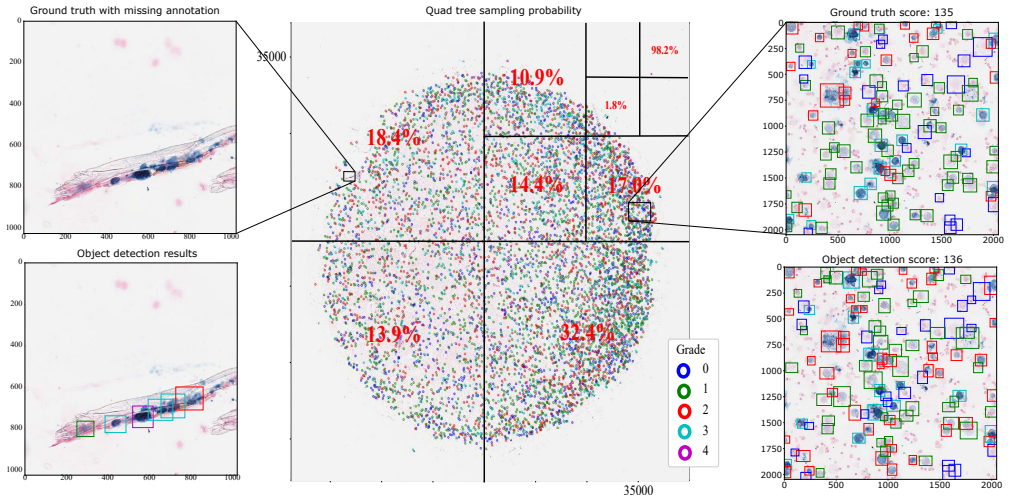
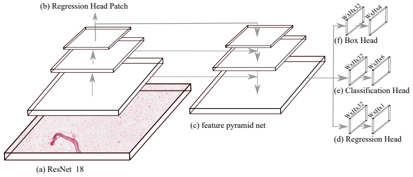
The carrying mechanism is a modified RetinaNet detector: a feature pyramid network built on a ResNet-18 backbone predicts object boxes and classes at multiple scales, and an added regression head predicts a continuous hemosiderin score per cell, while a patch regression head estimates the slide score directly. To train on rare high-grade cells, the paper introduces a quad-tree based patch sampler that allocates sampling probability according to cell density and grade, so grade-3 and grade-4 cells are not starved of training examples. The detector processes whole slides in about 101 seconds on a modern GPU.
What would settle it
Measure the iron content of bronchoalveolar lavage fluid from the same 17 cases by atomic absorption spectroscopy or a similar chemical assay and compare it with the automated Total Hemosiderin Score. A correlation near zero, or a case where the algorithm grades a high-iron sample as low-iron, would show the system measures visual staining patterns rather than the underlying hemorrhage.
Extended reading notes
Core claim
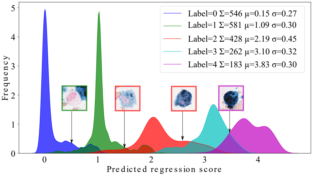
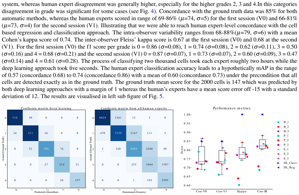
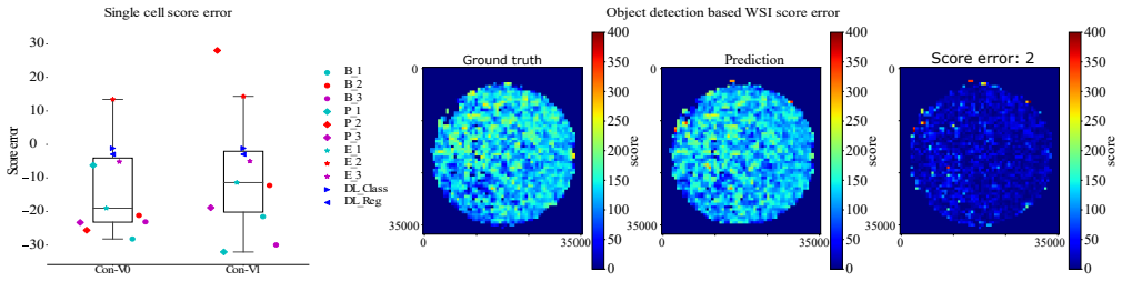
The central claim is that a single end-to-end object detection system, built on RetinaNet with a ResNet-18 backbone and an extra regression head, can both locate and grade hemosiderophages across gigapixel whole slide images. On a new, fully annotated dataset of 17 slides containing 78,047 cells, the system achieves a mean average precision of 0.66 over the five Golde grades and a cell-level concordance of 0.85 with the reference annotation, exceeding the mean human concordance of 0.73. The same pipeline estimates the whole-slide Total Hemosiderin Score with a mean error of about 9 points on a 0-400 scale, compared with 19 for deep regression and 21 for an SVM baseline. The paper also establishes that human grading is highly variable: inter-observer Fleiss' kappa is 0.67 and intra-observer concordance ranges from 0.68 to 0.88, which is why the authors propose automation as a more reliable route to EIPH scoring.
Load-bearing premise
The evaluation assumes that the single veterinary pathologist's annotations are a valid ground truth for grading, even though the paper reports only moderate inter-observer agreement (Fleiss' kappa = 0.67) and no independent gold standard such as a chemical iron measurement exists.
Editorial extensions
If this is right
- EIPH scoring can be fully automated on whole slide images, removing the need for a human to select and grade individual cells; this would make the test faster and cheaper in routine equine practice.
- Because the algorithm is deterministic, repeated runs give identical scores, directly addressing the observed 0.68-0.88 intra-observer variability and 0.67 inter-observer kappa of human raters.
- The system's mAP of 0.66 approaches the estimated human upper bound of 0.74 mAP given the measured concordance levels, so further gains are limited more by label noise than by detector architecture.
- Regression-based grading can reveal within-grade differences in iron load, offering a continuous measure of hemorrhage severity instead of the five discrete Golde grades.
- The released dataset and model provide a basis for building interactive annotation tools that flag regions missed by human experts.
Reading between the lines
- If the reference labels are treated as noisy rather than as truth, the model's 0.85 concordance could understate how well it matches an expert consensus; a panel majority-vote benchmark would be a stronger test.
- The missing independent validation the paper itself notes could be supplied by measuring iron content chemically in the same lavage samples; a strong correlation would confirm that the visual grades track actual hemorrhage.
- The same quad-tree sampling and regression-head detector should transfer to other rare-cell grading tasks, such as human pulmonary hemorrhage cytology, provided external datasets are used.
- Because human baseline variability is high, future comparisons should report agreement against a consensus ground truth rather than a single pathologist's labels.
Signed reviews
Editorial analysis
A structured set of objections, weighed in public.
Referee Report
Summary. This manuscript addresses automated scoring of exercise-induced pulmonary hemorrhage (EIPH) in equine bronchoalveolar lavage cytology whole slide images. The authors introduce a dataset of 17 fully annotated WSIs with 78,047 hemosiderophages annotated by one veterinary pathologist, evaluate nine human experts on single-cell classification with repeated sessions, and compare them with deep-learning classifiers and regressors. They also train RetinaNet-based object detectors with a novel quad-tree sampling strategy to detect and grade cells directly on gigapixel WSIs. The reported single-cell concordance is 0.85 for the deep-learning methods versus 0.68 to 0.86 for human experts, inter-observer Fleiss' kappa is 0.67, and the object detector reaches mAP 0.66 with a mean score error of 9 and inference under two minutes per slide. The paper concludes that the pipeline enables accurate, reproducible, and quick EIPH scoring.
Significance. If the central claim were appropriately bounded, the paper would be a useful contribution to veterinary digital pathology: it provides the largest fully annotated EIPH cytology dataset of which I am aware, a systematic human-variability study for this task, a novel quad-tree sampling strategy, and an open, fast object-detection pipeline. The reproducibility of the code and trained model, together with the quantitative documentation of inter- and intra-observer variability, are clear strengths. The main value is as a benchmark and as evidence that automated systems can match a specific expert's grading style; the claim of diagnostic accuracy is not supported by the evidence and needs revision.
major comments (5)
- [Material; Discussion and Outlook] The reference standard is a single veterinary pathologist's annotation, and the paper itself states that there is no true gold standard such as chemical measurement of iron content. Because the nine experts reach only moderate inter-observer agreement (Fleiss' kappa = 0.67) and intra-observer concordance of 0.68 to 0.88, the reported deep-learning concordance of 0.85 and the detection mAP of 0.66 quantify agreement with that one expert's grading style rather than diagnostic accuracy. The abstract and conclusion claim that the pipeline enables 'accurate' EIPH scoring, which is not supported by the present evidence; this claim must be reframed as agreement with an expert reference, or the model must be validated against an independent gold standard such as clinical outcome or chemical iron quantification.
- [Human Performance Evaluation; Results (Object Detection)] The human comparison is restricted to single-cell classification on pre-extracted cells, whereas the object-detection pipeline performs detection and classification on whole slides. The abstract's statement that the deep-learning approach 'partially exceeding human expert concordance' refers only to the single-cell task, not to the end-to-end WSI scoring task; no human expert scored whole slides, so the mAP of 0.66 and the score error of 9 have no human WSI-level comparator. The paper should either provide such a comparison, for example by having experts score the three test slides at the whole-slide level, or explicitly restrict the superiority claim to single-cell classification.
- [Results (Object Detection)] The key mAP and concordance values are reported without confidence intervals, and the mAP of 0.66 is computed from only three test slides. Given that the upper human concordance is 0.86 and the deep-learning concordance is 0.85, the claim of partially exceeding human performance depends on a difference well within plausible sampling variability. The paper should report per-slide results, confidence intervals, and ideally a statistical comparison with the human observers.
- [Object Detection Evaluation; Fig. 1] The ground truth is known to be incomplete: Fig. 1 shows a region missed by the human annotator but detected by RetinaNet. Consequently the reported mAP of 0.66 is a conservative lower bound, because some false-positive detections may be true hemosiderophages absent from the reference. The paper acknowledges this qualitatively but should quantify its potential impact, for example by estimating missed-cell prevalence on a small re-annotated subset or by reporting the sensitivity of the conclusions to label noise.
- [Results (Cell Classification)] The derived 'hypothetical mAP' of 0.57 to 0.74 for human experts assumes perfect detection and converts single-cell classification concordance into a detection metric. This is not a measured human detection baseline and should not be presented as one; the conversion needs a derivation or should be removed.
minor comments (8)
- [Abstract] The word 'Resultsf' is a typo and should read 'Results'.
- [Discussion and Outlook] The text contains typos including 'Unfortunatelly', 'Scince', and 'variablity'; these should be corrected.
- [Abstract; Methods] The acronym for the single-cell task is inconsistent: 'CoSH' appears in the abstract while 'CoCH' appears in Methods and Discussion; use one acronym throughout.
- [Methods, Eq. (1)] Equation (1) appears to contain the regression loss term twice with identical notation; clarify that one term corresponds to the cell regression head and the other to the patch regression head, and define c_i and ĉ_i separately for each head.
- [Table 2] The table caption says 'results per WSI', but the table rows are architectures rather than slides; provide a per-slide breakdown or change the caption accordingly.
- [Methods (Support vector machine)] The phrase 'a convexity value of 0.1' is ambiguous; this is presumably the RBF cost parameter C and should be named and defined.
- [Methods (Single Cell Classification)] The term 'maximal learning rate schedule' should be explained, for example by specifying whether a one-cycle policy or a similar schedule was used, so that the training protocol is reproducible.
- [Results (Cell Classification)] The concordance metric should be defined explicitly, and results should be reported separately for test set I and test set II because the two sets have different class distributions.
Circularity Check
No circularity: the deep-learning results are evaluated on held-out test slides, and the acknowledged single-pathologist ground-truth caveat is a validity limitation rather than a derivation loop.
full rationale
The claimed predictions—single-cell classification/regression concordance and whole-slide object-detection mAP—are obtained by training supervised models on one set of slides and evaluating on three separate hold-out test slides. No parameter is fitted to the reported test metrics, and the concordance/mAP numbers are empirical generalization results, not identities derivable from training labels. The main caveat is that all ground-truth annotations were made by a single veterinary pathologist (Discussion: 'all annotations were made by a single veterinary pathologist'; Material: 'the slides were completely annotated and scored by a veterinary pathologist'), and the paper explicitly notes the absence of a chemical gold standard. This limits what 'accuracy' means diagnostically—agreement with one expert's grading rather than with an independent truth—but this is a study-design/validity limitation, not a circular derivation: the model's outputs are not defined in terms of the evaluation labels, and the evaluation is on held-out data. The self-citations (SlideRunner [33], prior mitotic-count work [16,39]) are tooling or supportive context, not load-bearing premises that force the conclusions. Therefore no circularity step meeting the quoted-evidence standard is present.
Assumptions & free parameters
free parameters (5)
- Focal loss parameters (gamma, alpha_t) =
not specified
- Scaled sigmoid output range =
-0.5 to 4.5
- SVM RBF kernel parameters =
C=0.1 (convexity value), RBF kernel
- Quad-tree sampling depth and minimum cells per node =
depth=3, min 300 cells/node
- Learning rates and epochs =
0.01/0.001, 3+20 or 100 epochs
assumptions (4)
- domain assumption The Golde/Doucet hemosiderin scoring system is a valid ordinal measure of pulmonary hemorrhage severity.
- domain assumption Prussian Blue and Turnbull's Blue staining produce identical color patterns in the scanned images.
- domain assumption The single veterinary pathologist's annotations are reliable enough to serve as ground truth for training and evaluation.
- domain assumption The WSI annotations are complete (no missed cells).
Cite this review
Pith. "Pith review of Deep Learning-Based Quantification of Pulmonary Hemosiderophages in Cytology Slides." pith.science (2026). https://pith.science/paper/YV3GOUEB
@misc{pith2026190804767,
  author       = {Pith},
  title        = {Pith review of: Deep Learning-Based Quantification of Pulmonary Hemosiderophages in Cytology Slides},
  year         = {2026},
  howpublished = {\url{https://pith.science/paper/YV3GOUEB}},
  note         = {Machine review of arXiv:1908.04767}
}
abstract
Purpose: Exercise-induced pulmonary hemorrhage (EIPH) is a common syndrome in sport horses with negative impact on performance. Cytology of bronchoalveolar lavage fluid by use of a scoring system is considered the most sensitive diagnostic method. Macrophages are classified depending on the degree of cytoplasmic hemosiderin content. The current gold standard is manual grading, which is however monotonous and time-consuming. Methods: We evaluated state-of-the-art deep learning-based methods for single cell macrophage classification and compared them against the performance of nine cytology experts and evaluated inter- and intra-observer variability. Additionally, we evaluated object detection methods on a novel data set of 17 completely annotated cytology whole slide images (WSI) containing 78,047 hemosiderophages. Resultsf: Our deep learning-based approach reached a concordance of 0.85, partially exceeding human expert concordance (0.68 to 0.86, $\mu$=0.73, $\sigma$ =0.04). Intra-observer variability was high (0.68 to 0.88) and inter-observer concordance was moderate (Fleiss kappa = 0.67). Our object detection approach has a mean average precision of 0.66 over the five classes from the whole slide gigapixel image and a computation time of below two minutes. Conclusion: To mitigate the high inter- and intra-rater variability, we propose our automated object detection pipeline, enabling accurate, reproducible and quick EIPH scoring in WSI.
Figures
Figures from the paper (2 more)
Reference graph
Works this paper leans on
-
[1]
Ahmad, K. A., Bennett, M. M., Ahmad, S. F., Clark, R. H. & Tolia, V . N. Morbidity and mortality with early pulmonary haemorrhage in preterm neonates. Arch. Dis. Childhood-Fetal Neonatal Ed. 104, F63–F68 (2019)
work page 2019
-
[2]
Maldonado, F., Parambil, J. G., Yi, E., Decker, P. A. & Ryu, J. H. Haemosiderin-laden macrophages in the bronchoalveolar lavage fluid of patients with diffuse alveolar damage. Eur. Respir. J. 33, 1361–1366 (2009)
work page 2009
-
[3]
van Houten, J. et al. Pulmonary hemorrhage in premature infants after treatment with synthetic surfactant: an autopsy evaluation. The J. Pediatr. 120, S40–S44 (1992)
work page 1992
-
[4]
Golde, D. W., Drew, W. L., Klein, H. Z., Finley, T. N. & Cline, M. J. Occult pulmonary haemorrhage in leukaemia.Br Med J 2, 166–168 (1975)
work page 1975
-
[5]
Martínez-Martínez, M. U., Herrera-van Oostdam, D. A. & Abud-Mendoza, C. Diffuse alveolar hemorrhage in autoimmune diseases. Curr. rheumatology reports 19, 27 (2017)
work page 2017
-
[6]
Kahn, F. W., Jones, J. M. & England, D. M. Diagnosis of pulmonary hemorrhage in the immunocompromised host. Am. Rev. Respir. Dis. 136, 155–160 (1987)
work page 1987
-
[7]
Morley, P. S., Bromberek, J., Saulez, M. N., Hinchcliff, K. W. & Guthrie, A. J. Exercise-induced pulmonary haemorrhage impairs racing performance in t horoughbred racehorses. Equine V et. J.47, 358–365 (2015)
work page 2015
-
[8]
Hinchcliff, K. W. et al. Association between exercise-induced pulmonary hemorrhage and performance in thoroughbred racehorses. J. Am. V et. Med. Assoc. 227, 768–774 (2005)
work page 2005
Show all 39 references
-
[9]
K., Durando, M
Birks, E. K., Durando, M. M. & McBride, S. Exercise-induced pulmonary hemorrhage. V et. Clin. Equine Pract. 19, 87–100 (2003)
2003
-
[10]
Hinchcliff, K. et al. Exercise induced pulmonary hemorrhage in horses: American college of veterinary internal medicine consensus statement. J. V et. Intern. Med.29, 743–758 (2015)
2015
-
[11]
Hoffman, A. M. Bronchoalveolar lavage: sampling technique and guidelines for cytologic preparation and interpretation. V et. Clin. North Am. Equine Pract. 24, 423–435 (2008)
2008
-
[12]
Depecker, M. et al. Comparison of two cytological methods for detecting pulmonary haemorrhage in horses. V et. Rec.177, 305–305 (2015)
2015
-
[13]
& Seller, W
Denk, H., Künzele, H., Plenk, H., Rüschoff, J. & Seller, W. Romeis mikroskopische technik. Urban und Schwarzenberg, München-Wien. Baltim. 439–450 (1989)
1989
-
[14]
Waithe, D. et al. Object detection networks and augmented reality for cellular detection in fluorescence microscopy acquisition and analysis. bioRxiv 544833 (2019)
2019
-
[15]
E., Ersoz, S
Baykal, E., Dogan, H., Ercin, M. E., Ersoz, S. & Ekinci, M. Modern convolutional object detectors for nuclei detection on pleural effusion cytology images. Multimed. Tools Appl. 1–20 (2019). 10/12
2019
-
[16]
& Maier, A
Aubreville, M., Bertram, C., Klopfleisch, R. & Maier, A. Field Of Interest Proposal for Augmented Mitotic Cell Count: A Comparison of Two Networks. In SciTePress (ed.) Proceedings of the 12th International Joint Conference on Biomedical Engineering Systems and Technologies - V ...
2019 doi
-
[17]
Lowe, D. G. et al. Object recognition from local scale-invariant features. In ICCV, vol. 99, 1150–1157 (1999)
1999
-
[18]
& Harwood, D
Ojala, T., Pietikäinen, M. & Harwood, D. A comparative study of texture measures with classification based on featured distributions. PATTERN RECOGN 29, 51–59 (1996)
1996
-
[19]
& Triggs, B
Dalal, N. & Triggs, B. Histograms of oriented gradients for human detection. In CVPR, vol. 1, 886–893 (IEEE Computer Society, 2005)
2005
-
[20]
& Riess, C
Maier, A., Syben, C., Lasser, T. & Riess, C. A gentle introduction to deep learning in medical image processing. Zeitschrift für Medizinische Physik 29, 86–101 (2019)
2019
-
[21]
& Sun, J
Ren, S., He, K., Girshick, R. & Sun, J. Faster r-cnn: Towards real-time object detection with region proposal networks. In Adv Neural Inf Process Syst , 91–99 (2015)
2015
-
[22]
Liu, W. et al. Ssd: Single shot multibox detector. In ECCV, 21–37 (Springer, 2016)
2016
-
[23]
& Dollár, P
Lin, T.-Y ., Goyal, P., Girshick, R., He, K. & Dollár, P. Focal loss for dense object detection. InICCV, 2980–2988 (2017)
2017
-
[24]
K., Winn, J
Everingham, M., Van Gool, L., Williams, C. K., Winn, J. & Zisserman, A. The pascal visual object classes (voc) challenge. Int. journal computer vision 88, 303–338 (2010)
2010
-
[25]
Microsoft coco: Common objects in context
Lin, T.-Y .et al. Microsoft coco: Common objects in context. In ECCV, 740–755 (Springer, 2014)
2014
-
[26]
Zou, Z., Shi, Z., Guo, Y . & Ye, J. Object detection in 20 years: A survey.arXiv preprint arXiv:1905.05055 (2019)
2019 arXiv
-
[27]
N., Konjevod, G., Sakla, W
Mundhenk, T. N., Konjevod, G., Sakla, W. A. & Boakye, K. A large contextual dataset for classification, detection and counting of cars with deep learning. In ICCV, 785–800 (Springer, 2016)
2016
-
[28]
Litjens, G. et al. A survey on deep learning in medical image analysis. Med Imag Anal 42, 60–88 (2017)
2017
-
[29]
& Brox, T
Ronneberger, O., Fischer, P. & Brox, T. U-net: Convolutional networks for biomedical image segmentation. In Med Image Comput Comput Assist Interv , 234–241 (Springer, 2015)
2015
-
[30]
Ferlaino, M. et al. Towards deep cellular phenotyping in placental histology. arXiv preprint arXiv:1804.03270 (2018)
2018 arXiv
-
[31]
& Farhadi, A
Redmon, J., Divvala, S., Girshick, R. & Farhadi, A. You only look once: Unified, real-time object detection. In Proc IEEE Comput Soc Conf Comput Vis Pattern Recognit , 779–788 (2016)
2016
-
[32]
Huang, J. et al. Speed/accuracy trade-offs for modern convolutional object detectors. In Proc IEEE Comput Soc Conf Comput Vis Pattern Recognit, 7310–7311 (2017)
2017
-
[33]
& Maier, A
Aubreville, M., Bertram, C., Klopfleisch, R. & Maier, A. Sliderunner. In Bildverarbeitung für die Medizin 2018 , 309–314 (Springer, 2018)
2018
-
[34]
Doucet, M. Y . & Viel, L. Alveolar macrophage graded hemosiderin score from bronchoalveolar lavage in horses with exercise-induced pulmonary hemorrhage and controls. J V et Intern Med 16, 281–286 (2002)
2002
-
[35]
& Sun, J
He, K., Zhang, X., Ren, S. & Sun, J. Deep Residual Learning for Image Recognition. In CVPR, 770–778 (IEEE, 2016)
2016
-
[36]
Russakovsky, O. et al. Imagenet large scale visual recognition challenge. Int J Comput. Vis 115, 211–252 (2015)
2015
-
[37]
Feature pyramid networks for object detection
Lin, T.-Y .et al. Feature pyramid networks for object detection. In Proc IEEE Comput Soc Conf Comput Vis Pattern Recognit, 2117–2125 (2017)
2017
-
[38]
Paszke, A. et al. Automatic differentiation in PyTorch. In NIPS Autodiff Workshop (2017)
2017
-
[39]
Bertram, C. A. et al. Computerized calculation of mitotic distribution in canine cutaneous mast cell tumor sections: Mitotic count is area dependent. V et. Pathol.(2019). (in press). Acknowledgements C. B. gratefully acknowledges financial support received from the Dres. Jutta ...
2019
Reviewed August 14, 2026 · model on record in the stance chip above.
Discussion (0). Continue with ORCID to comment.