REVIEW 4 major objections 6 minor 1 references
Direct visualization of charge transport in suspended (or free-standing) DNA strands by low-energy electron microscopy
T0 review · 4 major / 6 minor · reviewed 2026-08-14 · deepseek-v4-flash
Pith's one-line read Low-energy electron microscopy reveals correlated charge redistribution in suspended DNA strands.
desk verdict A plausible new imaging observation undermined by an unexcluded mechanical-motion alternative; still deserves peer review to force the right controls. read the letter →
The pith
A machine-rendered reading of the paper's core claim, the machinery that carries it, and where it could break.
The reading
What carries the argument
The central object is the in-line low-energy electron hologram formed when electrons from a sharp field-emission tip (30–250 eV) pass through a suspended sample and interfere on a detector. Its charge sensitivity comes from the deflection of low-energy electron trajectories by local electric potentials: negative charge repels electrons and appears dark, positive charge attracts them and acts like a lens, creating bright regions and apparent bending of neighboring fibers (a biprism effect). The authors simulate this effect with a linear-charge model of the fiber to calibrate the magnitude of charge, and they use the cross-correlation function of normalized intensities at two sub-regions as the diagnostic: a dip at zero lag signals an anti-correlated time shift, interpreted as correlated charge redistribution.
What would settle it
Record holograms of the same suspended DNA network while holding the sample and electron source rigidly fixed, for example with a feedback-stabilized stage, and measure whether the intensity fluctuations persist or vanish. If they persist under rigid fixation, the charge interpretation survives; if they disappear or change character when mechanical vibration is damped, the fluctuations are motion artifacts. Alternatively, image an uncharged suspended fiber of the same geometry, such as a bare carbon nanotube, under identical conditions: if it shows the same intensity fluctuations, the effect is not specific to DNA charge.
Extended reading notes
Core claim
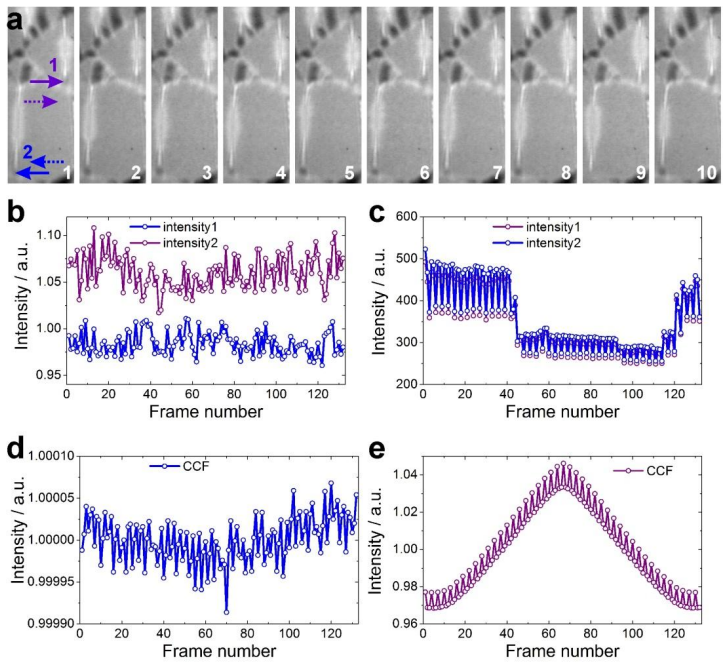
The central claim is that low-energy electron holography provides simultaneous imaging of macromolecular structure and its charge distribution, and that the charge redistribution in a suspended DNA network is not random but correlated. Specifically, the paper shows that a charged ssDNA fiber distorts the electron wavefront enough to visibly shift the holographic image of neighboring fibers by tens of nanometers, and that these distortions correspond to linear charges of order 0.05–0.1 e/nm. Tracking two sub-regions of a DNA network over 132 video-rate frames, the cross-correlation function of their normalized intensities shows a minimum at zero lag, indicating that the two intensity signals are shifted in time, as if the charges alternate between the regions in a coordinated way. The authors conclude that low-energy electron microscopy can visualize charge redistribution within DNA.
Load-bearing premise
The load-bearing premise is that the time-varying intensity fluctuations seen along the DNA fibers are caused by electric charge on the DNA, not by mechanical motion of the suspended fibers or by drift of the electron source relative to the sample; the carbon-only control regions do not eliminate this possibility because they sit on a stiffer, different substrate.
Editorial extensions
If this is right
- Low-energy electron holography can image the structure of a suspended biomolecule and the distribution of charge on it in the same measurement, at about 1 nm spatial resolution and sub-elementary charge sensitivity.
- The observed correlated intensity fluctuations imply that charge redistribution across a DNA network is coordinated over distances of tens of nanometers, rather than being a purely random local process.
- The same holographic distortions that bend the apparent positions of neighboring fibers provide a way to detect linear charges of order 0.05–0.1 e/nm on an individual fiber.
- Because the method works with electron doses far above the limits for high-energy electrons, it can record long time series (video-rate movies) of the same molecule without destroying it.
Reading between the lines
- If the antiphase cross-correlation reflects a genuine oscillation of charge between two DNA regions, then the technique could measure charge-transfer rates in single molecules; varying the DNA length or sequence in future experiments would test whether the oscillation period depends on molecular structure.
- The paper leaves open whether the correlated signal is internally generated by the DNA or driven by the electron beam charging the carbon support; imaging the same DNA network on a different support, such as a graphene membrane, would isolate the driver.
- The pronounced image 'bending' at 0.1 e/nm implies that low-energy electron holography could serve as a quantitative single-molecule electrometer, a use the authors mention only indirectly.
- A direct comparison of single-stranded and double-stranded DNA under identical imaging conditions would link these observations to the conflicting reports on DNA conductivity, potentially resolving whether the correlated fluctuations are a general property of charged biopolymers.
Signed reviews
Editorial analysis
A structured set of objections, weighed in public.
Referee Report
Summary. The manuscript reports low-energy electron holography of osmium-labeled single-stranded DNA strands stretched over holes in lacey carbon, claiming simultaneous structural imaging at about 1 nm resolution and detection of charge densities below one elementary charge per nanometer. The authors further claim that cross-correlation analysis of intensity time series in two sub-regions of a DNA network reveals correlated charge redistribution, which they interpret as evidence of charge transport. The evidence consists of time-dependent holograms, simulations of the biprism effect for charged fibers, and numerical reconstructions of fiber morphology.
Significance. If the central claim were established, the method would be significant: it would offer a non-destructive probe combining structural imaging with sub-elementary-charge sensitivity on individual biomolecules, with potential applications in molecular electronics and biosensing. The reported imaging of DNA with low-energy electrons and the explicit modeling of biprism distortions are useful contributions. However, the central charge-transport claim is not quantitatively supported in the present manuscript: the stated charge values are not derived from the data, and the interpretation of intensity fluctuations as charge redistribution is not distinguished from mechanical motion of the suspended fibers. As written, the paper's main new conclusion is therefore not demonstrated.
major comments (4)
- [Results, 'Quantitative estimation of charges' (Fig. 2(c)–(e))] The abstract claims simultaneous imaging of charging 'of the order of less than one elementary charge per nanometer,' but the manuscript does not show how this value is obtained from the experimental data. The simulations in Fig. 2(c)–(e) assume linear charges of 0.05 and 0.1 e/nm and illustrate that such charges displace holographic intensity extrema; no fit, calibration, or uncertainty analysis connects the experimental holograms to a charge value. The quantitative charge claim is therefore not evidenced.
- [Results, 'Correlated charge redistribution' (Fig. 3)] The central claim that charge redistribution between two DNA regions is correlated rests entirely on interpreting time-dependent intensity fluctuations in fixed 27×27 nm² sub-regions as charge dynamics. The paper itself states that a 1 Å lateral source/sample shift displaces the hologram by about 10 µm and that the suspended fibers are not mechanically rigid and deflect axially, changing the magnification. Such motion would move holographic fringes across the detector, producing intensity fluctuations in fixed sub-regions and an anti-correlated pattern between nearby sub-regions that can generate a cross-correlation minimum at zero lag. The carbon-only control is on the lacey carbon support rather than on suspended DNA spanning holes, so it does not rule out mechanical motion of the suspended strands. The correlated charge redistribution claim is therefore unsupported as presented.
- [Results, 'Visualisation of charge redistribution' and Fig. 4(a)] The interpretation of bent fiber images and of three fibers 'merging' into a bright spot as charge-induced trajectory distortions assumes that the fibers themselves remain fixed. Because the text attributes the blurry appearance to continuous lateral and axial mechanical variations of the source and sample, physical bending or drift of the non-rigid fibers would produce the same image distortions. The simulations of fixed charged fibers are not quantitatively compared with the experimental images, so they do not exclude a mechanical origin for the observed distortions. This ambiguity undermines the claim of direct visualization of charge distribution.
- [Fig. 3(d)–(e) and cross-correlation analysis] The cross-correlation analysis contains no significance testing or error bars. The 132-frame sequence is short and likely nonstationary given the admitted mechanical drift; the difference between the CCF and the reference CCF is described qualitatively as 'remarkable' without a statistical measure. A quantitative test (e.g., comparison with shuffled or simulated motion-corrupted intensity series) is needed to establish that the CCF minimum at zero lag is not a motion artifact.
minor comments (6)
- [Supplementary Information] The radiation-dose equation in the Supplementary Information is garbled, making the conversion to 4.58×10^11 Gray difficult to verify; please provide a clearly typeset derivation with all units.
- [Fig. 2] The text refers to 'intensity profiles ... shown in Fig 2(d)', while the caption labels the intensity profiles as panel (e); the panel numbering should be reconciled.
- [Abstract and Introduction] The phrase 'less than one elementary charge per nanometer' should explicitly distinguish the assumed simulation parameter from a measured experimental quantity; as written it is ambiguous.
- [Results, 'DNA molecules structure reconstruction'] The measured fiber widths of 4.57±0.51 nm and 6.60±0.51 nm indicate bundles rather than individual ssDNA molecules; this should be reconciled with the abstract's statement about imaging individual DNA molecules.
- [Fig. S1 caption] The figure caption cites 'Copyright (2019)' for a 2009 article; the copyright year appears to be a typo.
- [Results, general] The claimed resolution of about 1 nm is not demonstrated by a resolution metric such as Fourier ring correlation or an edge-profile measurement; a quantitative resolution estimate would strengthen the structural claims.
Circularity Check
No circular derivation: the correlated charge redistribution claim is an empirical inference from measured intensity fluctuations, not a fitted parameter or a self-citation chain.
full rationale
The central claim is an interpretation of time-dependent intensity fluctuations in measured holograms: the cross-correlation of normalized intensities at two sub-regions shows a minimum at zero lag, which the authors read as a time shift and hence correlated charge redistribution. This is an empirical inference, not a quantity defined in terms of itself. The simulation of charged fibers uses illustrative linear charge values (0.1 e/nm and 0.05 e/nm), explicitly not fitted to the data, so it does not smuggle the conclusion into the analysis. The reconstruction algorithm (ref 37) and the biprism charge model (ref 25) are methodological tools, and the graphene charge-imaging citations (refs 28 and 29) are prior experimental demonstrations by the same group rather than a derivation that assumes the present conclusion. The mechanical-motion alternative raised by the skeptic is a validity threat, not a circular reduction: the paper admits source/sample shifts and fiber deflection cause blurring, but admitting a confound is not the same as defining the prediction in terms of the input. No equation in the paper reduces the claimed charge correlation to a fitted parameter, and the conclusion is not forced by a self-citation chain. The paper's reasoning chain is therefore self-contained in the circularity sense, whatever its experimental robustness.
Assumptions & free parameters
free parameters (1)
- Linear charge Q per fiber in simulations =
0.05 e/nm and 0.1 e/nm (illustrative, not fitted)
assumptions (5)
- domain assumption Low-energy electrons in the 30-250 eV range cause no significant radiation damage to DNA, allowing long exposure imaging.
- domain assumption The biprism phase-shift model of a charged fiber, as described in ref 25, accurately describes the effect of small charges on low-energy electron holograms.
- ad hoc to paper Intensity fluctuations in holograms of suspended DNA reflect charge redistribution rather than mechanical motion of the sample.
- standard math The hologram reconstruction algorithm of ref 37 produces reliable sample distributions for the experimental parameters used.
- domain assumption The two reference sub-regions used for CCF_ref provide a valid baseline that is independent of the charge signal.
Cite this review
Pith. "Pith review of Direct visualization of charge transport in suspended (or free-standing) DNA strands by low-energy electron microscopy." pith.science (2026). https://pith.science/paper/C63MR27L
@misc{pith2026190809374,
  author       = {Pith},
  title        = {Pith review of: Direct visualization of charge transport in suspended (or free-standing) DNA strands by low-energy electron microscopy},
  year         = {2026},
  howpublished = {\url{https://pith.science/paper/C63MR27L}},
  note         = {Machine review of arXiv:1908.09374}
}
read the original abstract
Low-energy electrons offer a unique possibility for long exposure imaging of individual biomolecules without significant radiation damage. In addition, low-energy electrons exhibit high sensitivity to local potentials and thus can be employed for imaging charges as small as a fraction of one elementary charge. The combination of these properties makes low-energy electrons an exciting tool for imaging charge transport in individual biomolecules. Here we demonstrate the imaging of individual deoxyribonucleic acid (DNA) molecules at the resolution of about 1 nm with simultaneous imaging of the charging of the DNA molecules that is of the order of less than one elementary charge per nanometer. The cross-correlation analysis performed on different sections of the DNA network reveals that the charge redistribution between the two regions is correlated. Thus, low-energy electron microscopy is capable to provide simultaneous imaging of macromolecular structure and its charge distribution which can be beneficial for imaging and constructing nano-bio-sensors.
Figures
Reference graph
Works this paper leans on
-
[1]
Cryoprotection of protein crystals against radiation-damage in electron and x- ray dffraction
1 Henderson, R. Cryoprotection of protein crystals against radiation-damage in electron and x- ray dffraction. Proc. R. Soc. B 241, 6–8 (1990). 2 Germann, M., Latychevskaia, T., Escher, C. & Fink, H.-W. Nondestructive imaging of individual biomolecules. Phys. Rev. Lett. 104, 095501 (2010). 3 Howells, M. R. et al. An assessment of the resolution limitation...
work page 1990
Reviewed August 14, 2026 · model on record in the stance chip above.
Discussion (0). Continue with ORCID to comment.